Mast cell-cholinergic nerve interaction in mouse airways
- PMID: 19403609
- PMCID: PMC2727042
- DOI: 10.1113/jphysiol.2009.173054
Mast cell-cholinergic nerve interaction in mouse airways
Abstract
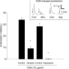
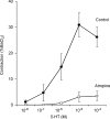
We addressed the mechanism by which antigen contracts trachea isolated from actively sensitized mice. Trachea were isolated from mice (C57BL/6J) that had been actively sensitized to ovalbumin (OVA). OVA (10 microg ml(-1)) caused histamine release (approximately total tissue content), and smooth muscle contraction that was rapid in onset and short-lived (t(1/2) < 1 min), reaching approximately 25% of the maximum tissue response. OVA contraction was mimicked by 5-HT, and responses to both OVA and 5-HT were sensitive to 10 microm-ketanserin (5-HT(2) receptor antagonist) and strongly inhibited by atropine (1microm). Epithelial denudation had no effect on the OVA-induced contraction. Histological assessment revealed about five mast cells/tracheal section the vast majority of which contained 5-HT. There were virtually no mast cells in the mast cell-deficient (sash -/-) mouse trachea. OVA failed to elicit histamine release or contractile responses in trachea isolated from sensitized mast cell-deficient (sash -/-) mice. Intracellular recordings of the membrane potential of parasympathetic neurons in mouse tracheal ganglia revealed a ketanserin-sensitive 5-HT-induced depolarization and similar depolarization in response to OVA challenge. These data support the hypothesis that antigen-induced contraction of mouse trachea is epithelium-independent, and requires mast cell-derived 5-HT to activate 5-HT(2) receptors on parasympathetic cholinergic neurons. This leads to acetylcholine release from nerve terminals, and airway smooth muscle contraction.
Figures


Similar articles
-
Modulation by 5-HT1A receptors of the 5-HT2 receptor-mediated tachykinin-induced contraction of the rat trachea in vitro.Br J Pharmacol. 1998 Apr;123(8):1571-8. doi: 10.1038/sj.bjp.0701771. Br J Pharmacol. 1998. PMID: 9605563 Free PMC article.
-
Non-adrenergic non-cholinergic excitatory innervation in the airways: role of neurokinin-2 receptors.Auton Autacoid Pharmacol. 2002 Aug;22(4):215-24. doi: 10.1046/j.1474-8673.2002.00262.x. Auton Autacoid Pharmacol. 2002. PMID: 12656947
-
The lack of compound 48/80-induced contraction in isolated trachea of mast cell-deficient Ws/Ws rats in vitro: the role of connective tissue mast cells.Eur J Pharmacol. 2000 Aug 25;402(3):297-306. doi: 10.1016/s0014-2999(00)00482-9. Eur J Pharmacol. 2000. PMID: 10958897
-
Potentiation by endothelin-1 of cholinergic nerve-mediated contractions in mouse trachea via activation of ETB receptors.Br J Pharmacol. 1995 Feb;114(3):563-9. doi: 10.1111/j.1476-5381.1995.tb17176.x. Br J Pharmacol. 1995. PMID: 7735683 Free PMC article.
-
Do mucosal mast cells contribute to the immediate asthma response?Jpn J Pharmacol. 2001 May;86(1):38-46. doi: 10.1254/jjp.86.38. Jpn J Pharmacol. 2001. PMID: 11430471
Cited by
-
Mast Cells and Their Progenitors in Allergic Asthma.Front Immunol. 2019 May 29;10:821. doi: 10.3389/fimmu.2019.00821. eCollection 2019. Front Immunol. 2019. PMID: 31191511 Free PMC article. Review.
-
Neural pathways in allergic inflammation.J Allergy (Cairo). 2010;2010:491928. doi: 10.1155/2010/491928. Epub 2011 Feb 9. J Allergy (Cairo). 2010. PMID: 21331366 Free PMC article.
-
House Dust Mite-Induced Allergic Airway Disease Is Independent of IgE and FcεRIα.Am J Respir Cell Mol Biol. 2017 Dec;57(6):674-682. doi: 10.1165/rcmb.2016-0356OC. Am J Respir Cell Mol Biol. 2017. PMID: 28700253 Free PMC article.
-
Mrgprs on vagal sensory neurons contribute to bronchoconstriction and airway hyper-responsiveness.Nat Neurosci. 2018 Mar;21(3):324-328. doi: 10.1038/s41593-018-0074-8. Epub 2018 Feb 5. Nat Neurosci. 2018. PMID: 29403029 Free PMC article.
-
Synaptic and membrane properties of parasympathetic ganglionic neurons innervating mouse trachea and bronchi.Am J Physiol Lung Cell Mol Physiol. 2010 Apr;298(4):L593-9. doi: 10.1152/ajplung.00386.2009. Epub 2010 Jan 29. Am J Physiol Lung Cell Mol Physiol. 2010. PMID: 20118300 Free PMC article.
References
-
- Adams GK, 3rd, Lichtenstein L. In vitro studies of antigen-induced bronchospasm: effect of antihistamine and SRS-A antagonist on response of sensitized guinea pig and human airways to antigen. J Immunol. 1979;122:555–562. - PubMed
-
- Aitken MM, Deline TR, Eyre P. Influence of the method of sensitization on some features of anaphylaxis in calves. J Comp Pathol. 1975;85:351–360. - PubMed
-
- Bjorck T, Dahlen SE. Leukotrienes and histamine mediate IgE-dependent contractions of human bronchi: pharmacological evidence obtained with tissues from asthmatic and non-asthmatic subjects. Pulm Pharmacol. 1993;6:87–96. - PubMed
-
- Chand N, Eyre P. Pharmacological study of chicken airway smooth muscle. J Pharm Pharmacol. 1978;30:432–435. - PubMed
-
- Chand N, Eyre P. Reactivity of isolated canine bronchus and pulmonary blood vessels to autonomic, autacoid agents and antigen. Agents Actions. 1979;9:4–8. - PubMed
Publication types
MeSH terms
Substances
LinkOut - more resources
Full Text Sources
Molecular Biology Databases

