. 2009 Jun 1;587(Pt 11):2623-34.
doi: 10.1113/jphysiol.2009.172312. Epub 2009 Apr 29.

Tumour necrosis factor alpha activates nuclear factor kappaB signalling to reduce N-type voltage-gated Ca2+ current in postganglionic sympathetic neurons

Affiliations

Tumour necrosis factor alpha activates nuclear factor kappaB signalling to reduce N-type voltage-gated Ca2+ current in postganglionic sympathetic neurons

Mohamed A Motagally et al. J Physiol. .

Abstract

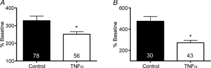
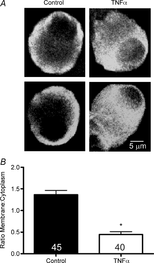
Inflammation has profound effects on the innervation of affected tissues, including altered neuronal excitability and neurotransmitter release. As Ca(2+) influx through voltage-gated Ca(2+) channels (VGCCs) is a critical determinant of excitation-secretion coupling in nerve terminals, the aim of this study was to characterize the effect of overnight incubation in the inflammatory mediator tumour necrosis factor alpha (TNFalpha; 1 nM) on VGCCs in dissociated neurons from mouse superior mesenteric ganglia (SMG). Voltage-gated Ca(2+) currents (I(Ca)) were measured using the perforated patch clamp technique and the VGCC subtypes present in SMG neurons were estimated based on inhibition by selective VGCC blockers: omega-conotoxin GVIA (300 nM; N-type), nifedipine (10 microM; L-type), and omega-conotoxin MVIIC (300 nM; N-, P/Q-type). We used intracellular Ca(2+) imaging with Fura-2 AM to compare Ca(2+) influx during depolarizations in control and TNFalpha-treated neurons. TNF receptor and VGCC mRNA expression were measured using PCR, and channel alpha subunit (CaV2.2) was localized with immunohistochemistry. Incubation in TNFalpha significantly decreased I(Ca) amplitude and depolarization-induced Ca(2+) influx. The reduction in I(Ca) was limited to omega-conotoxin GVIA-sensitive N-type Ca(2+) channels. Depletion of glial cells by incubation in cytosine arabinoside (5 microM) did not affect I(Ca) inhibition by TNFalpha. Preincubation of neurons with SC-514 (20 microM) or BAY 11-7082 (1 microM), which both inhibit nuclear factor kappaB signalling, prevented the reduction in I(Ca) by TNFalpha. Inhibition of N-type VGCCs following TNFalpha incubation was associated with a decrease in CaV2.2 mRNA and reduced membrane localization of CaV2.2 immunoreactivity. These data suggest that TNFalpha inhibits I(Ca) in SMG neurons and identify a novel role for NF-kappaB in the regulation of neurotransmitter release during inflammatory conditions with elevated circulating TNFalpha, such as Crohn's disease and Guillain-Barré syndrome.


Figures

Figure 1
Figure 1. Tumour necrosis factor α(TNFα) inhibited ICa in superior mesenteric ganglia (SMG) neurons
A, expression of TNFα receptor 1 (TNFR1) in the SMG of mice, as detected by RT-PCR. B and C, comparison of ICa from an untreated control neuron (A) and a neuron incubated overnight in 1 nm TNFα (B). Voltage clamp protocol is illustrated in inset. D, mean ±s.e.m.IV relations from a group of control neurons and TNFα-treated neurons. TNFα significantly reduced ICa. *P < 0.05, two-way ANOVA with Bonferroni's post hoc test.
Figure 2
Figure 2. TNFα selectively inhibited N-type current
Examples of ICa elicited by a step to 0 mV from a holding potential of −100 mV from an untreated (A) and a TNFα-treated (C) neuron before and during superfusion of 300 nmω-conotoxin GVIA (CTX) and 10 μm nifedipine. B, percentage inhibition of ICa in control neurons by ω-conotoxin GVIA (300 nm), the N- and P/Q-type blocker ω-conotoxin MVIIC (300 nm) and nifedipine (10 μm), indicating that ICa in control SMG neurons is primarily carried by N-type channels. *P < 0.05, Kruskal–Wallis test with Dunn's multiple comparison test. D, current density (pA pF−1) inhibition by 300 nmω-conotoxin GVIA (estimate of N-type current) and 10 μm nifedipine (estimate of L-type current) in control and TNFα-treated neurons (ω-conotoxin GVIA). *P < 0.05 versus untreated controls, unpaired 2-tailed t test. Number of neurons is indicated within bars.
Figure 3
Figure 3. Voltage dependence of activation and inactivation of ICa was not altered by TNFα
A, Boltzmann fits of mean normalized conductances showing the voltage dependence of steady-state inactivation of ICa in control neurons (n= 10) and TNFα-treated neurons (n= 12). The V0.5 was −72.6 ± 0.8 and −70 ± 2.1 mV in control and TNFα-treated neurons, respectively. The slope factor was −17.1 ± 0.7 and −18.4 ± 1.4 in control and TNFα-treated neurons, respectively. B, voltage-dependent activation curves for control neurons (n= 14) and 1 nm TNFα-treated neurons (n= 9). The V0.5 was −20.9 ± 0.6 and −20.8 ± 1.3 mV in control and TNFα-treated neurons, respectively. The slope factor was −3.9 ± 0.6 and −5.1 ± 1.3 in control and TNFα-treated neurons, respectively.
Figure 4
Figure 4. SC-514 blocked the inhibition of ICa by TNFα
A, sample recordings of ICa from a control neuron, a TNFα-treated neuron and a neuron that was pretreated with 20 μm SC-514 and subsequently exposed to SC-514 and TNFα. B, mean ±s.e.m. peak ICa amplitude at 0 mV in untreated controls, SC-514-treated controls, TNFα-treated neurons, and SC-514-pretreated neurons that subsequently were exposed to SC-514 and TNFα overnight. *P < 0.05, one-way ANOVA with Bonferroni's post hoc test. Number of neurons is indicated within bars.
Figure 5
Figure 5. Inhibition of NF-κB blocked the reduction in depolarization-induced Ca2+ transients by TNFα
A, representative Ca2+ imaging recording of an isolated SMG neuron displaying a peak response following superfusion with a 40 mm K+-containing saline solution. B, percentage changes in F340 : F380 during high K+ depolarization in control, 1 nm TNFα-treated neurons, neurons treated with 20 μm SC-514 and TNFα, neurons treated with 1 μm BAY 11 - 7082 and TNFα, and neurons treated with the p38 mitogen-activated protein kinase (MAPK) inhibitor PD 169316 (30 μm) and TNFα. *P < 0.001 versus TNFα-treated neurons, Kruskal–Wallis test and Dunn's multiple comparisons test. Number of neurons is indicated within bars.
Figure 6
Figure 6. Depletion of glial cells did not affect the reduction of Ca2+ influx by TNFα
A, mean ±s.e.m. percentage increase in Ca2+ influx in response to 40 mm K+ in cells cultured for 4 days, with TNFα added to some cultures for the final day. B, neurons cultured in the presence of cytosine arabinoside (ara-C; 5 μm) for 3 days exhibited the same reduction in Ca2+ influx following overnight incubation in TNFα.
Figure 7
Figure 7. Overnight incubation in TNFα reduced mRNA of the N-type channel α subunit
Real-time PCR analysis of CaV2.2 mRNA expression relative to GAPDH in neurons incubated overnight in control medium containing TNFα. *P= 0.05, Mann–Whitney test. Number of wells is indicated within bars, with each well containing neurons dissociated from two SMG.
Figure 8
Figure 8. TNFα reduced membrane localization of CaV2.2 protein
A, sample confocal micrographs of CaV2.2 immunoreactivity in dissociated SMG neurons incubated overnight in the absence or presence of TNFα. In untreated controls, channel immunoreactivity was localized to the plasmalemma and sub-membrane space, whereas in TNFα-treated neurons, membrane localization was reduced relative to cytoplasmic immunoreactivity. B, quantification of the ratio of membrane to cytoplasmic CaV2.2 immunoreactivity in control and TNFα-treated neurons. *P < 0.001, Mann–Whitney test. Number of neurons examined is indicated within bars.

References

    1. Baker DM, Santer RM, Blaggan AS. Morphometric studies on the microvasculature of pre- and paravertebral sympathetic ganglia in the adult and aged rat by light and electron microscopy. J Neurocytol. 1989;18:647–660. - PubMed
    1. Baud V, Karin M. Signal transduction by tumor necrosis factor and its relatives. Trends Cell Biol. 2001;11:372–377. - PubMed
    1. Besirli CG, Deckwerth TL, Crowder RJ, Freeman RS, Johnson EM., Jr Cytosine arabinoside rapidly activates Bax-dependent apoptosis and a delayed Bax-independent death pathway in sympathetic neurons. Cell Death Differ. 2003;10:1045–1058. - PubMed
    1. Blandizzi C, Fornai M, Colucci R, Baschiera F, Barbara G, De Giorgio R, De Ponti F, Breschi MC, Del Tacca M. Altered prejunctional modulation of intestinal cholinergic and noradrenergic pathways by α2-adrenoceptors in the presence of experimental colitis. Br J Pharmacol. 2003;139:309–320. - PMC - PubMed
    1. Borgland SL, Connor M, Ryan RM, Ball HJ, Christie MJ. Prostaglandin E2 inhibits calcium current in two sub-populations of acutely isolated mouse trigeminal sensory neurons. J Physiol. 2002;539:433–444. - PMC - PubMed

Publication types

MeSH terms