. 2009 Jun 15;587(Pt 12):3045-54.
doi: 10.1113/jphysiol.2009.169417. Epub 2009 Apr 29.

Direct evidence of nitric oxide release from neuronal nitric oxide synthase activation in the left ventricle as a result of cervical vagus nerve stimulation

Affiliations

Direct evidence of nitric oxide release from neuronal nitric oxide synthase activation in the left ventricle as a result of cervical vagus nerve stimulation

Kieran E Brack et al. J Physiol. .

Abstract

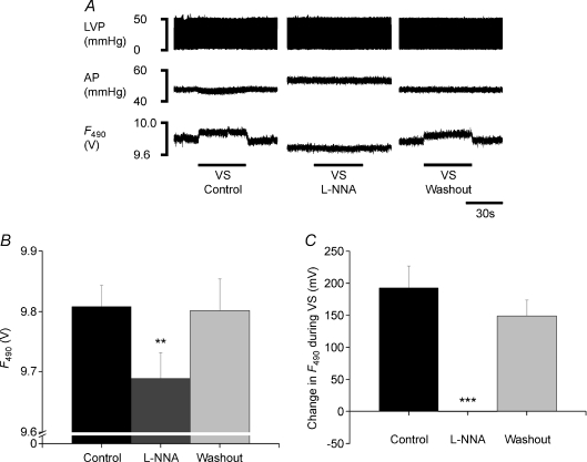
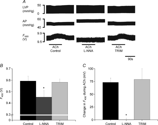
Information regarding vagal innervation in the cardiac ventricle is limited and the direct effect of vagal stimulation on ventricular myocardial function is controversial. We have recently provided indirect evidence that the anti-fibrillatory effect of vagus nerve stimulation on the ventricle is mediated by nitric oxide (NO). The aim of this study was to provide direct evidence for the release of nitric oxide in the cardiac ventricle during stimulation of the efferent parasympathetic fibres of the cervical vagus nerve. The isolated innervated rabbit heart was employed with the use of the NO fluorescent indicator 4,5-diaminofluorescein diacetate (DAF-2 DA) during stimulation of the cervical vagus nerves and acetylcholine perfusion in the absence and presence of the non-specific NO synthase inhibitor NG-nito-L-arginine (L-NNA) and the neuronal NO synthase selective inhibitor 1-(2-trifluormethylphenyl)imidazole (TRIM). Using the novel fluorescence method in the beating heart, we have shown that NO-dependent fluorescence is increased by 0.92 +/- 0.26, 1.20 +/- 0.30 and 1.91 +/- 0.27% (during low, medium and high frequency, respectively) in the ventricle in a stimulation frequency-dependent manner during vagus nerve stimulation, with comparable increases seen during separate stimulation of the left and right cervical vagus nerves. Background fluorescence is reduced during perfusion with L-NNA and the increase in fluorescence during high frequency vagal stimulation is inhibited during perfusion with both L-NNA (1.97 +/- 0.35% increase before L-NNA, 0.00 +/- 0.02% during L-NNA) and TRIM (1.78 +/- 0.18% increase before TRIM, -0.11 +/- 0.08% during TRIM). Perfusion with 0.1 microM acetylcholine increased NO fluorescence by 0.76 +/- 0.09% which was blocked by L-NNA (change of 0.00 +/- 0.03%) but not TRIM (increase of 0.82 +/- 0.21%). Activation of cardiac parasympathetic efferent nerve fibres by stimulation of the cervical vagus is associated with NO production and release in the ventricle of the rabbit, via the neuronal isoform of nitric oxide synthase.


Figures

Figure 1
Figure 1. Frequency dependent changes in NO fluorescence during VS
A, raw data illustrating continuous left ventricular pressure (LVP), aortic perfusion pressure (AP) and DAF-2 fluorescence at the excitation wavelength of 490 nm (F490) at varying intensities of vagus nerve stimulation (VS). B, mean data representing F490 at baseline, during steady state VS and post-stimulation for low, medium and high frequency left and right VS. C, mean data representing the actual change in F490 during low, medium and high frequency left and right VS. Data are mean ±s.e.m., n= 7, and analysed using 2-factor repeated measured ANOVA with Bonferroni post test analysis indicating *P < 0.05, **P < 0.01 and ***P < 0.001 steady state vs baseline or the data comparisons indicated by the horizontal bars. For B: 2-factor ANOVA analysis was performed using baseline and steady state values for left and right VS separately.
Figure 3
Figure 3. Inhibition of VS-dependent change in NO fluorescence with TRIM
A, raw data illustrating continuous left ventricular pressure (LVP), aortic perfusion pressure (AP) and DAF-2 fluorescence at the excitation wavelength of 490 nm (F490) during high frequency (15 Hz) vagus nerve stimulation (VS) during control, during perfusion with 1-(2-trifluormethylphenyl)imidazole (TRIM, 200 μm) and after washout. B, basal F490 and C, the change in F490 with VS during control, TRIM and washout. Data are mean ±s.e.m., n= 7, and analysed using single-factor repeated measures ANOVA with Bonferroni post test indicating ***P < 0.001 TRIM vs control and washout.
Figure 2
Figure 2. Inhibition of VS-dependent change in NO fluorescence with L-NNA
A, raw data illustrating continuous left ventricular pressure (LVP), aortic perfusion pressure (AP) and DAF-2 fluorescence at the excitation wavelength of 490 nm (F490) during high frequency (15 Hz) vagus nerve stimulation (VS) during control, during perfusion with NG-nitro-l-arginine (l-NNA, 200 μm) and after washout. B, basal F490 and C, the change in F490 during VS during control, l-NNA and washout. Data are mean ±s.e.m., n= 7, and analysed using single-factor repeated measures ANOVA with Bonferroni post test indicated by **P < 0.01 and ***P < 0.001 l-NNA vs control and washout.
Figure 4
Figure 4. Change in NO fluorescence with acetylcholine and NOS inhibition
A, raw data illustrating continuous left ventricular pressure (LVP), aortic perfusion pressure (AP) and DAF-2 fluorescence at the excitation wavelength of 490 nm (F490) with 0.1 μm acetylcholine (ACh) during control, during perfusion with NG-nitro-l-arginine (l-NNA, 200 μm) and 1-(2-trifluormethylphenyl)imidazole (TRIM, 200 μm). Mean data representing B, basal F490 and C, the change in F490 with ACh during control and perfusion with l-NNA and TRIM. Data are mean ±s.e.m.n= 4, and analysed using single-factor repeated measures with Bonferroni post tests for comparisons of individual groups of data as indicated by *P < 0.05 l-NNA vs control and TRIM.
Figure 5
Figure 5. Correlation of the changes in NO fluorescence with corresponding changes in perfusion pressure
Data illustrating the change in aortic perfusion pressure (ΔAP) with the change in DAF-2 fluorescence at the excitation of 490 nm (ΔF) during high frequency vagus nerve stimulation (VS, protocol I, n= 7), 0.1 μm acetylcholine (ACh, protocol II, n= 4) and perfusion with NG-nitro-l-arginine (l-NNA, 200 μm, protocol I (n= 7) + protocol III (n= 8)), sodium nitroprusside (SNP, 100 μm, protocol III, n= 8) and bradykinin (BK, 100 μm, protocol III, n= 8). l-NNA:L, non-innervated Langendorff-perfused isolated heart. l-NNA:N, innervated isolated heart preparation. Data are mean ±s.e.m. Dotted line represents a linear fitted line for data representing l-NNA:L, l-NNA:N, SNP, BK and ACh.

Comment in

  • Cardiac defibrillator neurones.
    Jones JF. Jones JF. J Physiol. 2009 Jun 15;587(Pt 12):2715. doi: 10.1113/jphysiol.2009.174466. J Physiol. 2009. PMID: 19525555 Free PMC article. No abstract available.

References

    1. Akiyama T, Yamazaki T. Effects of right and left vagal stimulation on left ventricular acetylcholine levels in the cat. Acta Physiol Scand. 2001;172:11–16. - PubMed
    1. Amezcua JL, Dusting GJ, Palmer RMJ, Moncada S. Acetylcholine induces vasodilatation in the rabbit heart through the release of nitric oxide, the endogenous nitrovasodilator. Br J Pharmacol. 1988;95:830–834. - PMC - PubMed
    1. Amezcua JL, Palmer RMJ, de Souza BM, Moncada S. Nitric oxide synthesized from l-arginine regulates vascular tone in the coronary circulation of the rabbit. Br J Pharmacol. 1989;97:1119–1124. - PMC - PubMed
    1. Armour JA, Randall WC. Functional anatomy of the canine cardiac nerves. Acta Anat. 1975;91:510–528. - PubMed
    1. Brack KE, Coote JH, Ng GA. The effect of direct autonomic nerve stimulation on left ventricular force in the isolated innervated Langendorff perfused rabbit heart. Auton Neurosci. 2006;124:69–80. - PubMed

Publication types

LinkOut - more resources