. 2009 Jun 15;587(Pt 12):2987-3000.
doi: 10.1113/jphysiol.2009.168765. Epub 2009 Apr 29.

Mechanisms underlying short-term modulation of transmitter release by presynaptic depolarization

Affiliations

Mechanisms underlying short-term modulation of transmitter release by presynaptic depolarization

Tetsuya Hori et al. J Physiol. .

Abstract

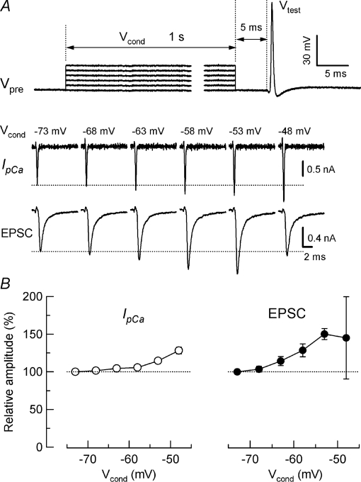
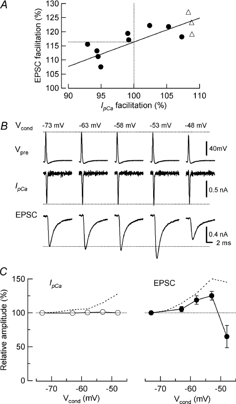
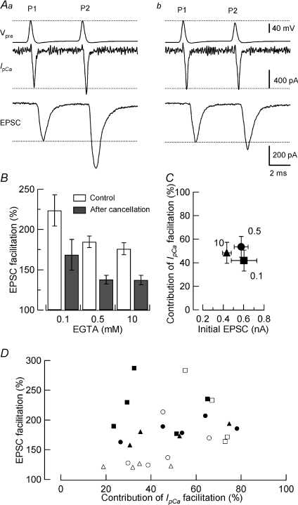
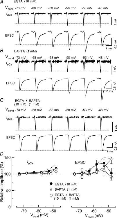
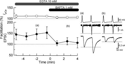
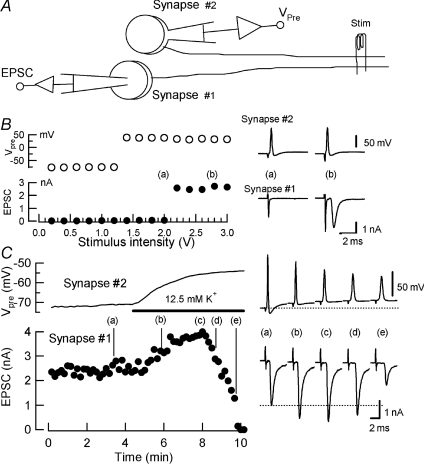
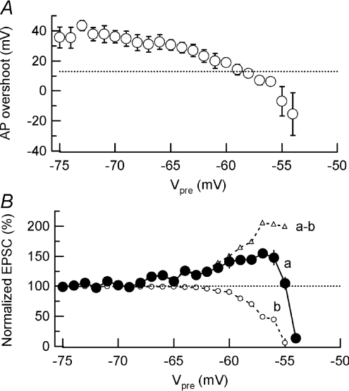
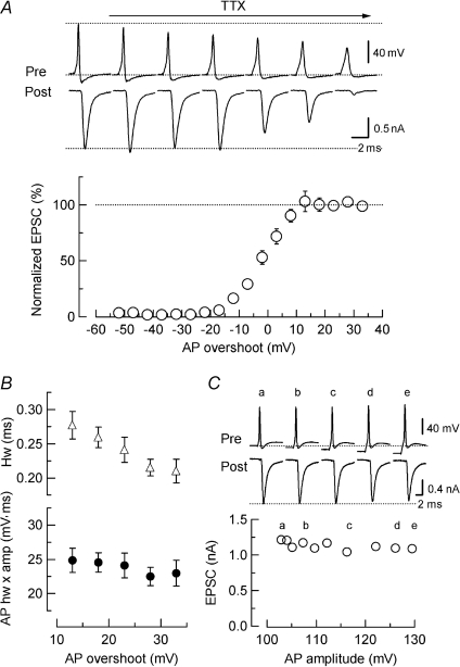
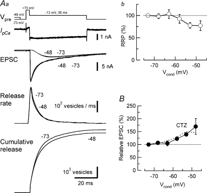
Presynaptic terminal depolarization modulates the efficacy of transmitter release. Residual Ca2+ remaining after presynaptic depolarization is thought to play a critical role in facilitation of transmitter release, but its downstream mechanism remains unclear. By making simultaneous pre- and postsynaptic recordings at the rodent calyx of Held synapse, we have investigated mechanisms involved in the facilitation and depression of postsynaptic currents induced by presynaptic depolarization. In voltage-clamp experiments, cancellation of the Ca2+-dependent presynaptic Ca2+ current (I(pCa)) facilitation revealed that this mechanism can account for 50% of postsynaptic current facilitation, irrespective of intraterminal EGTA concentrations. Intraterminal EGTA, loaded at 10 mM, failed to block postsynaptic current facilitation, but additional BAPTA at 1 mM abolished it. Potassium-induced sustained depolarization of non-dialysed presynaptic terminals caused a facilitation of postsynaptic currents, superimposed on a depression, with the latter resulting from reductions in presynaptic action potential amplitude and number of releasable vesicles. We conclude that presynaptic depolarization bidirectionally modulates transmitter release, and that the residual Ca2+ mechanism for synaptic facilitation operates in the immediate vicinity of voltage-gated Ca2+ channels in the nerve terminal.


Figures

Figure 1
Figure 1. Facilitation of Ca2+-dependent presynaptic Ca2+ current (IpCa) and EPSCs by sustained presynaptic depolarization
Simultaneous pre- and postsynaptic voltage-clamp recordings of IpCa and EPSCs in the presence of TTX at the calyx of Held of mice. A, top traces show presynaptic command voltage protocol (Vpre), comprising 1 s depolarizing conditioning pulse (Vcond) followed by an action potential (AP) waveform test pulse (Vtest). Sample records of IpCa (middle traces) and EPSCs (bottom traces) elicited by Vtest, after depolarizing calyces from holding potential (−73 mV) to various membrane potentials (Vcond indicated above the traces). Dotted lines indicate control amplitudes of IpCa and EPSCs elicited by Vtest without Vcond. B, summary data from 7 synapses. Relative changes of IpCa (left panel, open circles) and EPSCs (right panel, filled circles) produced by Vcond (abscissae). Ordinate (relative amplitude) indicates the relative amplitudes of IpCa and EPSCs compared with those without Vcond. Essentially the same results were obtained from calyces of Held of rats (data not shown).
Figure 2
Figure 2. Experimental dissection of small depolarization-dependent enhancement (SDE) into IpCa facilitation-dependent and -independent components
A, protocol used for cancellation of IpCa facilitation. AP waveform command pulse was gradually diminished and a null point of IpCa facilitation was determined by the least squares method from a linear regression line for data points (filled circles) including those in control (open triangles). B, sample records of AP command pulses (Vpre), IpCa and EPSCs after cancelling IpCa facilitation. C, EPSC facilitation and depression (right panel) after cancelling IpCa facilitation (left panel). Summary data from 8 synapses. Dashed lines are data in Fig. 1B (without IpCa cancellation) for comparison.
Figure 4
Figure 4. IpCa facilitation-dependent and -independent components of paired-pulse facilitation (PPF) of EPSCs
Aa, PPF of EPSCs evoked by a pair of AP waveform-induced IpCa (P1, P2), with 5 ms ISI, in the presence of botulinum toxin E (BoNT/E; 20 nm) and 0.5 mm EGTA in the presynaptic pipette. Ab, IpCa facilitation was cancelled by adjusting the second command voltage (P2). B, PPF of EPSCs before (open columns) and after (filled columns) IpCa cancellation, in the presence of 0.1 mm, 0.5 mm, or 10 mm EGTA in the presynaptic pipette. C, the relationship between the mean percentage contribution of IpCa facilitation to PPF (ordinate) and mean amplitude of the initial EPSCs (P1, abscissa) in the presence of EGTA at 0.1 mm (square), 0.5 mm (circle) and 10 mm (triangle). D, all-points plot showing percentage contribution of IpCa facilitation (abscissa) vs. the magnitude of EPSC facilitation (ordinate). Open symbols represent SDE data at −53 mV Vcond (Fig. 2). Filled symbols represent PPF data. Presynaptic pipettes contained 0.5 mm EGTA (circles), 10 mm EGTA (triangles), 0.1 mm EGTA (filled squares), or 25 μm BAPTA (open squares). For SDE data, but not for PPF data, a weak correlation was observed between the SDE magnitude and percentage contribution of IpCa facilitation (r= 0.68, P < 0.01, Spearman's rank correlation test).
Figure 3
Figure 3. Blocking effects of EGTA and BAPTA on the facilitation of IpCa and EPSCs
IpCa and EPSCs elicited by AP waveform Vtest following different Vcond. Calyceal terminals had been loaded with 10 mm EGTA (A), 1 mm BAPTA (B) or 1 mm BAPTA plus 10 mm EGTA (C), via presynaptic patch pipettes. D, summary data from 6–10 synapses. Relative changes of IpCa (left panel) and EPSCs (right panel) produced by Vcond (abscissa). Data with EGTA (10 mm, A), BAPTA (1 mm, B) and EGTA (10 mm) plus BAPTA (1 mm, C) are indicated by filled circles, open triangles and open circles, respectively. Dashed lines represent data with 0.5 mm EGTA (Fig. 1B) for comparison.
Figure 5
Figure 5. Block of PPF by intraterminal infusion of BAPTA BAPTA
(1 mm) was infused into presynaptic terminals, using pipette perfusion, during whole-cell recording. The presynaptic patch pipette contained 10 mm EGTA and BoNT/E (20 nm). Infusion of BAPTA blocked PPF (filled circles) and IpCa facilitation (open circles). Circles and bars represent mean amplitudes and s.e.m. (n = 5) of three data sampled every 30 s. Data points and bars represent mean and s.e.m. from 5 terminals. Sample records before (a) and after (b) BAPTA infusion are shown in the right panel.
Figure 6
Figure 6. The effect of sustained presynaptic depolarization on synaptic transmission at intact synapses
A, recording and stimulation scheme. EPSCs were evoked by afferent fibre stimulation (Stim), and recorded from a medial nucleus of trapezoid body (MNTB) principal cell (Synapse #1). Presynaptic membrane potential (Vpre) was monitored from a nearby calyceal terminal of a different synapse (Synapse #2). B, all-or-none generation of presynaptic action potentials (APs, Vpre, open circles, Synapse #2) and EPSCs (filled circles, Synapse #1) at different thresholds (stimulus intensities in abscissa). Sample traces show presynaptic APs (Synapse #2) and EPSCs (Synapse #1) evoked by different stimulus intensities (a and b). C, bath application of 12.5 mm[K+] solution (bar) depolarized presynaptic terminal (Synapse #2) and caused an increase (sample records at b and c), followed by a decrease (d and e) of the EPSC amplitude (Synapse #1).
Figure 8
Figure 8. Simultaneous occurrence of facilitation and depression of transmitter release during sustained presynaptic depolarization
A, the AP overshoot during presynaptic depolarization (Vpre, presynaptic membrane potentials, abscissa) at Synapse #2. A dotted line indicates the level at which EPSCs start to decline during TTX application (Fig. 7A). B, the relative EPSC amplitude (Synapse #1, ordinate) during K+-induced presynaptic depolarization (Vpre at Synapse #2 in abscissa). Summary data from 5 experiments (curve a). A dashed line represents control EPSC amplitude before K+ application. Open circles (curve b) indicate EPSC amplitudes estimated from AP size reduction (Fig. 8). Open triangles indicate EPSC amplitudes obtained by subtracting data b from data a, representing components of EPSCs facilitated by presynaptic depolarization.
Figure 7
Figure 7. The relationship between presynaptic AP amplitude and EPSC amplitude
In simultaneous pre- and postsynaptic recordings, EPSCs were evoked by presynaptic APs (elicited by 1 ms depolarizing current injection). Presynaptic APs and EPSCs sampled from different epochs after bath application of TTX (10 nm) are shown in the upper panel. The lower panel shows the relationship between the relative EPSC amplitude (ordinate, normalized to the amplitude before TTX application, open circles) and absolute amplitude of presynaptic AP overshoot (abscissa, mV) during TTX application. Data derived from 7 synapses. The EPSC amplitude remained unchanged (initial amplitude indicated by a dashed line) until the AP overshoot declined below 10 mV. Presynaptic resting potential was −78 ± 1 mV (n= 7). B, half-width (open triangles) and half-width multiplied by amplitude (filled circles) of APs having 13–33 mV overshoot. C, no effect of presynaptic hyperpolarization on the EPSC amplitude (ordinate) at a calyx of Held synapse. Presynaptic AP amplitude (abscissa) increased when the nerve terminal was hyperpolarized by DC current injection. The presynaptic resting potential before current injection was −78 mV. The initial levels of presynaptic membrane potential (Pre, a) and peak EPSC amplitude (Post, a) are indicated by dotted lines. Essentially the same results were obtained at another synapse in mice and at 8 synapses in rats.
Figure 9
Figure 9. Pre- and postsynaptic mechanisms underlying EPSC depression caused by presynaptic depolarization
Aa, estimation of the readily releasable pool (RRP) size of synaptic vesicles before (−73 mV) and after depolarizing Vcond (1 s, voltage protocol in the top row). Sample records on the second and third rows show IpCa and EPSCs after Vcond to −48 mV and −73 mV (superimposed). Traces in the fourth row show release rates, calculated from the deconvolution method at different Vcond (superimposed). In the bottom row, RRP size was estimated from the cumulative number of quanta released during a test pulse to −13 mV (50 ms, top trace) before (−73 mV) and after Vcond to −48 mV for 1 s (superimposed). The aCSF solution contained kynurenate (1 mm), but did not contain cyclothiazide (CTZ). Dotted lines superimposed on EPSC traces indicate AMPA receptor currents caused by residual glutamate in the synaptic cleft (Sakaba & Neher, 2001) at −48 mV and −73 mV Vcond. B, contribution of postsynaptic AMPA receptor desensitization to EPSC depression assessed by CTZ (100 μm) at various Vcond. Ab, presynaptic depolarization-induced reduction in the RRP size at various Vcond. Data points derived from 4 synapses. Control SDE without CTZ (Fig. 1B, dashed line) is superimposed on the data with CTZ.

References

    1. Alle H, Geiger JRP. Combined analog and action potential coding in hippocampal mossy fibers. Science. 2006;311:1290–1293. - PubMed
    1. Atluri PP, Regehr WG. Determinants of the time course of facilitation at the granule cell to Purkinje cell synapse. J Neurosci. 1996;16:5661–5671. - PMC - PubMed
    1. Awatramani GB, Price GD, Trussell LO. Modulation of transmitter release by presynaptic resting potential and background calcium levels. Neuron. 2005;48:109–121. - PubMed
    1. Barnes-Davies M, Forsythe ID. Pre- and postsynaptic glutamate receptors at a giant excitatory synapse in rat auditory brainstem slices. J Physiol. 1995;488:387–406. - PMC - PubMed
    1. Bertram R, Sherman A, Stanley EF. Singledomain/bound calcium hypothesis of transmitter release and facilitation. J Neurophysiol. 1996;75:1919–1931. - PubMed

Publication types

LinkOut - more resources