. 2009 Jun;136(11):1899-907.
doi: 10.1242/dev.034629. Epub 2009 Apr 29.

Multiple roles for Sox2 in the developing and adult mouse trachea

Affiliations

Multiple roles for Sox2 in the developing and adult mouse trachea

Jianwen Que et al. Development. 2009 Jun.

Abstract

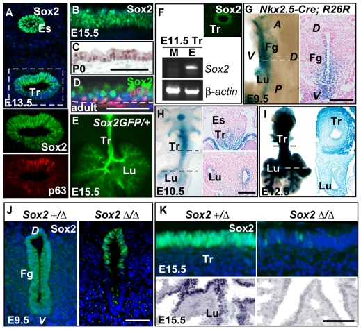
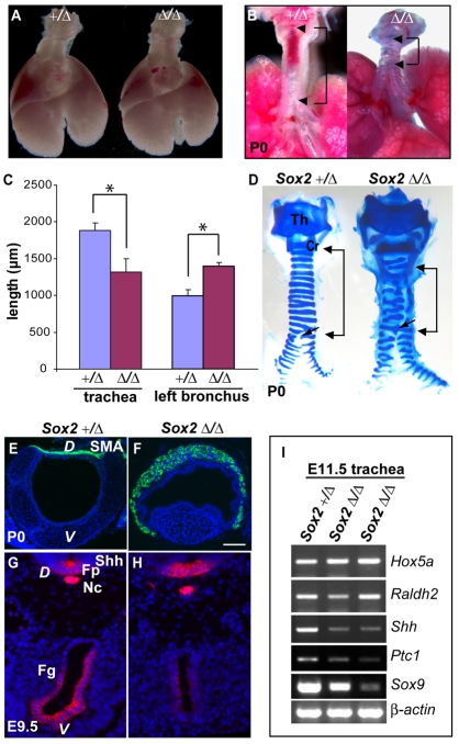
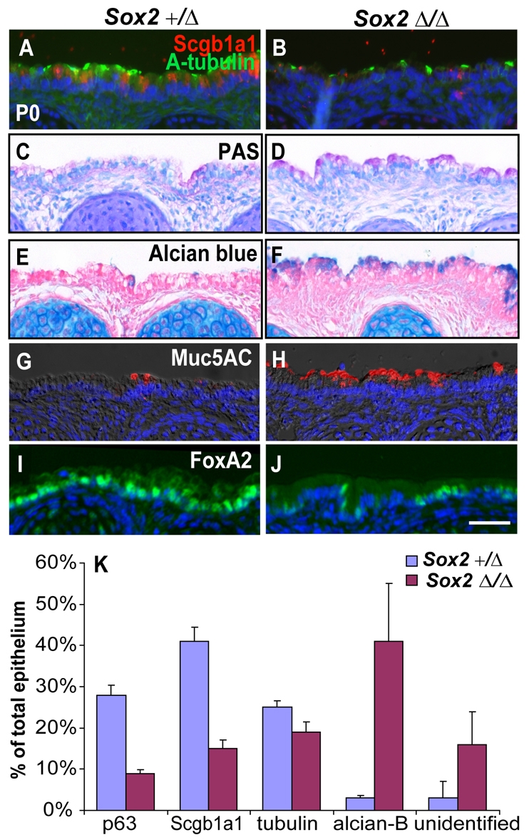
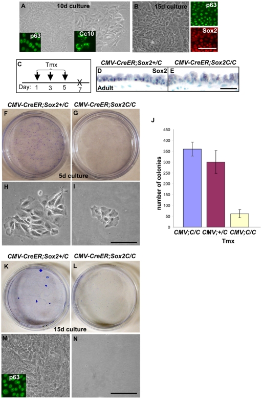
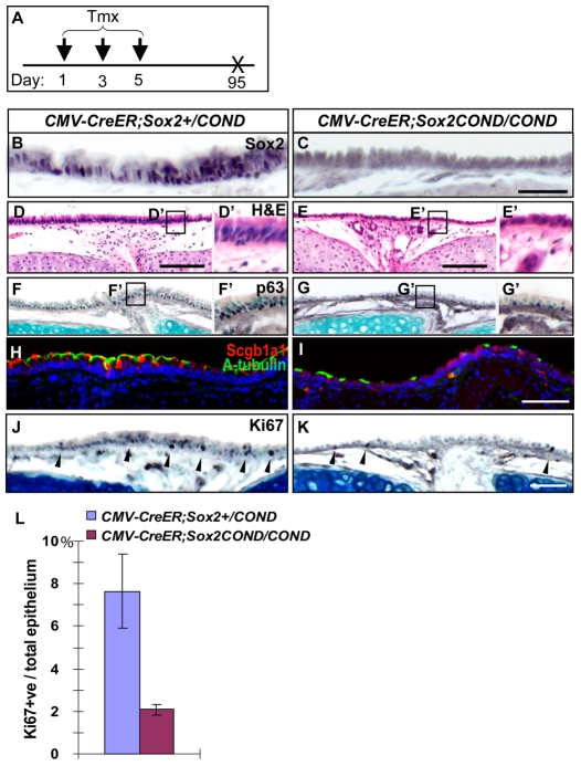
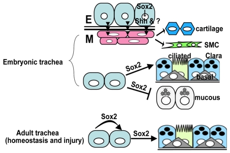
The esophagus, trachea and lung develop from the embryonic foregut, yet acquire and maintain distinct tissue phenotypes. Previously, we demonstrated that the transcription factor Sox2 is necessary for foregut morphogenesis and esophagus development. We show that Sox2 is also required for the normal development of the trachea and lung. In both the embryo and adult, Sox2 is exclusively expressed in the epithelium of the trachea and airways. We use an Nkx2.5-Cre transgene and a Sox2 floxed allele to conditionally delete Sox2 in the ventral epithelial domain of the early anterior foregut, which gives rise to the future trachea and lung buds. All conditional mutants die of respiratory distress at birth, probably due to abnormal differentiation of the laryngeal and tracheal cartilage as a result of defective epithelial-mesenchymal interaction. About 60% of the mutants have a short trachea, suggesting that the primary budding site of the lung shifts anteriorly. In the tracheal epithelium of all conditional mutants there are significantly more mucus-producing cells compared with wild type, and fewer basal stem cells, ciliated and Clara cells. Differentiation of the epithelium lining the conducting airways in the lung is abnormal, suggesting that Sox2 also plays a role in the differentiation of embryonic airway progenitors into specific lineages. Conditional deletion of Sox2 was then used to test its role in adult epithelium maintenance. We found that epithelial cells, including basal stem cells, lacking Sox2 show a reduced capacity to proliferate in culture and to repair after injury in vivo. Taken together, these results define multiple roles for Sox2 in the developing and adult trachea.


Figures

Fig. 1.
Fig. 1.
Expression of Sox2 in the trachea and use of Nkx2.5-Cre to delete Sox2. (A-F) Sox2 expression in the developing and adult trachea. (A) Immunohistochemical localization of Sox2 protein in the esophagus and trachea at E13.5. Lower panels are magnified views of trachea stained with anti-Sox2 and anti-p63 antibodies. (B-D) Immunohistochemical localization of Sox2 in the trachea at E15.5 (B), P0 (C) and adult (D) stages. In D, p63 and Sox2 colocalize in basal cells. (E) Fluorescence microscopy of the trachea and lung at E15.5. (F) RT-PCR shows that Sox2 is exclusively expressed in the epithelium, and insert shows an E11.5 trachea immunostained with anti-Sox2 antibody. (G-I) Nkx2.5-Cre expression in the early foregut (stages E9.5-12.5) as detected in R26R embryos through X-gal staining. Left and right panels show the whole-mount X-gal staining and a section of these stained samples, respectively. The dashed line delineates where the section is cut. (J,K) Immunostaining with anti-Sox2 antibody on early foregut and its derivatives in Nkx2.5-Cre;Sox2+/COND (Sox2+/Δ) controls and Nkx2.5-Cre;Sox2COND/COND (Sox2 Δ/Δ) mutants. Nuclei are counterstained with DAPI (blue). Es, esophagus; Tr, trachea; Lu, lung; Fg, foregut; A, anterior; P, posterior; D, dorsal; V, ventral; M, mesenchyme; E, epithelium. Scale bars: 100 μm.
Fig. 2.
Fig. 2.
Abnormal development of larynx and trachea in Nkx2.5-Cre;Sox2COND/COND mutants. (A) Gross morphology of lung and trachea in control (left) and mutant (right) mice. (B,C) Short trachea (bracket) and long main bronchus in P0 mutants (n=5; *P<0.01). (D-F) Abnormal differentiation of mesenchymal cells in the mutant trachea. (D) Alcian blue staining reveals abnormal laryngeal and tracheal cartilage (bracket). Note the reduced number of incomplete cartilage rings in mutants (arrows). (E,F) Immunostaining with anti-SMA antibody to show ectopic expression of smooth muscle actin in the ventral side of the trachea in mutants. (G-I) Downregulated Shh signaling in the early foregut of mutants. (G,H) Immunohistochemical localization of Shh protein in the unseparated foregut. Nuclei are counterstained with DAPI (blue). (I) Semi-quantitative RT-PCR for expression in control and mutant tracheas of several genes involved in the development of laryngeal and tracheal cartilage. Th, thyroid; Cr, cricoid; Fg, foregut; Fp, floor plate; Nc, notochord; D, dorsal; V, ventral. Scale bar: 100 μm.
Fig. 3.
Fig. 3.
Decreased number of p63-positive cells in Nkx2.5-Cre;Sox2COND/COND mutants at different developmental stages. (A-D) Immunohistochemical staining with anti-p63 antibody in control (A,C) and mutant (B,D) tracheas at E15.5 (A,B) and E18.5 (C,D). Nuclei are counterstained with methyl green. (E,F) Immunostaining with anti-keratin 5 (green) and anti-p63 (red) antibodies in the P0 trachea. (G) Quantification of p63-positive cells in tracheas at E13.5, E15.5, E18.5 and P0. At each stage, three individual tracheas were included. Scale bars: 50 μm.
Fig. 4.
Fig. 4.
Abnormal differentiation of tracheal and lung epithelial cells at P0. (A,B) Decreased number of ciliated (acetylated α-tubulin-positive, green) and Clara (Scgb1a1-positive, red) cells in mutant trachea (B), compared with control (A). (C-H) Increased number of mucus-producing cells in mutant tracheas visualized by PAS (C,D), alcian blue (E,F) and anti-Muc5ac (G,H) staining. (I,J) Immunostaining with anti-Foxa2 antibody. (K) Quantification of the percentage of the different cell lineage to total epithelium in the trachea. Numbers are obtained from analyzing ten sections at the same level from each of three individual tracheas from both Nkx2.5-Cre;Sox2+/COND controls and Nkx2.5-Cre;Sox2COND/COND mutants. PAS, periodic acid-Schiff. Scale bar: 50 μm.
Fig. 5.
Fig. 5.
Sox2 regulates the proliferation of adult tracheal epithelium in culture. (A) Representative colonies present on culture day 10 express either p63 or Scgb1a1 (CC10; inserts). (B) Representative colony present on day 15 that co-expresses p63 and Sox2. (C-E) Efficient deletion of Sox2 in the adult trachea of CMV-CreER;Sox2COND/COND mutants. (C) Timeline of the Tamoxifen (Tmx) treatment protocol. (D,E) Immunohistochemistry of Sox2 in wild-type (D) and mutant (E) tracheas. (F-J) Sox2 deletion reduces proliferation of tracheal epithelium isolated from wild type (F,H) or conditional mutants (G,I) and cultured for 5 days. H and I are representative colonies from F and G, respectively. (J) Number of colonies per 35 mm culture dish. Data are from three independent experiments. (K-N) Basal cell colonies present in controls (K), but not in mutants (L), in 15-day cultures. M is a representative colony from K. N is from L, showing no surviving colony in this 35 mm culture dish. Insert in M shows representative colony stained with anti-p63. Colonies in F,G and K are stained with Wright blue. Scale bars: 50 μm.
Fig. 6.
Fig. 6.
Sox2 is required for homeostasis of the adult tracheal epithelium. (A) Timeline of the Tamoxifen treatment. (B,C) Immunohistochemistry with anti-Sox2 antibody. (D-E′) Hematoxylin and eosin (H&E) staining of adult trachea. D′ and E′ are magnified views of boxed region in D and E, respectively. (F-G′) Immunohistochemistry staining with anti-p63 antibody. (H,I) Immunostaining with anti-Scgb1a1 (red) and anti-acetylated α-tubulin (green) antibodies. (J-L) Decreased proliferation of tracheal epithelium after loss of Sox2 protein. (J,K) Immunohistochemistry staining with anti-Ki67 antibody. (L) Ratio of Ki67-positive epithelium along total tracheal epithelium. Four mutant and control tracheas are included. Scale bars: 100 μm.
Fig. 7.
Fig. 7.
Sox2 is required for proliferation and differentiation of the regenerating tracheal epithelium in a SO2-injured mouse model. (A) Timeline of the Tamoxifen and SO2 treatment protocol. (B-C″) H&E staining of regenerated trachea. B′ (ventral), B″ (dorsal) and C′ (ventral), C″ (dorsal) are magnified views of the boxed region in B and C, respectively. Arrowheads indicate epithelium. Note that in C there are numerous inflammatory cells within the lumen, asterisks in C indicate smooth muscles bulging into the lumen. (D-I) Immunohistochemistry of the regenerated epithelium with antibodies against Ki67 shows more proliferative cells (arrowheads) in control (D) versus mutant (E) tracheas. D′ and E′ are magnified views of the boxed region in D and E, respectively. (F,G) Anti-keratin 5 (green) and anti-p63 (red); (H,I) anti-Scgb1a1 (red) and anti-acetylated α-tubulin (green). D, dorsal; V, ventral. Scale bars: 50 μm.
Fig. 8.
Fig. 8.
Model for the roles of Sox2 in developing and adult trachea. At embryonic stage, Sox2 is required for the differentiation of tracheal mesenchyme and epithelium. Conditional deletion of Sox2 results in a decreased number of basal, Clara and ciliated cells, and more mucus-producing cells in the developing trachea. In the adult, Sox2 regulates both proliferation and differentiation at steady state and following injury. Sox2 deletion leads to reduced proliferation of tracheal epithelium both in vivo and in vitro. Loss of Sox2 also impairs the regeneration process of the trachea after inhaled SO2-induced injury.

References

    1. Aubin, J., Lemieux, M., Tremblay, M., Berard, J. and Jeannotte, L. (1997). Early postnatal lethality in Hoxa-5 mutant mice is attributable to respiratory tract defects. Dev. Biol. 192, 432-445. - PubMed
    1. Cardoso, W. V. and Lu, J. (2006). Regulation of early lung morphogenesis: questions, facts and controversies. Development 133, 1611-1624. - PubMed
    1. Cardoso, W. V. and Whitsett, J. A. (2008). Resident cellular components of the lung: developmental aspects. Proc. Am. Thorac. Soc. 5, 767-771. - PMC - PubMed
    1. Chen, F., Desai, T. J., Qian, J., Niederreither, K., Lu, J. and Cardoso, W. V. (2007). Inhibition of Tgf beta signaling by endogenous retinoic acid is essential for primary lung bud induction. Development 134, 2969-2979. - PubMed
    1. Chen, X., Qin, R., Liu, B., Ma, Y., Su, Y., Yang, C. S., Glickman, J. N., Odze, R. D. and Shaheen, N. J. (2008a). Multilayered epithelium in a rat model and human Barrett's esophagus: expression patterns of transcription factors and differentiation markers. BMC Gastroenterol. 8, 1. - PMC - PubMed

Publication types

MeSH terms

Substances