. 2009 Jul;83(13):6578-90.
doi: 10.1128/JVI.00548-09. Epub 2009 Apr 29.

Human immunodeficiency virus type 1 Nef protein targets CD4 to the multivesicular body pathway

Affiliations

Human immunodeficiency virus type 1 Nef protein targets CD4 to the multivesicular body pathway

Luis L P daSilva et al. J Virol. 2009 Jul.

Abstract

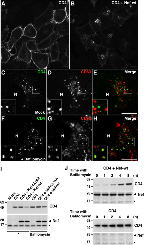
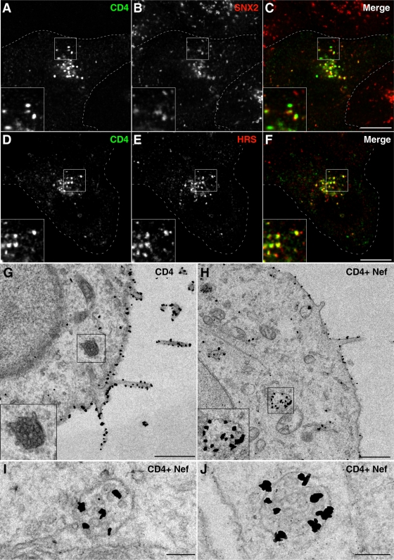
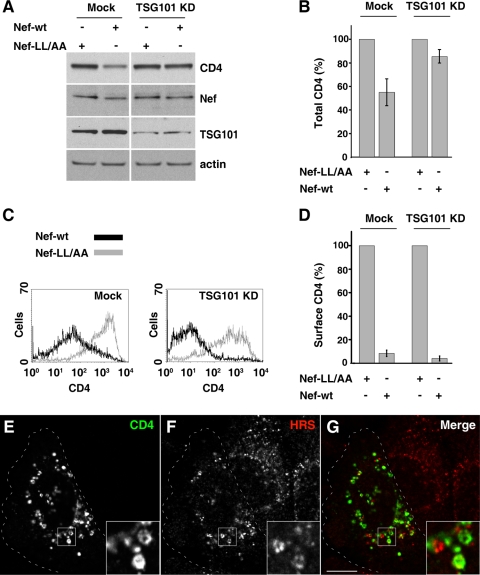
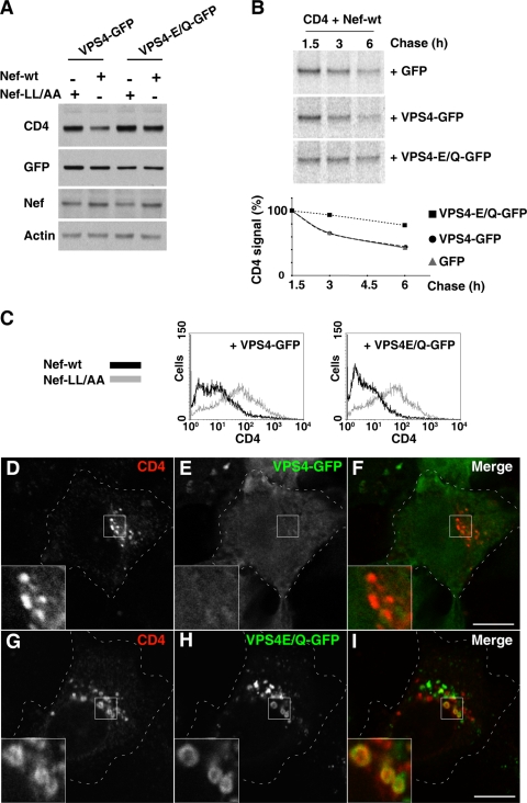
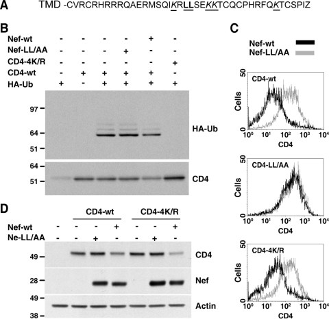
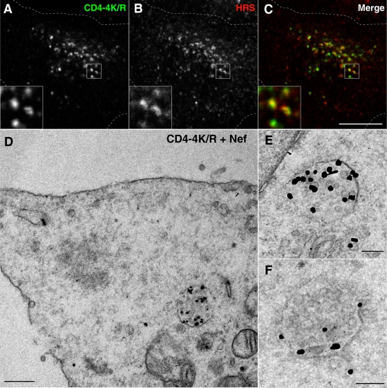
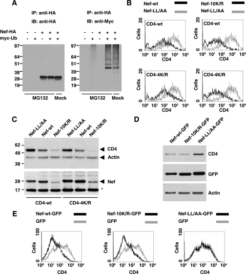
The Nef protein of human immunodeficiency virus type 1 downregulates the CD4 coreceptor from the surface of host cells by accelerating the rate of CD4 endocytosis through a clathrin/AP-2 pathway. Herein, we report that Nef has the additional function of targeting CD4 to the multivesicular body (MVB) pathway for eventual delivery to lysosomes. This targeting involves the endosomal sorting complex required for transport (ESCRT) machinery. Perturbation of this machinery does not prevent removal of CD4 from the cell surface but precludes its lysosomal degradation, indicating that accelerated endocytosis and targeting to the MVB pathway are separate functions of Nef. We also show that both CD4 and Nef are ubiquitinated on lysine residues, but this modification is dispensable for Nef-induced targeting of CD4 to the MVB pathway.


Figures

FIG. 1.
FIG. 1.
Nef targets CD4 to lysosomes. (A and B) HeLa cells grown on coverslips were transfected with pCMV-CD4 (A) or pCMV-CD4 plus pCIneo-Nef-wt (B). After 16 h cells were fixed, permeabilized, and stained with mouse monoclonal antibody to CD4 followed by Alexa-488-conjugated donkey polyclonal antibody to mouse IgG. (C to H) HeLa cells were cotransfected with pCMV-CD4 and pCIneo-Nef-wt and, 16 h later, were incubated for an additional 5 h with no additions (C to E) or with 1 μM bafilomycin A1 (F to H). Cells were then fixed, permeabilized, and stained with Alexa-488-conjugated mouse monoclonal antibody to CD4 (green channel) and mouse IgG1 monoclonal antibody to CD63. This was followed by incubation with Alexa-594-conjugated goat polyclonal antibody specific to mouse IgG1 isotype (red channel). Cells were imaged by confocal laser scanning microscopy. Yellow in the merged images indicates colocalization. Bar, 10 μm. Insets are images of the boxed areas at a magnification of ×2. N, nucleus. (I) HeLa cells were transfected with pCMV-CD4 together with either pCIneo, pCIneo-Nef-wt, or pCIneo-Nef-LL/AA. After transfection, cells were incubated for 16 h and then for an additional 3 h either in the absence (−) or presence of 1 μM bafilomycin A1. Total cell extracts were analyzed by SDS-PAGE and immunoblotting with antibodies to CD4 (upper panel) and Nef (lower panel). The antibody to Nef detects a nonspecific band (asterisk) that is also present in untransfected cell extracts and serves as an internal loading control. (J) HeLa cells were transfected with pCMV-CD4 (lower panel) or pCMV-CD4 plus pCIneo-Nef-wt (upper panel) and, after 16 h, were incubated in the absence or presence of 1 μM bafilomycin A1 for the different periods indicated in the figure. Total cell extracts were analyzed by SDS-PAGE and immunoblotting as in panel I. Molecular mass (in kDa) markers are indicated on the left.
FIG. 2.
FIG. 2.
Nef induces redistribution of CD4 to multivesicular bodies. (A to F) HeLa cells transfected with pCMV-CD4 and pCIneo-Nef-wt were fixed 16 h later and costained with mouse monoclonal antibody to CD4 and rabbit polyclonal antibodies to either SNX2 (A to C) or HRS (D to F), followed by Alexa-488-conjugated donkey antibody to mouse IgG (green channel) and Alexa-594-conjugated donkey antibody to rabbit IgG (red channel). Cells were imaged by confocal laser scanning microscopy. Cell outlines are indicated by dashed lines. Yellow in the merged images indicates colocalization. Bar, 10 μm. The insets represent the boxed areas at a magnification of ×2. (G to J) HeLa cells were transfected with pCMV-CD4 (G) or pCMV-CD4 plus pCIneo-Nef-wt (H to J). After 16 h, cells were fixed and processed for immunoelectron microscopy as described in the Materials and Methods section. Examples of MVBs are shown in the insets of panels G and H and in panels I and J. The insets in panels G and H show the boxed areas at a magnification of×2. Bars, 1 μm (G and H) 0.2 μm (I and J).
FIG. 3.
FIG. 3.
Depletion of TSG101 inhibits Nef-induced CD4 degradation. (A) Cells were either mock transfected or transfected with siRNA for TSG101 (a component of ESCRT-I) twice at 48-h intervals. After the second round of siRNA transfection, cells were transfected with pCMV-CD4 together with pCIneo-Nef-wt or pCIneo-Nef-LL/AA. After 16 h, equivalent amounts of cell lysates were subjected to SDS-PAGE and immunoblotting with antibodies to CD4, Nef, TSG101, and actin (loading control). (B) The CD4 signal intensity for each condition shown in panel A was determined and used to calculate the relative amount of CD4 in either mock- or siRNA-treated cells. Bars represent the means ± standard deviations from three independent experiments. (C) Cells treated in the presence or absence of siRNA for TSG101 as in panel A were subsequently transfected with pCMV-CD4 together with pNef-wt.IRES.GFP or pNef-LL/AA.IRES.GFP and analyzed for cell surface CD4 by FACS, as described in Materials and Methods. (D) Bar graphs represent surface CD4 levels (median values from FACS histogram plots) in cells transfected with pNef-wt.IRES.GFP relative to levels in cells transfected with pNef-LL/AA.IRES.GFP (100%). Results are shown for either mock- or TSG101 siRNA-treated cells as in panel A and represent the means ± standard deviations (n = 3). (E to G) Cells depleted of TSG101 as in panel A were cotransfected with pCMV-CD4 and pCIneo-Nef-wt, fixed, permeabilized, and costained with mouse monoclonal antibody to CD4 and rabbit polyclonal antibody to HRS, followed by incubation with secondary antibodies as described in the legends of Fig. 2A to F. Cells were imaged by confocal laser scanning microscopy. Cell outlines are indicated by dashed lines. The insets show the boxed areas at a magnification of ×3. Yellow in the merged images indicates colocalization. Bar, 10 μm.
FIG. 4.
FIG. 4.
Expression of dominant-negative VPS4 inhibits Nef-induced CD4 degradation. (A) HeLa cells were transfected with pCMV-CD4 and pCIneo-Nef-wt or pCIneo-Nef-LL/AA in combination with pGFP-based constructs encoding VPS4 or VPS4-E/Q. After 16 h, cell lysates were prepared, and equivalent amounts were analyzed by SDS-PAGE and immunoblotting with antibodies to CD4, GFP, Nef, and actin (loading control), as shown at the left of each panel. (B) HeLa cells were transfected with the above indicated plasmids encoding CD4, Nef-wt, and either GFP, VPS4-GFP, or VPS4-E/Q-GFP, as shown at the right of each panel. After 16 h cells were pulse-labeled with [35S]methionine-cysteine for 15 min, followed by incubation in chase buffer for the times indicated above each lane. Cell extracts were used for immunoprecipitation with antibody to CD4, and the resulting immunoprecipitates were analyzed by SDS-PAGE. CD4 signal intensity was quantified using the ImageQuant software and is expressed as a percentage of the total amount of CD4 immunoprecipitated at time 1.5 h. (C) HeLa cells transfected as in panel A were analyzed for surface CD4 by FACS, as described in the Materials and Methods section. (D to I) HeLa cells grown on coverslips were transfected with the above indicated plasmids encoding CD4 and Nef-wt, plus either VPS4-GFP (D to F) or VPS4-E/Q-GFP (G to I). After 16 h, cells were fixed, permeabilized, and stained with mouse monoclonal antibody to CD4 followed by Alexa-594-conjugated donkey antibody to mouse IgG (red channel). Cells were imaged by confocal laser scanning microscopy. Cell outlines are indicated by dashed lines. The insets show the boxed areas at a magnification of ×3. Bar, 10 μm.
FIG. 5.
FIG. 5.
Nef-induced downregulation does not require CD4 ubiquitination. (A) Sequence of the CD4 C-terminal cytosolic tail with key residues underlined. The dileucine motif involved in CD4 internalization is highlighted in bold, and the four lysine residues are indicated in italics. (B) HeLa cells were cotransfected with pCMV-based vectors encoding either CD4-wt or CD4-4K/R and pCIneo-based plasmids encoding HA-tagged ubiquitin, Nef-wt, or Nef-LL/AA, as indicated on top. After 16 h, equivalent amounts of cell lysates were subjected to immunoprecipitation with antibody to CD4. The resulting immunoprecipitates were analyzed by SDS-PAGE, followed by immunoblotting with antibodies to HA (upper panel) or CD4 (lower panel). (C) FACS analysis of HeLa cells transfected with pCMV-CD4 (CD4-wt, CD4-4K/R, or CD4-LL/AA) and pNef.IRES.GFP (Nef-wt or Nef-LL/AA). After 16 h, cell surface CD4 was labeled, and cells were analyzed by FACS, as described in Materials and Methods. (D) HeLa cells were transfected with pCMV-CD4 (CD4-wt or CD4-4K/R) and pCIneo-Nef (Nef-wt or Nef-LL/AA). After 16 h, equivalent amounts of cell lysates were subjected to SDS-PAGE and immunoblotting with antibodies to CD4, Nef, and actin (loading control). Molecular mass (in kDa) markers are indicated on the left.
FIG. 6.
FIG. 6.
CD4 ubiquitination is not required for delivery to MVBs. (A to C) HeLa cells transfected with pCMV-CD4-4K/R mutant and pCIneo-Nef-wt were fixed, permeabilized, and double labeled with mouse monoclonal antibody to CD4 and rabbit polyclonal antibody to HRS, followed by incubation with secondary antibodies as described in the legends of Fig. 2D to F. Cells were imaged by confocal laser scanning microscopy. Cell outlines are indicated by dashed lines. The insets show the boxed areas at a magnification of ×3. Bar, 10 μm. (D to F) HeLa cells were transfected with the above indicated plasmids encoding CD4-4K/R and Nef-wt and, 16 h later, were fixed and prepared for immunoelectron microscopy as described in Materials and Methods. Higher magnifications of MVBs labeled for CD4 are shown in panels E and F. Bars, 0.5 μm (D) and 0.2 μm (E and F).
FIG. 7.
FIG. 7.
Nef ubiquitination is not required for CD4 downregulation in HeLa or T cells. (A) HeLa cells were transfected with pCIneo-based plasmids encoding the proteins shown on top. Cells were incubated for 16 h and then for an additional 6 h in either the presence or absence of 10 μM MG132, as indicated below the lanes. After incubation, equivalent amounts of cell lysates were subjected to immunoprecipitation with antibody to HA. The resulting immunoprecipitates were analyzed by SDS-PAGE followed by immunoblotting with antibodies to HA (left panel) or myc (right panel). (B) FACS analysis of HeLa cells transfected with pCMV-CD4 (CD4-wt or CD4-4K/R) and pNef.IRES.GFP (Nef-wt, Nef-LL/AA, or Nef-10K/R). After 16 h, cells were analyzed for surface CD4 by FACS, as described in the Materials and Methods section. (C) HeLa cells were transfected with pCMV-CD4 (CD4-wt or CD4-4K/R) and pCIneo-Nef (Nef-wt, Nef-LL/AA, or Nef-10K/R). After 16 h, equivalent amounts of cell lysates were subjected to SDS-PAGE and immunoblotting with antibodies to CD4, Nef, and actin (loading control). A nonspecific band detected by the Nef antibody is indicated by an asterisk. Molecular mass (in kDa) markers are indicated on the left. (D) Human JM T cells endogenously expressing CD4 were transfected with pNef-GFP (Nef-wt-GFP, Nef-LL/AA-GFP, or Nef-10K/R-GFP). After 16 h, equivalent amounts of cell lysates were subjected to SDS-PAGE and immunoblotting with antibodies to CD4, GFP, and actin (loading control). (E) JM T-cells were transfected with pGFP-based plasmids encoding either GFP or one of the Nef-GFP fusion proteins indicated above each panel. After 16 h, cells were stained with APC-conjugated antibody to CD4 and analyzed by FACS as described in Materials and Methods.

References

    1. Acres, R. B., P. J. Conlon, D. Y. Mochizuki, and B. Gallis. 1986. Rapid phosphorylation and modulation of the T4 antigen on cloned helper T cells induced by phorbol myristate acetate or antigen. J. Biol. Chem. 26116210-16214. - PubMed
    1. Aiken, C., J. Konner, N. R. Landau, M. E. Lenburg, and D. Trono. 1994. Nef induces CD4 endocytosis: requirement for a critical dileucine motif in the membrane-proximal CD4 cytoplasmic domain. Cell 76853-864. - PubMed
    1. Aiken, C., L. Krause, Y. L. Chen, and D. Trono. 1996. Mutational analysis of HIV-1 Nef: identification of two mutants that are temperature-sensitive for CD4 downregulation. Virology 217293-300. - PubMed
    1. Anderson, S. J., M. Lenburg, N. R. Landau, and J. V. Garcia. 1994. The cytoplasmic domain of CD4 is sufficient for its down-regulation from the cell surface by human immunodeficiency virus type 1 Nef. J. Virol. 683092-3101. - PMC - PubMed
    1. Atkins, K. M., L. Thomas, R. T. Youker, M. J. Harriff, F. Pissani, H. You, and G. Thomas. 2008. HIV-1 Nef binds PACS-2 to assemble a multikinase cascade that triggers major histocompatibility complex class I (MHC-I) down-regulation: analysis using short interfering RNA and knock-out mice. J. Biol. Chem. 28311772-11784. - PMC - PubMed

Publication types

Substances