. 2009 Jun;20(12):2963-78.
doi: 10.1091/mbc.e08-12-1251. Epub 2009 Apr 29.

Obscurin interacts with a novel isoform of MyBP-C slow at the periphery of the sarcomeric M-band and regulates thick filament assembly

Affiliations

Obscurin interacts with a novel isoform of MyBP-C slow at the periphery of the sarcomeric M-band and regulates thick filament assembly

Maegen A Ackermann et al. Mol Biol Cell. 2009 Jun.

Abstract

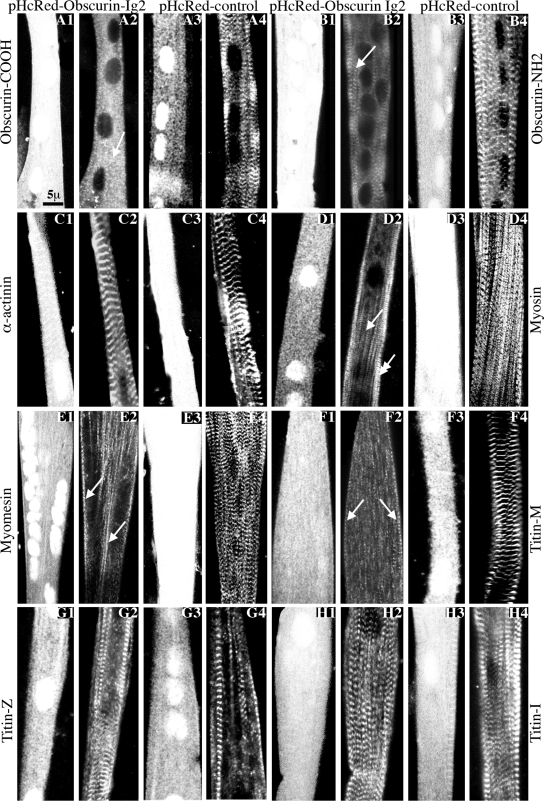
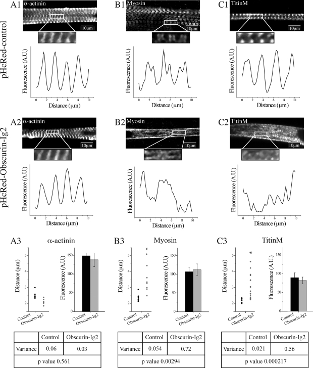
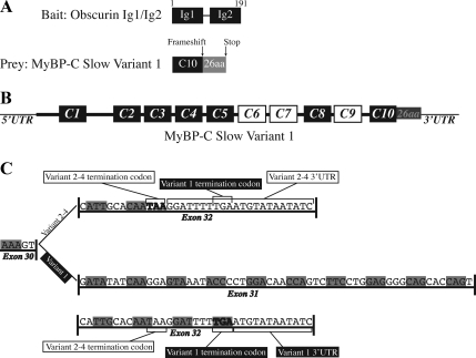
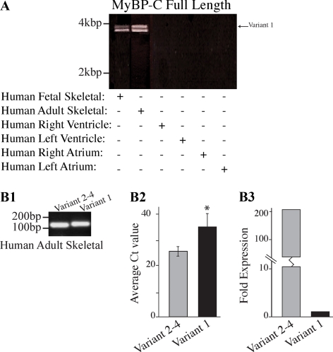
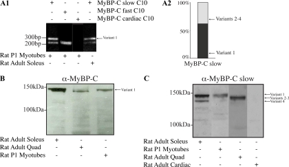
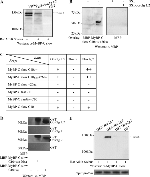
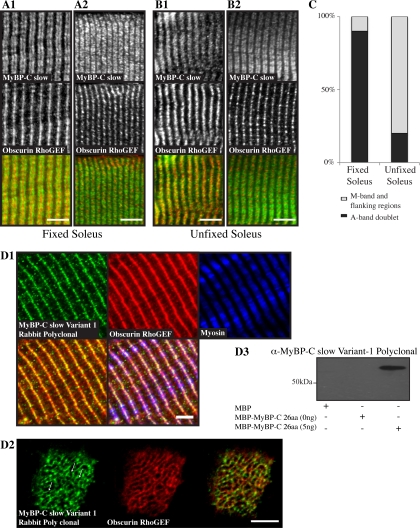
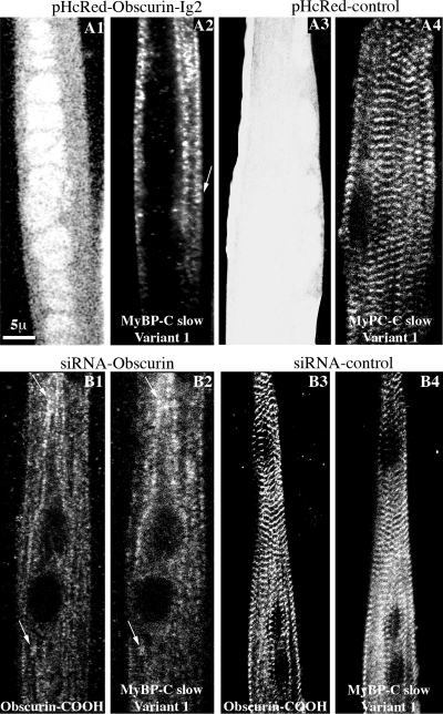
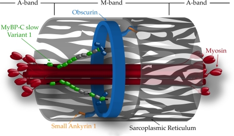
Obscurin is a multidomain protein composed of adhesion and signaling domains that plays key roles in the organization of contractile and membrane structures in striated muscles. Overexpression of the second immunoglobulin domain of obscurin (Ig2) in developing myotubes inhibits the assembly of A- and M-bands, but not Z-disks or I-bands. This effect is mediated by the direct interaction of the Ig2 domain of obscurin with a novel isoform of myosin binding protein-C slow (MyBP-C slow), corresponding to variant-1. Variant-1 contains all the structural motifs present in the known forms of MyBP-C slow, but it has a unique COOH terminus. Quantitative reverse transcription-polymerase chain reaction indicated that MyBP-C slow variant-1 is expressed in skeletal muscles both during development and at maturity. Immunolabeling of skeletal myofibers with antibodies to the unique COOH terminus of variant-1 demonstrated that, unlike other forms of MyBP-C slow that reside in the C-zones of A-bands, variant-1 preferentially concentrates around M-bands, where it codistributes with obscurin. Overexpression of the Ig2 domain of obscurin or reduction of expression of obscurin inhibited the integration of variant-1 into forming M-bands in skeletal myotubes. Collectively, our experiments identify a new ligand of obscurin at the M-band, MyBP-C slow variant-1 and suggest that their interaction contributes to the assembly of M- and A-bands.


Figures

Figure 1.
Figure 1.
Adenoviral overexpression of the Ig2 domain of obscurin in primary cultures of rat skeletal myotubes resulted in disorganized M- and A-bands but not Z-disks or I-bands. (A1–A4 and B1–B4) The striated distribution of endogenous obscurin was severely altered after overexpression of pHcRed-Obscurin-Ig2 virus (A1 and A2 and B1 and B2), but not control pHcRed virus (A3 and A4 and B3 and B4) as shown by immunostaining with antibodies to the COOH terminus (A2, arrow; and A4) or the NH2 terminus (B2, arrow; and B4) of obscurin (odd numbered panels show pHcRed fluorescence). (C1–C4) The regular organization of α-actinin at Z-disks was unaffected after overexpression of pHcRed-Obscurin-Ig2 (C1 and C2) or control pHcRed (C3 and C4) virus. (D1–D4) Sarcomeric myosin exhibited adiffuse cytoplasmic distribution (single arrow) with residual accumulation in striated structures at the cell periphery (double arrow) in cultures infected with pHcRed-Obscurin-Ig2 virus (D1-D2) but not in cultures infected with pHcRed virus where it assumed its typical periodic organization at A-bands (D3 and D4). (E1-E4 and F1-F4) Similar to sarcomeric myosin, myomesin (E1 and E2, arrows) and COOH-terminal epitopes of titin present at the M-band (F1 and F2, arrows) were primarily detected along fibrils showing occasional periodicity after overexpression of pHcRed-Obscurin-Ig2 but not control virus (E3 and E4 and F3 and F4, respectively). (G1-G4 and H1-H4) Strong periodic labeling of the NH2 terminus (G1-G4) and middle portion (H1-H4) of titin at the Z-disk and I-band, respectively, was observed in myotubes infected with either pHcRed-Obscurin-Ig2 (G1 and G2 and H1 and H2) or control pHcRed virus (G3 and G4 and H3 and H4).
Figure 2.
Figure 2.
The striated organization, but not the expression levels, of sarcomeric myosin and the COOH terminus of titin were significantly altered after overexpression of the Ig2 of obscurin in primary cultures of rat myotubes. (A1–C2) Representative examples of myotubes infected with control pHcRed (A1–C1) or pHcRed-Obscurin-Ig2 (A2–C2) viruses that were used to generate fluorescence profiles (A1–C2, bottom). α-Actinin, a Z-disk protein, shows regular fluorescent peaks in control (A1, bottom) and experimental (A2, bottom) samples; however, sarcomeric myosin at A-bands (B1 and B2) and the COOH terminus of titin at M-bands (C1 and C2) show complete lack of organization in cells infected with the obscurin-Ig2 virus (B2 and C2, bottom, respectively), compared with cells infected with control virus (B1 and C1, bottom, respectively). (A3–C3) Measurements of the average ”peak-to-peak“ distances for α-actinin (A3, left) indicated that they were slightly, yet significantly, decreased in myotubes treated with the obscurin-Ig2 virus (∼2.1 μm) compared with cells treated with control virus (∼2.5 μm); however, the variances between the two cell populations were not statistically different (A3, bottom). Likewise, ”trough-to-trough“ and peak-to-peak measurements for myosin (B3) and the COOH terminus of titin (C3) demonstrated significantly different distances (left) and variances (bottom) between experimental and control samples. Interestingly, the average fluorescent intensities for α-actinin (A3, right), myosin (B3, right), and the COOH terminus of titin (C3, right) were similar between cells treated with pHcRed-obscurin-Ig2 or pHcRed virus.
Figure 3.
Figure 3.
Yeast two-hybrid analysis identified MyBP-C slow variant-1 as a major ligand of the extreme NH2 terminus of obscurin. (A) Schematic representation of the NH2-terminal immunoglobulin domains Ig1 and Ig2 of obscurin that were used as bait to screen a human skeletal muscle library. Three of the five positive preys encoded part of the COOH-terminal immunoglobulin domain of MyBP-C Slow (C10; amino acids 1071-1119; NM_002465) followed by a novel sequence of 26 amino acids that results from a frameshift mutation and use of a novel termination codon. (B) Schematic representation of the structural organization of MyBP-C slow variant-1; the Ig and FnIII domains are shown as black and white boxes, respectively. (C) Sequence comparison of the COOH termini of the different MyBP-C slow isoforms. Variant-1 contains an additional exon, exon 31, between exons 30 and 32, which introduces a frameshift mutation that results in the addition of 26 new residues and an alternative stop codon.
Figure 4.
Figure 4.
Molecular characterization of MyBP-C slow variant-1 in human skeletal muscle during development and at maturity. (A) RT-PCR was used to amplify full-length MyBP-C slow transcripts. Adult and fetal skeletal muscle cDNA of human origin was amplified using primers, forward-8 and reverse-8 that flanked the 5′ and 3′ UTR regions, respectively. Two major bands of ∼4 and ∼3.8 kbp were obtained in both samples. Notably, no amplification product was detected when cDNA from right or left ventricles and atria was used. Sequence analysis of the fetal and adult products indicated that the top band corresponds to MyBP-C slow variant-1 (NM_002456), whereas the lower band corresponds to a mixed population of variants 2–4 (NM_206819, NM_206820, and NM_206821). (B1) RT-PCR using human skeletal muscle RNA and primer sets designed to specifically amplify the unique COOH terminus of variant-1 (primers F9/R9) or the common COOH terminus of variants 2–4 (primers F9/R10). (B2) Quantification of the mRNA levels of MyBP-C slow variant-1 and variants 2–4 expressed as average Ct values (n = 5; p < 0.01). (B3) Average Ct values from five independent experiments were used to calculate the -fold difference of the expression levels of variant-1 compared with variants 2–4; these indicated that variant-1 is expressed in lower amounts (∼200-fold difference) compared with variants 2–4.
Figure 5.
Figure 5.
Molecular characterization of MyBP-C slow variant-1 in rat skeletal muscle during development and at maturity. (A1) RT-PCR was performed using rat cDNA from adult soleus muscle or cultures of P1 skeletal myotubes and primers forward-5 and reverse-8 located in the C10 domain and the 3′ UTR, respectively. Two PCR products were generated with sizes of ∼325 and ∼250 nt. Sequence analysis indicated that the longer product (∼325 nt) included the C10 domain together with the unique COOH-terminal 79 nucleotides, corresponding to variant-1, whereas the smaller product (∼250 nt) contained the C10 domain followed by the 12-nucleotide-long COOH terminus found in variants 2–4. cDNA prepared from primary cultures of skeletal myotubes also contained transcripts of the fast isoform (∼220 nt) but not of the cardiac isoform (for both the fast and cardiac isoforms the primers sets were designed to anneal to sequences present in the respective C10 repeats and their 3′ UTRs). (A2) Semiquantitative analysis, using five independent experiments, of the relative expression levels of MyBP-C slow variants in cultures of rat skeletal myotubes demonstrated that variant-1 composes ∼60% of the total MyBP-C slow population. (B) Protein homogenates of rat origin were prepared from adult soleus and quadriceps skeletal muscles and P1 skeletal myotubes cultured for 7 d. These were immunoprobed with a MyBP-C antibody that recognizes all forms of MyBP-C (slow, fast, and cardiac; pan MyBP-C; B) or an antibody specific for the slow isoforms (C). Two closely migrating bands of ∼126–129 and ∼132 kDa were detected in homogenates prepared from slow twitch soleus muscle whereas a main band of ∼126–129 kDa was detected in homogenates prepared from fast twitch quadriceps muscle. Notably, homogenates prepared from P1 myotubes contained a main band of ∼132 kDa. (C) Use of a different gel system (see Materials and Methods) that allowed better separation of the closely migrating bands detected with the pan-MyBP-C antibody, and of an antibody that specifically recognizes the slow forms of MyBP-C also demonstrated the presence of a main band of ∼132 kDa in soleus and P1 myotube homogenates and a less prominent band of ∼126 kDa in soleus, which correspond to variant-1 and variant-4, respectively. To the contrary, a prominent immunoreactive band of ∼128–129 kDa was detected in quadriceps homogenates, which was absent from soleus and P1 lysates, and may represent a mixed population of variants 2 and 3. No immunoreactive band was detected in lysates prepared from heart muscle, which confirmed the specificity of the antibody used.
Figure 6.
Figure 6.
Characterization of the interaction between the NH2 terminus of obscurin and the COOH terminus of MyBP-C slow variant-1. (A) Equivalent amounts of GST-Obscurin-Ig1/2 and GST were bound to glutathione matrices and incubated with protein homogenates prepared from adult rat soleus muscle. Total lysates were also included in the Western blot for comparison purposes. Recombinant GST-Obscurin-Ig1/2, but not GST, efficiently adsorbed native MyBP-C slow variant-1 (top band), and to a lesser degree, variant-4 (bottom band), as shown by SDS-PAGE and immunoblotting with the MyBP-C slow antibody. (B) The specific and direct interaction between the NH2 terminus of obscurin and the COOH terminus of MyBP-C slow variant-1 was further verified in vitro by overlay assays. Equal amounts of bacterially expressed GST and GST-obscurin-Ig1/2 were separated by SDS-PAGE and overlaid with recombinant MBP-MyBP-C slow-C10Y2H+26aa or control MBP. MBP-MyBP-C slow-C10Y2H+26aa, but not MBP, bound to GST-Obscurin-Ig1/2, but not to GST. (C) The minimal regions of obscurin and MyBP-C slow variant-1 involved in binding were identified using the yeast system. A series of deletion constructs were generated for both obscurin and MyBP-C slow variant-1 and introduced into the bait and prey vector, respectively. Yeast two-hybrid analysis indicated that the Ig2 domain of obscurin and the C10 domain of MyBP-C slow are both necessary and sufficient to support their direct association in vitro. This interaction is specific for the C10 domain of MyBP-C slow because neither the C10 domain of the fast isoform nor the C10 domain of the cardiac isoform were able to interact with the NH2 terminus of obscurin in the yeast system. (D) To confirm the ability of the minimal interacting domains of obscurin and MyBP-C slow variant-1 identified in the yeast system to support their direct association, we also used an in vitro pull-down assay. Equivalent amounts of bacterially expressed GST-Obscurin-Ig1/2, GST-Obscurin-Ig1, GST-Obscurin-Ig2, and GST were bound to glutathione matrices and tested for their ability to retain recombinant MBP, MBP-MyBP-C slow-C10Y2H+26aa, and MBP-MyBP-C slow C10Y2H in a pull-down assay. GST-Obscurin-Ig1/2 and GST-Obscurin-Ig2, but not GST-Obscurin-Ig1 or GST, were able to efficiently pull down MBP-MyBP-C slow-C10Y2H+26 and MBP-MyBP-C slow-C10Y2H. (E) GST fusion proteins of obscurin (GST-Obscurin-Ig1, GST-Obscurin-Ig2, and GST-Obscurin-Ig3) were incubated with protein homogenates prepared from adult rat soleus muscle and examined for their ability to adsorb endogenous MyBP-C slow variant-1; GST-Obscurin-Ig2 adsorbed native MyBP-C slow variant-1, whereas neither GST-Obscurin-Ig1 nor GST-Obscurin-Ig3 was able to precipitate endogenous MyBP-C slow variant-1. Coomassie Blue staining of the input protein is shown in the bottom panel to indicate equal loading.
Figure 7.
Figure 7.
MyBP-C slow variant-1 preferentially accumulates at the M-band. Fixed (A1 and A2) and unfixed (B1 and B2) adult rat soleus fibers were costained with antibodies specific for the MyBP-C slow isoforms (A1–B2, top, green) and the Rho-GEF domain of obscurin (A1–B2, middle, red), which specifically labels M-bands. (A1 and A2) In ∼90% of myofibers in fixed soleus muscle, MyBP-C slow assumed its typical staining at A-bands (top) and obscurin at M-bands (middle), as clearly shown in the overlay (bottom). In ∼10% of myofibers, however, MyBP-C slow was also present midway of A-bands, at M-bands (top), as shown by colabeling with antibodies to the Rho-GEF domain of obscurin (middle); areas of overlap between MyBP-C slow and obscurin look yellow in the overlay (bottom). (B1 and B2) By contrast, in unfixed soleus muscle, in ∼80% of myofibers the MyBP-C slow antibody stained the entire A-band, including the central M-band (top), as shown by costaining with antibodies to the Rho-GEF domain of obscurin (middle); areas of coincident distribution between MyBP-C slow and obscurin look yellow in the overlay (bottom). In the remaining ∼20% of myofibers, MyBP-C slow assumed its typical organization at A-bands (top) as no overlapping staining with obscurin (middle) was observed in the color overlay (bottom). Bar (A and B), 10 μm. (C) Graph presenting the percentage of cells showing labeling at A- or A- and M-bands in fixed and unfixed soleus muscle. (D1) Labeling of rat adult soleus muscle with affinity-purified rabbit antibodies raised against the unique COOH terminus of MyBP-C slow-variant-1 (top left, green) and antibodies to the Rho-GEF domain of obscurin (top middle, red) that label the M-band and to the slow isoform of sarcomeric myosin (top right, blue) revealed the presence of MyBP-C slow variant-1 in the middle of A-bands (bottom right, triple staining overlay), at M-bands and possibly flanking regions (bottom left, double staining overlay). (D2) Cross sections of adult rat soleus muscle were costained with antibodies to the unique COOH terminus of MyBP-C slow variant-1 (left, green) and the Rho-GEF domain of obscurin (middle, red). Similarly to obscurin, MyBPC slow variant-1 is present in a reticular pattern at the level of the M-band, as shown in the color overlay (right; and arrows). Bar, 5 μm. (D3) Western blot analysis indicated that the affinity-purified antibodies for MyBP-C slow variant-1, used in D1, specifically recognize recombinant MBP-MyPB-C-26aa.
Figure 8.
Figure 8.
MyBP-C slow variant-1 failed to assemble into M-bands in primary cultures of skeletal myotubes after manipulation of the expression of obscurin. (A1–A4) Confocal images of P1 myotubes treated with pHcRed-Obscurin-Ig2 virus (A1) and stained with antibodies specific to MyBP-C slow variant-1 (A2); MyBP-C slow variant-1 failed to organize at M-bands, after overexpression of the Ig2 repeat of obscurin and concentrated along fibrillar structures showing occasional periodicity (A2, arrow). This was not the case in cells infected with control pHcRed virus (A3) in which MyBP-C slow variant-1 showed a regular distribution at M-bands. (B1–B4) Confocal images of P1 myotubes treated with a siRNA virus that specifically targets obscurin and labeled with antibodies to the COOH terminus of obscurin (B1) and antibodies specific for MyBP-C slow variant-1 (B2); MyBP-C slow variant-1 failed to assemble into M-bands when obscurin was knocked down and concentrated in the same structures as residual obscurin did (arrows). In cells infected with control siRNA virus, both obscurin (B3) and MyBP-C slow variant-1 (B4) assumed their typical organization at M-bands.
Figure 9.
Figure 9.
Model of MyBP-C slow variant-1 at the M-band. MyBP-C slow variant-1 (shown as an array of green, Ig, and gray, FnIII, ovals) is located at the periphery of the M-band where it interacts with the NH2-terminal Ig2 domain of obscurin (shown in blue) via its C10 repeat. Variant-1 may also associate with the S2 portion of superficial myosin filaments (shown in red) through its MyBP-C motif (shown as a black line), located between repeats C1 and C2. Although the NH2-terminal region of obscurin is oriented to facilitate its interactions with sarcomeric proteins at the surface of the M-band, like variant-1, its COOH-terminal region faces the surrounding SR membranes (shown as light gray structure with a series of fenestrae), in which it can interact with sAnk1 (shown in orange), an integral component of the SR. Myosin head groups are shown to denote the boundaries between A- and M-bands. For reasons of simplicity, other ligands of obscurin at the M-band (e.g., titin and myomesin; Fukuzawa et al., 2008) are not depicted.

References

    1. Alyonycheva T. N., Mikawa T., Reinach F. C., Fischman D. A. Isoform-specific interaction of the myosin-binding proteins (MyBPs) with skeletal and cardiac myosin is a property of the C-terminal immunoglobulin domain. J. Biol. Chem. 1997;272:20866–20872. - PubMed
    1. Arimura T., Matsumoto Y., Okazaki O., Hayashi T., Takahashi M., Inagaki N., Hinohara K., Ashizawa N., Yano K., Kimura A. Structural analysis of obscurin gene in hypertrophic cardiomyopathy. Biochem. Biophys. Res. Commun. 2007;362:281–287. - PubMed
    1. Armani A., Galli S., Giacomello E., Bagnato P., Barone V., Rossi D., Sorrentino V. Molecular interactions with obscurin are involved in the localization of muscle-specific small ankyrin1 isoforms to subcompartments of the sarcoplasmic reticulum. Exp. Cell Res. 2006;312:3546–3558. - PubMed
    1. Auerbach D., Bantle S., Keller S., Hinderling V., Leu M., Ehler E., Perriard J. C. Different domains of the M-band protein myomesin are involved in myosin binding and M-band targeting. Mol. Biol. Cell. 1999;10:1297–1308. - PMC - PubMed
    1. Bagnato P., Barone V., Giacomello E., Rossi D., Sorrentino V. Binding of an ankyrin-1 isoform to obscurin suggests a molecular link between the sarcoplasmic reticulum and myofibrils in striated muscles. J. Cell Biol. 2003;160:245–253. - PMC - PubMed

Publication types

MeSH terms