. 2009 Jul;20(13):3012-24.
doi: 10.1091/mbc.e08-09-0977. Epub 2009 Apr 29.

Clustering of syndecan-4 and integrin beta1 by laminin alpha 3 chain-derived peptide promotes keratinocyte migration

Affiliations

Clustering of syndecan-4 and integrin beta1 by laminin alpha 3 chain-derived peptide promotes keratinocyte migration

Eri Araki et al. Mol Biol Cell. 2009 Jul.

Abstract

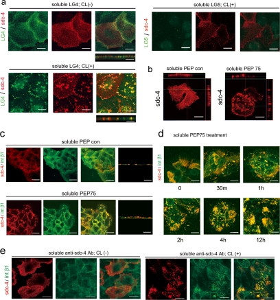
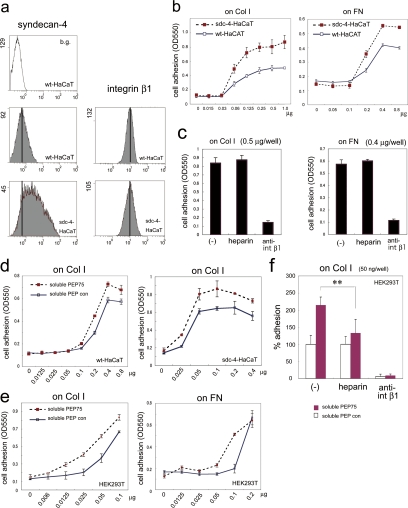
Syndecans function as receptors for extracellular matrix (ECM) with integrins in cell spreading. However, the molecular mechanism of their specific involvement in cell migration or in wound healing has not been elucidated yet. Here, we report that a synthetic peptide, PEP75, which contains the syndecan-binding sequence of the laminin alpha 3LG4 module, induces keratinocyte migration in in vitro and in vivo. Soluble PEP75 induced the clustering of syndecan-4 and conformation-modified integrin beta1 colocalized with syndecan-4 in soluble PEP75-induced clusters. Treatment of cells in solution with PEP75 resulted in the exposure of the P4G11 antibody epitope of integrin beta1 in immunostaining as well as in flow cytometry and augmented integrin beta1-dependent cell adhesion to ECM. Pulldown assays demonstrated that PEP75 bound to syndecan-4, but not to integrin beta1. A siRNA study revealed a role for syndecan-4 in PEP75-induced up-regulation of P4G11 antibody binding and migration of HaCaT cells. We conclude that binding of soluble PEP75 to syndecan-4 induces the coupling of integrin beta1, which is associated with integrin beta1-conformational changes and activation, and leads to keratinocyte migration. To activate integrin function through syndecans could be a novel therapeutic approach for chronic wound.


Figures

Figure 1.
Figure 1.
PEP75 induces integrin β1–dependent keratinocyte migration. (a) Scattering assay of HaCaT cells in the presence or absence of soluble PEP75 in 5% FBS/DMEM. Cells were exposed to peptide in solution for 48 h. Immunofluorescence revealed the disruption of focal contacts (vinculin staining) and stress fibers (F-actin staining). Scale bars, 20 and 250 μm. (b) Scratch wound migration assays with HaCaT cells in 1%FBS/Ca2+-free EMEM were performed for 24 h in the presence of 10 μg/ml soluble PEP75 or PEP con in the presence of heparin or anti-integrin β1 neutralizing antibodies. Cells that migrated from the starting wound edge were counted in randomly selected and nonoverlapping fields, and the results were expressed as means ± SD of three independent experiments. Scale bar, 250 μm. (c) Colloidal gold phagokinetic assay using primary keratinocytes. Cells were incubated with increasing doses of peptide for 12 h or with 15 μg/ml soluble PEP75 for the indicated periods of time. After incubation, three randomly selected and nonoverlapping fields were analyzed using the NIH ImageJ 1.60 program. The migration index (pixels) is expressed as the area consumed by cell migration tracks. Data represent the means ± SD (n = 3). Scale bar, 200 μm. Experiment was performed two times, and representative data are shown. (d) Colloidal gold phagokinetic assays with primary keratinocytes were carried out using the indicated anti-integrin α chain or β1 chain neutralizing antibodies. The migration index (%) is expressed as a percentage relative to cells treated with control IgG, which was set as 100%. Data represents the means ± SD of three independent experiments performed in triplicate. **p = 0.00068 compared with those of IgG.
Figure 2.
Figure 2.
PEP75 promotes wound healing in vivo. (a) A 6-mm full-thickness wound was made on the back of a C57BL/6J mouse, and 15 μl of a 1.0 μg/μl solution of PEP75 was topically applied to the wound twice, on days 1 and 4, with an occlusive dressing. Unclosed areas were photographed and analyzed using the NIH ImageJ 1.60 program. Results are expressed as percent wound healing at day 8 relative to the wound area at day 1, which was set as 100%. Data represents the means ± SD of six animals. **p = 0.014. (b) PEP75 or PEP con (50 μl of a 0.2 μg/μl solution) was applied to a rabbit earlobe skin wound with occlusive dressing once, on day 1. Paraffin-embedded sections were stained with hematoxylin-eosin and then examined by microscopy. Reepithelialization was calculated as the ratio of the diameter of the unclosed (ω) area to that of original wound (W) and is expressed as a percentage of the ratio of the healed wound to that of the original wound, which was set as 100%. Data represents the means ± SD of three animals. **p = 0.0011 at 4 d and 0.0139 at 8 d.
Figure 3.
Figure 3.
Soluble ligands induce syndecan-4 clustering and the colocalization of syndecan-4 and integrin β1. (a) HaCaT cells cultured on glass slides were incubated with soluble recombinant laminin α3LG4- or α3LG5-human Fc chimeric proteins (Momota et al., 2005; 2.0 μg/ml) for 30 min at 4°C, followed by anti-human IgG Fc antibody (20 μg/ml) for 30 min at 37°C for cross-linking (CL+). As a control, anti-human IgG Fc was omitted (CL−). Recombinant α3LG4, but not α3LG5 bound to cell surface syndecan-4. Cross-linking changed the syndecan-4 distribution from fine dots to clusters at cell surface. Side views were also shown. (b) Treatment of a single HaCaT cell with PEP con or PEP75 (10 μg/ml) clearly demonstrated that the clustering of syndecan-4 was located at the apical cell surface. (c) HaCaT cells were treated with soluble PEP con or PEP75 for 2 h in 0.1% BSA/DMEM. Syndecan-4 clustering induced by soluble PEP75 colocalized with integrin β1 (P4G11 antibody). (d) Time-course study of syndecan-4 and integrin β1 clustering that was observed from 30 min to 12 h after the addition of soluble PEP75. (e) Anti-syndecan-4 mAb (2.0 μg/ml) was incubated with HaCaT cells for 30 min at 4°C, followed by cross-linking with anti-mouse IgG (20 μg/ml) for 30 min at 37°C. Antibody-induced syndecan-4 clustering also colocalized with integrin β1. Data are representative of at least two experiments. Scale bar, 20 μm.
Figure 4.
Figure 4.
Integrin β1 that colocalizes with clustered syndecan-4 displays conformational changes. HaCaT cells grown on coverslips were treated with soluble PEP75 or PEP con (10 μg/ml), and the codistribution of syndecan-4 or of heparan sulfate (HS) and integrin β1 was examined by immunostaining. Anti-HS antibody (IgM) was used instead of anti-syndecan-4 antibody when no appropriate fluorescence-conjugated anti-integrin β1 antibodies were available. The colocalization of integrin β1 at sites of syndecan-4 clustering was demonstrated with P4G11 and K20 antibodies. However, unlike P4G11, JB1A, mAb13, and 12G10 antibodies failed to detect integrin β1 at sites of HS clustering. The data shown is representative of at least three experiments. Scale bar, 20 μm.
Figure 5.
Figure 5.
Integrin β1 conformational changes are induced by PEP75 in solution. Cells prepared in suspension (106 cells) were incubated with soluble PEP75 (100 μg/ml) for 30 min at 37°C, followed by incubation with the indicated antibodies on ice. (a) PEP75 significantly increased of the fluorescence intensity (F.I.; x-axis) of HaCaT cells and fibroblasts when P4G11 mAb was used as a probe, and heparin completely blocked this increase. The cells were collected after flow cytometry and examined by confocal laser scanning microscopy. Scale bar, 20 μm. (b) JB1A, K20, and 12G10 antibodies failed to detect changes in the F.I. after treatment with soluble PEP75. The faint gray color represents background fluorescence of JB1A. (c) In the presence of 2 mM Mn2+ for 30 min at 37°C, P4G11 associated F.I. increased, whereas a AG89-associated F.I. was almost completely lost. PEP75 and (Mn2+) showed a similar response pattern. (d) Anti-integrin β1 antibody (JB1A) or control mouse IgG was incubated with cells for 30 min at 0°C, followed by treatment with (CL+) or without cross-linking antibody (CL−) for 30 min at 37°C. Then, P4G11 was incubated with cells on ice for flow cytometry. Antibody-mediated cross-linking of integrin β1 using anti-JB1A antibody increased the P4G11-associated F.I. The data are representative of at least two experiments.
Figure 6.
Figure 6.
Overexpression of syndecan-4 enhances integrin β1–dependent cell adhesion to ECM proteins and PEP75 treatment in solution augments integrin β1–dependent cell adhesion. (a) Syndecan-4 and integrin β1 expression in stable HaCaT transformants that overexpressed syndecan-4 (sdc-4-HaCaT) and parental HaCaT cells (wt-HaCaT) was analyzed by flow cytometry. JB1A antibody was used for integrin β1. b.g., background. (b) Adhesion assay of sdc-4-HaCaT and wt-HaCaT cells on 96-well plates coated with various amounts of collagen I (Col I) and fibronectin (FN). y-axis, cell number shown as OD550; x-axis, coated substrates: μg/well. (c) Sdc-4-HaCaT adhesion to Col I- or FN-coated dishes was dependent on integrin β1 and was not affected by heparin. Cells were seeded onto Col 1 (0.5 μg/well) or FN (0.4 μg/well). Cells were allowed to adhere for 45 min. For inhibition assays, cells were preincubated with heparin (10 μg/ml) or anti-integrin β1 antibody (mAb13, 12 μg/ml) for 15 min before seeding. Heparin and mAb13 were also present during the assay. (d) Incubation with soluble PEP75 (10 μg/ml) for 30 min in solution enhanced cell adhesion to collagen I in both wt-HaCaT and sdc-4-HaCaT cells. (e) Adhesion of HEK293T cells to Col I and FN was also enhanced by soluble PEP75 treatment in solution. (f) The soluble PEP75-induced adhesion of HEK293T cells to Col I was reduced by heparin and was entirely dependent on integrin β1. (b–e) Data represents the means ± SD of at least three experiments performed in triplicate. For the inhibition experiments in panel f, the percentage of adherent cells is expressed relative to cells treated with PEP con in the absence of inhibitor, which was set as 100%. **p = 0.0069.
Figure 7.
Figure 7.
Specific involvement of syndecan-4 in integrin β1 conformational changes and cell migration. (a) Pulldown assay with peptide-conjugated Sepharose beads. HaCaT cell lysates were incubated with peptide beads. For syndecans, bound materials were digested with heparitinase I/chondrotinase ABC and were then subjected to 10% SDS-PAGE and Western blotting. Syndecan-4, but not -1 specifically bound to PEP75. For integrin β1, bound materials were directly eluted by boiling in SDS-buffer and separated by 7% SDS-PAGE. Immunoprecipitation was also done to indicate the input amount of integrin β1 (input). **Note that syndecan-1 (∼90 kDa) bound only at a background level to both peptide-beads. (b) siRNA-mediated knockdown is shown by flow cytomeric analysis for syndecan-4 (siRNA sdc-4 RSS340730) or by Western blotting for syndecan-1 (siRNA sdc-1 RSS303157). Flow cytomeric analysis revealed that integrin β1 conformational changes after soluble PEP75 treatment were dependent on stndecan-4, but not on syndecan-1. The total fluorescence intensity is indicated in parenthesis. In Western blotting of syndecan-1, optical densities of the bands were obtained by NIH image and are shown in parentheses. The siRNA assays were performed using three different siRNAs. The data are representative of at least two independent experiments with each siRNA. Knockdown of syndecan-4, but not -1 resulted in severely decreased cell mobility in a scratch wound assay of HaCaT cells in 1%FBS/Ca2+-free EMEM. Data are representative of at least two experiments. The number of cells that migrated from the starting wound edge was counted in randomly selected and nonoverlapping fields. Data are shown as means ± SD of two independent experiments. The results obtained with syndecan-4 siRNA (RSS340730) and syndecan-1 siRNA (RSS303157) are shown for both flow cytometry analysis and scratch wound assay.
Figure 8.
Figure 8.
Analysis of the signaling molecules involving in PEP75-induced biological activities. Chemical inhibitors to signal molecules were examined in PEP75-induced activities by scratch wound healing assays (a), flow cytometry analysis of P4G11 binding (b), and augmented cell adhesion assay (c). (a) HaCaT cells were incubated with soluble PEP75 or PEP con (10 μg/ml) in 1%FBS/Ca2+-free EMEM in the presence or absence of chemical inhibitor. Scale bar, 250 μm. The number of cells that migrated from the starting wound edge was counted in randomly selected and nonoverlapping fields and the data are shown as means ± SD of two independent experiments. (b) HaCaT cells in suspension were incubated with chemical inhibitors for 15 min, and followed by addition of peptide (100 μg/ml) for 30 min at 37°C. None of chemical inhibitors blocked the increase of P4G11 binding induced by PEP75 treatment. The data are representative of two independent experiments. (c) Soluble PEP75 (10 μg/ml augmentation of cell adhesion of HEK293T cells on collagen I was examined in the presence or absence of chemical inhibitors. The assay was performed in sextuplicate, and the data represents the means ± SD. Data are representative of at least two experiments. The number of attached cells in the presence of PEP con was set as 100%. **p = 0.00022. For inhibition studies, cells were preincubated for 15 min with the indicated chemical inhibitors and then incubated with soluble peptide for 30 min at 37°C. The Rac1 inhibitor (NSC23766) was used at concentrations of 100 μM in scratch assay and flow cytometric analysis or 100 and 10 μM in augmented cell adhesion assays.

References

    1. Alexopoulou A. N., Multhaupt H. A., Couchman J. R. Syndecans in wound healing, inflammation and vascular biology. Int. J. Biochem. Cell Biol. 2007;39:505–528. - PubMed
    1. Bass M. D., Roach K. A., Morgan M. R., Mostafavi-Pour Z., Schoen T., Muramatsu T., Mayer U., Ballestrem C., Spatz J. P., Humphries M. J. Syndecan-4-dependent Rac1 regulation determines directional migration in response to the extracellular matrix. J. Cell Biol. 2007;177:527–538. - PMC - PubMed
    1. Beauvais D. M., Burbach B. J., Rapraeger A. C. The syndecan-1 ectodomain regulates alphavbeta3 integrin activity in human mammary carcinoma cells. J. Cell Biol. 2004;167:171–181. - PMC - PubMed
    1. Berrier A. L., Yamada K. M. Cell-matrix adhesion. J. Cell Physiol. 2007;213:565–573. - PubMed
    1. Carey D. J., Stahl R. C., Tucker B., Bendt K. A., Cizmeci-Smith G. Aggregation-induced association of syndecan-1 with microfilaments mediated by the cytoplasmic domain. Exp. Cell Res. 1994;214:12–21. - PubMed

Publication types

MeSH terms

LinkOut - more resources