

9-O-acetylated sialoglycoproteins are important immunomodulators in Indian visceral leishmaniasis
- PMID: 19403782
- PMCID: PMC2691061
- DOI: 10.1128/CVI.00453-08
9-O-acetylated sialoglycoproteins are important immunomodulators in Indian visceral leishmaniasis
Erratum in
-
Correction for Ghoshal et al., "9-O-Acetylated Sialoglycoproteins Are Important Immunomodulators in Indian Visceral Leishmaniasis".mSphere. 2024 Jun 25;9(6):e0015724. doi: 10.1128/msphere.00157-24. Epub 2024 May 21. mSphere. 2024. PMID: 38771096 Free PMC article. No abstract available.
Abstract
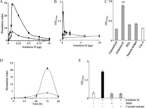
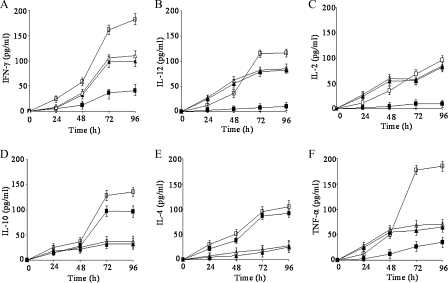
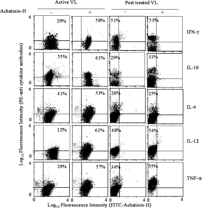
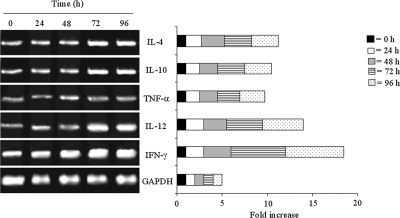
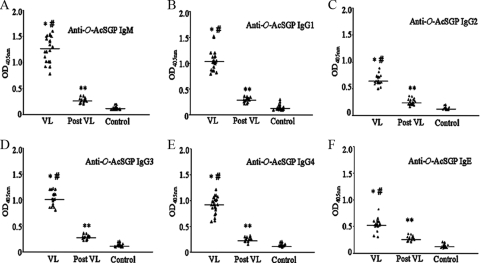
Overexpression of disease-associated 9-O-acetylated sialoglycoproteins (9-O-AcSGPs) on peripheral blood mononuclear cells (PBMC) of visceral leishmaniasis (VL) patients (PBMC(VL)) compared to their levels of expression in healthy individuals has been demonstrated using a lectin, achatinin-H, with specificity toward 9-O-acetylated sialic acid derivatives alpha2-6 linkage with subterminal N-acetylgalactosamine (9-O-AcSAalpha2-6GalNAc). The decreased presence of disease-associated 9-O-AcSGPs on different immune cells of parasitologically cured individuals after successful treatment relative to the levels in patients with active VL prior to treatment was demonstrated. However, their contributory role as immunomodulatory determinants on PBMC(VL) remained unexplored. Accordingly, 9-O-AcSGPs on PBMC(VL) were sensitized with achatinin-H, leading to their enhanced proliferation compared to that observed with different known mitogens or parasite antigen. This lymphoproliferative response was characterized by evaluation of the TH1/TH2 response by intracellular staining and enzyme-linked immunosorbent assay for secreted cytokines, and the results were corroborated by their genetic expression. Sensitized PBMC(VL) evidenced a mixed TH1/TH2 cellular response with a predominance of the TH1 response, indicating the ability of 9-O-AcSGPs to modulate the host cell toward a favorable response. Interestingly, the humoral and cellular responses showed a good correlation. Further, high levels of anti-9-O-AcSGP antibodies with an order of distribution of immunoglobulin M (IgM) > IgG1 = IgG3 > IgG4 > IgG2 > IgE could be explained by a mixed TH1/TH2 response. A good correlation of enhanced 9-O-AcSGPs with both the cell-mediated (r = 0.98) and humoral (r = 0.99) response was observed. In summary, it may be concluded that sensitization of 9-O-AcSGPs on PBMC(VL) may provide a basis for the modulation of the host's immune response by their controlled expression, leading to a beneficial immune response and influencing the disease pathology.
Figures
Similar articles
-
Identification of 9-O-acetylated sialoglycans on peripheral blood mononuclear cells in Indian Visceral Leishmaniasis.Glycoconj J. 2004;20(9):531-6. doi: 10.1023/B:GLYC.0000043289.86611.44. Glycoconj J. 2004. PMID: 15454691
-
Immunoglobulin subclass distribution and diagnostic value of Leishmania donovani antigen-specific immunoglobulin G3 in Indian kala-azar patients.Clin Diagn Lab Immunol. 1999 Mar;6(2):231-5. doi: 10.1128/CDLI.6.2.231-235.1999. Clin Diagn Lab Immunol. 1999. PMID: 10066659 Free PMC article.
-
Cytosolic tryparedoxin of Leishmania donovani modulates host immune response in visceral leishmaniasis.Cytokine. 2018 Aug;108:1-8. doi: 10.1016/j.cyto.2018.03.010. Epub 2018 Mar 16. Cytokine. 2018. PMID: 29554571
-
The immunology of post-kala-azar dermal leishmaniasis (PKDL).Parasit Vectors. 2016 Aug 23;9(1):464. doi: 10.1186/s13071-016-1721-0. Parasit Vectors. 2016. PMID: 27553063 Free PMC article. Review.
-
Immunotherapy for visceral leishmaniasis: A trapeze of balancing counteractive forces.Int Immunopharmacol. 2022 Sep;110:108969. doi: 10.1016/j.intimp.2022.108969. Epub 2022 Jun 20. Int Immunopharmacol. 2022. PMID: 35738089 Review.
Cited by
-
Highly sialylated mucin-type glycopeptide from porcine intestinal mucosa after heparin extraction: O-glycan profiling and immunological activity evaluation.Glycoconj J. 2021 Oct;38(5):527-537. doi: 10.1007/s10719-021-10014-y. Epub 2021 Sep 4. Glycoconj J. 2021. PMID: 34480673
-
Glycosylation of erythrocyte spectrin and its modification in visceral leishmaniasis.PLoS One. 2011;6(12):e28169. doi: 10.1371/journal.pone.0028169. Epub 2011 Dec 2. PLoS One. 2011. PMID: 22164239 Free PMC article.
-
Sialic acids siglec interaction: a unique strategy to circumvent innate immune response by pathogens.Indian J Med Res. 2013 Nov;138(5):648-62. Indian J Med Res. 2013. PMID: 24434319 Free PMC article. Review.
-
A perspective on the emergence of sialic acids as potent determinants affecting leishmania biology.Mol Biol Int. 2011;2011:532106. doi: 10.4061/2011/532106. Epub 2011 Jul 25. Mol Biol Int. 2011. PMID: 22091406 Free PMC article.
-
The Role of Sialic Acids in the Establishment of Infections by Pathogens, With Special Focus on Leishmania.Front Cell Infect Microbiol. 2021 May 13;11:671913. doi: 10.3389/fcimb.2021.671913. eCollection 2021. Front Cell Infect Microbiol. 2021. PMID: 34055669 Free PMC article. Review.
References
-
- Abbas, A. K., K. M. Murphy, and A. Sher. 1996. Functional diversity of helper T lymphocytes. Nature 383787-793. - PubMed
-
- Atta, A. M., D'Oliveira, J. Correa, M. L. Atta, R. P. Almeida, and E. M. Carvalho. 1998. Anti-leishmanial IgE antibodies: a marker of active disease in visceral leishmaniasis. Am. J. Trop. Med. Hyg. 59426-430. - PubMed
-
- Bacellar, O., C. Brodskyn, J. Guerreiro, M. Barral-Netto, C. H. Costa, R. L. Coffman, W. D. Johnson, and E. M. Carvalho. 1996. Interleukin 12 restores IFN-gamma production and cytotoxic responses in visceral leishmaniasis. J. Infect. Dis. 1731515-1518. - PubMed
-
- Bandyopadhyay, S., M. Chatterjee, S. Sundar, and C. Mandal. 2004. Identification of 9-O-acetylated sialoglycans on peripheral blood mononuclear cells in Indian visceral leishmaniasis. Glycoconj. J. 20531-536. - PubMed
-
- Bandyopadhyay, S., M. Chatterjee, T. Das, S. Bandyopadhyay, S. Sundar, and C. Mandal. 2004. Antibodies directed against O-acetylated sialoglycoconjugates accelerate complement activation in Leishmania donovani promastigotes. J. Infect. Dis. 1902010-2019. - PubMed
Publication types
MeSH terms
Substances
LinkOut - more resources
Full Text Sources
