

In vivo measurement of axon diameter distribution in the corpus callosum of rat brain
- PMID: 19403788
- PMCID: PMC2677796
- DOI: 10.1093/brain/awp042
In vivo measurement of axon diameter distribution in the corpus callosum of rat brain
Abstract
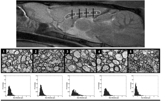
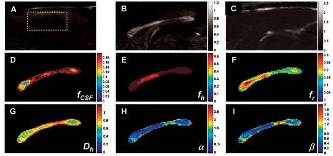
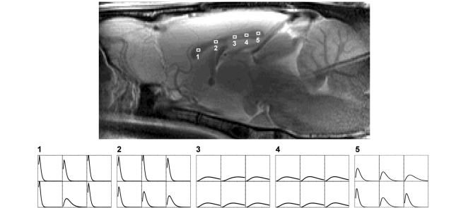
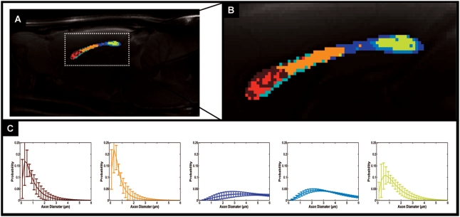
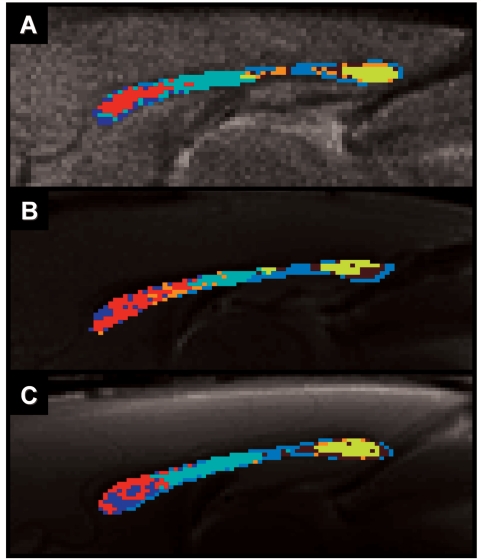
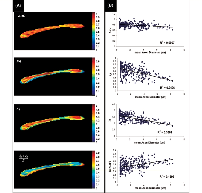
Here, we present the first in vivo non-invasive measurement of the axon diameter distribution in the rat corpus callosum. Previously, this measurement was only possible using invasive histological methods. The axon diameter, along with other physical properties, such as the intra-axonal resistance, membrane resistance and capacitance etc. helps determine many important functional properties of nerves, such as their conduction velocity. In this work, we provide a novel magnetic resonance imaging method called AxCaliber, which can resolve the distinct signatures of trapped water molecules diffusing within axons as well as water molecules diffusing freely within the extra-axonal space. Using a series of diffusion weighted magnetic resonance imaging brain scans, we can reliably infer both the distribution of axon diameters and the volume fraction of these axons within each white matter voxel. We were able to verify the known microstructural variation along the corpus callosum of the rat from the anterior (genu) to posterior (splenium) regions. AxCaliber yields a narrow distribution centered approximately 1 microm in the genu and splenium and much broader distributions centered approximately 3 microm in the body of the corpus callosum. The axon diameter distribution found by AxCaliber is generally broader than those usually obtained by histology. One factor contributing to this difference is the significant tissue shrinkage that results from histological preparation. To that end, AxCaliber might provide a better estimate of the in vivo morphology of white matter. Being a magnetic resonance imaging based methodology, AxCaliber has the potential to be used in human scanners for morphological studies of white matter in normal and abnormal development, and white matter related diseases.
Figures
References
-
- Aboitiz F, Montiel J. One hundred million years of interhemispheric communication: the history of the corpus callosum. Braz J Med Biol Res. 2003;36:409–20. - PubMed
-
- Aboitiz F, Scheibel AB, Fisher RS, Zaidel E. Fiber composition of the human corpus callosum. Brain Res. 1992;598:143–53. - PubMed
-
- Assaf Y, Basser PJ. Composite hindered and restricted model of diffusion (CHARMED) MR imaging of the human brain. Neuroimage. 2005;27:48–58. - PubMed
-
- Assaf Y, Basser PJ. Non parametric approach for axon diameter distribution estimation from diffusion measurements. Proc Intl Soc Magn Reson Med. 2007;15:1536.
-
- Assaf Y, Ben-Bashat D, Chapman J, Peled S, Biton IE, Kafri M, et al. High b-value q-space analyzed diffusion-weighted MRI: application to multiple sclerosis. Magn Reson Med. 2002a;47:115–26. - PubMed
Publication types
MeSH terms
Grants and funding
LinkOut - more resources
Full Text Sources
Other Literature Sources
