Comparative Study
. 2009 Apr 29;29(17):5389-401.
doi: 10.1523/JNEUROSCI.5129-08.2009.

Protracted withdrawal from alcohol and drugs of abuse impairs long-term potentiation of intrinsic excitability in the juxtacapsular bed nucleus of the stria terminalis

Affiliations
Comparative Study

Protracted withdrawal from alcohol and drugs of abuse impairs long-term potentiation of intrinsic excitability in the juxtacapsular bed nucleus of the stria terminalis

Walter Francesconi et al. J Neurosci. .

Abstract

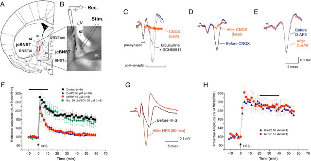
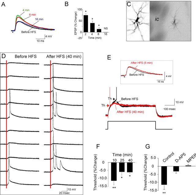
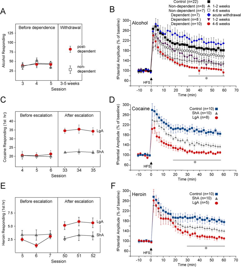
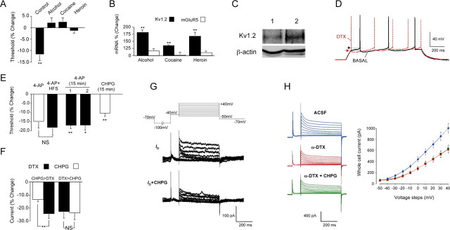
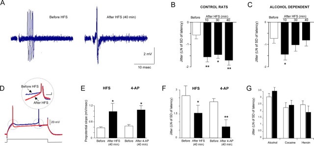
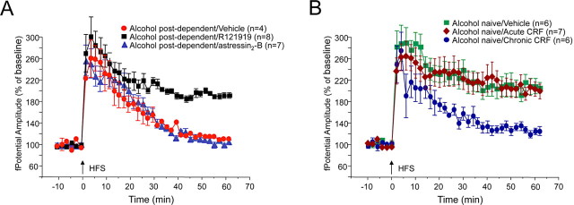
The juxtacapsular bed nucleus of the stria terminalis (jcBNST) is activated in response to basolateral amygdala (BLA) inputs through the stria terminalis and projects back to the anterior BLA and to the central nucleus of the amygdala. Here we show a form of long-term potentiation of the intrinsic excitability (LTP-IE) of jcBNST neurons in response to high-frequency stimulation of the stria terminalis. This LTP-IE, which was characterized by a decrease in the firing threshold and increased temporal fidelity of firing, was impaired during protracted withdrawal from self-administration of alcohol, cocaine, and heroin. Such impairment was graded and was more pronounced in rats that self-administered amounts of the drugs sufficient to maintain dependence. Dysregulation of the corticotropin-releasing factor (CRF) system has been implicated in manifestation of protracted withdrawal from dependent drug use. Administration of the selective corticotropin-releasing factor receptor 1 (CRF(1)) antagonist R121919 [2,5-dimethyl-3-(6-dimethyl-4-methylpyridin-3-yl)-7-dipropylamino-pyrazolo[1,5-a]pyrimidine)], but not of the CRF(2) antagonist astressin(2)-B, normalized jcBNST LTP-IE in animals with a history of alcohol dependence; repeated, but not acute, administration of CRF itself produced a decreased jcBNST LTP-IE. Thus, changes in the intrinsic properties of jcBNST neurons mediated by chronic activation of the CRF system may contribute to the persistent emotional dysregulation associated with protracted withdrawal.


Figures

Figure 1.
Figure 1.
Field potential in the jcBNST. A, Coronal brain slices were obtained from the rostral cerebrum of Wistar rats at the level indicated in the diagram (modified from Paxinos and Watson, 1998). st, Stria terminalis; jcBNST, juxtacapsular bed nucleus of the stria terminalis (shown in red); BNSTdl, dorsolateral bed nucleus of the stria terminalis; BNSTdm, dorsomedial bed nucleus of the stria terminalis; BNSTv, ventral bed nucleus of the stria terminalis; ac, anterior commissure; LV, lateral ventricle; ic, internal capsule. The boundary of the jcBNST is in good agreement with Dong et al. (2000) and was operationally defined by the area in which a glutamatergic field potential was readily evoked by stimulation of the stria terminalis. B, Photomicrograph of a brain slice at the level of the BNST demonstrating electrode placements for stimulation (Stim.) and electrophysiological recordings (Rec.). C, Field potential evoked in the jcBNST by stimulation of the stria terminalis in coronal brain slices is characterized by two fast negative components followed by a more variable slow positive deflection (blue trace). The second negative component of the field potential and the positive deflection were abolished by application of the AMPA glutamate receptor inhibitor CNQX in bath (20 μm) (red trace). Blocking GABAA and GABAB receptors with bicuculline (30 μm) and SCH50911 (20 μm), respectively, increased the size of the postsynaptic components of the field potential without altering the field morphology (black trace). D, Local application of CNQX by diffusion of the inhibitor included in the recording electrode (25 mm) blocked the second negative and the positive components of the field potential (red trace), suggesting they are postsynaptic responses that originate locally in the jcBNST. E, Bath application of the NMDA inhibitor d-AP-5 (50 μm) had no effect on the field potential (red trace). Stimuli artifacts were removed, and the asterisk indicates their location. F, Delivery of HFS (5 trains at 100 Hz for 1 s at 10 s intervals) resulted in a protracted potentiation of the amplitude of the postsynaptic component of the field potential (sample traces are shown in G). A transient application of either d-AP-5 or the mGluR5 inhibitor MPEP around the time of delivery of HFS (horizontal bar) prevented potentiation of the amplitude of the field potential. However, tetanization during continuous application of the GABAA and GABAB inhibitors bicuculline and SCH50911, respectively, resulted in the same degree of potentiation as controls (baseline renormalized). H, Application of either d-AP-5 or the mGluR5 inhibitor MPEP to an established LTP of field potential (horizontal bar) was ineffective.
Figure 2.
Figure 2.
LTP-IE in the jcBNST. A, HFS of the stria terminalis induced only a transient EPSP potentiation that reverted to basal levels within 16 min. Stimuli artifacts were removed, and the asterisk indicates their location. B, Quantification of EPSP amplitude changes in cells of the jcBNST at the indicated times after HFS of the stria terminalis (*p < 0.05 from baseline; NS, t(25) = 0.123). C, jcBNST neurons visualized by intracellular injection of biocytin after recording demonstrate a multipolar morphology (ic, internal capsula). D, HFS caused increased probability of firing in response to single stimuli (red line) applied to the stria terminalis. Traces shown are 10 sweeps representing evoked responses before and 40 min after HFS. E, Threshold (arrowheads) for action potential generation in response to a depolarizing current pulse (500 ms, 0.07 nA) was shifted to more hyperpolarized membrane potentials for a protracted time after HFS (red trace). Inset, Such a shift also could be observed even in the absence of potentiation of EPSP after HFS (red trace), as in the representative neuron shown (same neuron as in D). F, The shift of the threshold for action potential generation after HFS was significant for over 40 min after HFS (**p < 0.01 from baseline, n = 14, at 10 min, t = 4.26; *p < 0.05 from baseline at 25 min, t = 2.72, and at 40 min, t = 6.66). G, HFS-induced shift of the action potential threshold was prevented by a transient application for 20 min around the time of delivery of HFS (as in Fig. 1F) of either the NMDA inhibitor d-AP-5 (50 μm) or the mGluR inhibitor MPEP (10 μm) in the perfusion bath (t = −8.491,*p < 0.05 from control, n = 15; for d-AP-5, t = −1.376, n = 8, NS; and for MPEP, t = 1.044, n = 6, NS).
Figure 3.
Figure 3.
Disruption of LTP-IE in the jcBNST during protracted withdrawal from self-administration of alcohol, cocaine, and heroin. A, Operant responding for ethanol over the last three self-administration sessions before induction of dependence (left panel) and 3–5 weeks after withdrawal (right panel). The average number of presses for ethanol in the post-dependent group during protracted abstinence (right panel) was significantly increased over the nondependent group (p < 0.01). B, In dependent rats, potentiation of the amplitude of the field potential in the jcBNST, which is a manifestation of LTP-IE, as shown in Figure 2, progressively declined over time and was completely abolished at 4–6 weeks after withdrawal 60 min after HSF. In dependent rats tested during acute withdrawal (3–6 h), the potentiation of the field potential was not impaired. In nondependent rats, potentiation of the field potential was impaired at 1–2 weeks after alcohol self-administration but was readily observed at 4–6 weeks after cessation of alcohol self-administration. Two-way ANOVA results partitioned for condition (F(5,1680,0.05) = 195.33, p < 0.001) and for time (F(29,1680,0.05) = 19.0, p < 0.001), with no significant interaction between these factors. *Dependent at 4–6 weeks different from control, dependent during acute withdrawal, and non-dependent at both 1–2 and 4–6 weeks (p < 0.01); dependent at 1–2 weeks different from control and dependent during acute withdrawal (p < 0.05); non-dependent at 1–2 weeks different from control and non-dependent at 4–6 weeks (p < 0.05). C, Rats were trained to self-administer cocaine on a fixed-ratio 1 schedule and allowed access to cocaine self-administration for either 1 h (ShA) or 6 h (LgA) per day; the latter induces an escalating pattern of cocaine intake. In the first hour of cocaine self-administration during the last three self-administration sessions (right panel), LgA rats averaged 35.0 ± 0.8 cocaine infusions versus 22.4 ± 0.3 in ShA rats (p < 0.01). D, Potentiation of field potential in the jcBNST of brain slices from LgA rats was impaired 1–2 weeks after cessation of cocaine self-administration. ShA rats showed a level of potentiation that was between that of control and LgA rats. Two-way ANOVA results partitioned for access to self administration (F(2,750,0.05) = 269.0, p < 0.001) and for time (F(29,750,0.05) = 13.5, p < 0.001), with no significant interaction between these factors. *LgA different from control and ShA (p < 0.01); ShA different from control (p < 0.01). E, Rats were similarly trained to self-administer heroin on a fixed-ratio 1 schedule and allowed access for either 1 h (ShA) or 23 h (LgA) per day. In the first hour of heroin self-administration during the last three self-administration sessions (right panel), LgA rats averaged 5.54 ± 0.2 infusions versus 3.05 ± 0.2 for ShA rats (p < 0.01). F, Also in LgA heroin self-administering rats, potentiation of the field potential in the jcBNST was impaired 1–2 weeks after withdrawal. ShA rats showed a partial level of impairment at the same time after cessation of self-administration. Two-way ANOVA results partitioned for access to self administration (F(2,690,0.05) = 114.42, p < 0.001) and for time (F(29,690,0.05) = 14.15, p < 0.001), with no significant interaction between these factors. *LgA different from control (p < 0.01) and ShA (p < 0.05); ShA different from control (p < 0.01).
Figure 4.
Figure 4.
The threshold for action potential generation in jcBNST neurons is regulated by the ID current. A, HFS-induced shift toward hyperpolarized threshold for action potential generation was not seen in brain slices from ethanol (n = 8), cocaine (n = 8), or heroin (n = 8) post-dependent rats, unlike in controls (n = 14) measured 10 min after HFS (t = 4.14, **p < 0.01 for baseline; alcohol, t = −0.99, NS; cocaine, t = −1.44, NS; heroin, t = 1.10, NS). B, Combined microarray and qPCR analyses demonstrated that expression of Kv1.2, a member of the Kv family of K+ channels implicated in mediating the ID current, was significantly increased in post-dependent rats, whereas the expression of mGluR5 and other genes potentially involved in ID regulation was unaltered. Bar graph represents a quantification of the qPCR validation of the microarray results. Kv1.2 (black bars): From control, t = −7.76, n = 15; for alcohol, t = −4.25, n = 5; and cocaine, t = −3.92, n = 5; n = 7 for heroin; **p < 0.01). mGluR5 (white bars): For alcohol, t = −0.621; for cocaine, t = −0.178; for heroin, t = −0.854. C, Kv1.2 was significantly increased in the jcBNST of ethanol post-dependent (2) rats versus control (1) rats by Western blotting (n = 5, one-tail t test, t = 2.07, n = 5–6, *p < 0.05). D, Pharmacological inhibition of the ID current by bath application of the ID blocker α-DTX (1 μm) shifted the action potential threshold toward more hyperpolarized membrane potentials (arrowheads indicate action potential threshold as in Fig. 2D), and the frequency of firing was increased. Patch whole-cell recordings were performed in current-clamp mode. Current pulses of 1 s duration were applied at the resting membrane potential (in this case, approximately −80 mV) to elicit action potentials. E, Application of 4-AP (40 μm) also induced a protracted shift of the threshold for action potential generation toward hyperpolarization (from −51.9 ± 2.6 mV before 4-AP application to −59.1 ± 2.4 mV 40 min after 4-AP application, n = 9, white bar labeled 4-AP) similarly to an LTP-IE-inducing HFS. Delivery of HFS in the presence of 4-AP (4-AP + HFS) did not further shift the action potential threshold (t = 0.21, NS, n = 9), suggesting that HFS acts at least in part by reducing ID. A transient (15 min) application of 4-AP (40 μm) also induced a persistent shift of the threshold for action potential generation from baseline that was significant for over 40 min after washout (column marked 1 in the graph; t = 3.379, n = 6, p = 0.010, at 40 min after washout). In a separate set of cells, synaptic transmission was blocked by application of the AMPA inhibitor CNQX (10 μm), the NMDA inhibitor d-AP-5 (50 μm), the mGluR5 inhibitor MPEP (10 μm), the GABAA inhibitor bicuculline (30 μm), and the GABAB inhibitor SCH50911 (20 μm) 15 min before the transient (15 min) application of 4-AP (40 μm). Again, a persistent shift of the threshold for action potential generation from baseline was observed that was significant for over 40 min after washout (column marked 2 in the graph; t = 2.92, n = 6, p = 0.017, at 40 min after washout). A persistent shift of the threshold for action potential generation was also induced by a transient (15 min) application of the specific mGluR5 agonist CHPG (200 μm) (t = 6.292, n = 5, p = 0.002, at 40 min after washout). F, Activation of mGluR5 by 200 μm CHPG reduced the whole-cell current by 13.2 ± 1.96% compared with control. Subsequent application of 1 μm α-DTX to block the ID current significantly reduced the whole-cell current to 23.9 ± 3.73% compared with control (t = 3.43, **p < 0.01, n = 8). When α-DTX was applied first, it reduced the whole-cell current by 22.7 ± 5.14%, and subsequent application of CHPG no longer had an effect on the whole-cell current (23.3 ± 5.18%, t = 1.04, NS, n = 6). Sample recordings are shown in G. G, Top panel shows the voltage protocol used in patch whole cell to obtain the records shown below and in I. Cells were held in voltage-clamp mode at −70 mV, and a 2000 ms pulse to −100 mV was applied so that a maximal amount of ID was available for activation during subsequent voltage steps. This was followed by a 100 ms pulse to −40 mV to inactivate the A-current. Voltage steps from −50 to +40 mV then were applied for 500 ms, and these were used for analysis of ID. Middle panel demonstrates ID by subtracting the recording made in the presence of α-DTX (1 μm) from the control recording (Bekkers and Delaney, 2001). Bottom panel shows ID during application of CHPG (200 μm) in the same cell as the panel above. CHPG blocked 56.9 ± 4.8% (n = 8) of the ID. H, CHPG had no effect on whole-cell current (left) and I–V relationship (right) when applied after application of α-DTX, indicating that CHPG did not affect currents other than the ID. Blue traces and blue line (diamond) in the graph represent total K+ whole-cell current in aCSF; red traces and red line (triangles) in the graph represent K+ whole-cell current in the presence of α-DTX; green traces and green line (squares) in the graph represent K+ whole-cell current in the presence of α-DTX and CHPG.
Figure 5.
Figure 5.
Activity-dependent increase of temporal fidelity of firing of jcBNST neurons. Extracellular single unit (A–C) and intracellular recordings (D–G) were used to investigate temporal fidelity in the jcBNST. A, Five successive extracellularly recorded action potentials locked to the stimulus applied to the stria terminalis were superimposed. Delivery of HFS reduced the latency to the first spike and jitter of the spike in the jcBNST for a protracted time. B, Quantification of the jitter in jcBNST neurons after HFS in control rats. The natural logarithm of the SD of the latency of action potential was used as a measure of the jitter of spike. In the jcBNST, the jitter was significantly reduced from control for up to 40 min after HFS (after 10 min, t = 6.11, p < 0.01; after 30 min, t = 2.18, p < 0.05; and after 40 min, t = 6.49, p < 0.01; n = 7). C, Conversely, rats with a history of alcohol dependence had only a transient reduction of jitter after HFS (after 10 min, t = 3.68, p < 0.05; after 30 min, t = 1.33, NS from before HFS, and after 40 min, t = 0.53, NS from before HFS; n = 5). D, Sample traces showing action potentials evoked by a 350 ms depolarizing pulse in jcBNST neurons by intracellular recording in control rats before and 40 min after HFS of the stria terminalis demonstrating HFS induced shortening of spike latency. Inset shows superimposed traces from the same neuron showing increased depolarizing prepotential slope after HFS (40 min). E, Average depolarizing prepotential slope was significantly increased 40 min after HFS in control rats (t = −1.89, p < 0.05; n = 9) (left bars). Similarly, bath application of the ID blocker 4-AP (40 μm) mimicked the HFS-induced increase in depolarizing prepotential slope (t = 2.06, p < 0.05, n = 9) (right bars). F, The jitter was significantly decreased in the same recordings 40 min after HFS (t = 2.12, p < 0.05; n = 9) (left bars). Similarly, a decrease of the jitter from was also observed 40 min after 4-AP application (t = 2.97, p < 0.01; n = 9) (right bars). G, In rats with history of alcohol, cocaine, or heroine dependence no significant decreases of jitter were observed 40 min after HFS (t = −1.37, t = −0.83, t = 1.04, NS, respectively, for n = 8, n = 10, n = 9, respectively); white bars indicate jitter of spike before HFS, and black bars indicate jitter of the spike after HFS. *p < 0.05, **p < 0.01.
Figure 6.
Figure 6.
Double in situ hybridization for GAD65 and GAD67 and for CRF in the dorsal BNST. A, A large number of GAD-containing neurons (shown in purple) are present in the jcBNST and in the dorsolateral BNST in general. In situ hybridization signal for CRF (brown grains) was seen in the dorsolateral BNST, including the jcBNST, and were primarily colocalized with GAD65 and GAD67. LV, Lateral ventricle. B, Higher magnification of the area in the dotted box in A shows high level of colocalization of CRF with GAD65 and GAD67 in the midsection of the jcBNST. C, Higher magnification of the two neurons containing CRF and GAD65 and GAD67 signal marked by the arrows in B.
Figure 7.
Figure 7.
Disruption of jcBNST LTP in protracted abstinence is attributable to activation of the CRF system. A, Subcutaneous administration of the selective CRF1 receptor antagonist R121919 (three 20 mg/kg injections, 12 h apart) restored normal jcBNST LTP in alcohol post-dependent rats 4 weeks after withdrawal, significantly increasing jcBNST LTP compared with vehicle (20% hydroxypropyl β-cyclodextrin, pH 4.5). Conversely, intracerebroventricular injection for 3 d of the selective CRF2 receptor antagonist A2-B (4 μg/μl in 2 μl every 12 h) did not restore capacity for potentiation of the amplitude of the field potential in alcohol post-dependent rats 4 weeks after withdrawal. BNST slices were prepared 1 h after the final antagonist injection. B, Repeated intracerebroventricular administration of CRF (2 daily injections of 1 μg for 10 d) induced a significant impairment of jcBNST LTP in drug-naive rats, whereas a single administration of CRF (1 μg 2 h before the animals were killed) had no effect.

Similar articles

Cited by

References

    1. Ahmed SH, Kenny PJ, Koob GF, Markou A. Neurobiological evidence for hedonic allostasis associated with escalating cocaine use. Nat Neurosci. 2002;5:625–626. - PubMed
    1. Ahmed SH, Lutjens R, van der Stap LD, Lekic D, Romano-Spica V, Morales M, Koob GF, Repunte-Canonigo V, Sanna PP. Gene expression evidence for remodeling of lateral hypothalamic circuitry in cocaine addiction. Proc Natl Acad Sci U S A. 2005;102:11533–11538. - PMC - PubMed
    1. Aizenman CD, Linden DJ. Rapid, synaptically driven increases in the intrinsic excitability of cerebellar deep nuclear neurons. Nat Neurosci. 2000;3:109–111. - PubMed
    1. Armano S, Rossi P, Taglietti V, D'Angelo E. Long-term potentiation of intrinsic excitability at the mossy fiber-granule cell synapse of rat cerebellum. J Neurosci. 2000;20:5208–5216. - PMC - PubMed
    1. Bekkers JM, Delaney AJ. Modulation of excitability by alpha-dendrotoxin-sensitive potassium channels in neocortical pyramidal neurons. J Neurosci. 2001;21:6553–6560. - PMC - PubMed

Publication types

LinkOut - more resources