Comparative Study
. 2009 Apr 29;29(17):5402-10.
doi: 10.1523/JNEUROSCI.4699-08.2009.

Early deficits in spatial memory and theta rhythm in experimental temporal lobe epilepsy

Affiliations
Comparative Study

Early deficits in spatial memory and theta rhythm in experimental temporal lobe epilepsy

Laetitia Chauvière et al. J Neurosci. .

Abstract

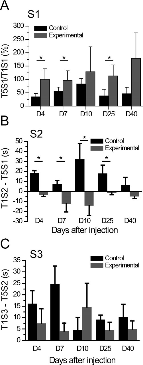
Patients with temporal lobe epilepsy (TLE), the most common form of epilepsy in adults, often display cognitive deficits. The time course and underlying mechanisms of cognitive decline remain unknown during epileptogenesis (the process leading to epilepsy). Using the rat pilocarpine model of TLE, we performed a longitudinal study to assess spatial and nonspatial cognitive performance during epileptogenesis. In parallel, we monitored interictal-like activity (ILA) in the hippocampal CA1 region, as well as theta oscillations, a brain rhythm central to numerous cognitive processes. Here, we report that spatial memory was altered soon after pilocarpine-induced status epilepticus, i.e., already during the seizure-free, latent period. Spatial deficits correlated with a decrease in the power of theta oscillations but not with the frequency of ILA. Spatial deficits persisted when animals had spontaneous seizures (chronic stage) without further modification. In contrast, nonspatial memory performances remained unaffected throughout. We conclude that the reorganization of hippocampal circuitry that immediately follows the initial insult can affect theta oscillation mechanisms, in turn, resulting in deficits in hippocampus-dependent memory tasks. These deficits may be dissociated from the process that leads to epilepsy itself but could instead constitute, as ILA, early markers in at-risk patients and/or provide beneficial therapeutic targets.


Figures

Figure 1.
Figure 1.
Spatial and nonspatial performances before treatment. Top, Experimental paradigm. During the S1 session, animals explore the two new objects (5 trials of 2 min each). One object is moved to a different quadrant, and animals are allowed to explore (S2 session, 5 trials of 2 min each). One object is then replaced (S3 session, 5 trials of 2 min each). Bottom, Mean duration (sec.) spent exploring the familiar object (black squares) and the other object (red circles) during session 1 (left histogram), session 2 (central histogram), and session 3 (right histogram). There was a significant habituation for both objects between the first (T1S1) and the last (T5S1) trial (p < 0.0001; n = 18). Object displacement led to renewed exploration, as compared with the time spent exploring the object in the last trial of S1 (T5S1; T1S2/T5S1: p = 0.009, n = 18). Object replacement led to renewed exploration, but it did not reach significance during T1S3 (T5S2/T1S3: p = 0. 225, n = 18), because the animals were still displaying increased interest in the displaced object during T5S2. However, the exploration time of the familiar object was significantly smaller than the time spent exploring the new object, showing that animals had detected the novelty (p = 0.049; n = 18). Data are expressed as means ± SEM and *p < 0.05.
Figure 2.
Figure 2.
Early and persistent deficit in spatial memory task in experimental TLE. A, Ratio of the time spent exploring both objects during T5S1 versus the time spent exploring both objects during T1S1, during the latent and chronic periods. A ratio <100% indicates less exploration, i.e., habituation. Experimental animals (n = 12) did not present any habituation of exploration of the two objects, between the first and the fifth trial for all testing days after SE (D4, D7, D25, and D40; p > 0.05) compared with the control group (n = 6), except at D10 and D40, where there was instead a significant decrease of exploration between T3S1 and T5S1 for the control group, caused by a delayed reaction to the objects. There was no significant effect (p > 0.05) all along the five testing days, for both groups. B, Difference between the time (s) spent exploring the displaced object in T1S2 and the time (s) spent exploring the same object in T5S1 (T1S2–T5S1) 4, 7, 10, 25, and 40 d after injection. Positive values indicate renewed exploration, hence spatial memory function (control/experimental, *p < 0.05). Experimental animals (n = 12) did not present any significant interest for the spatial change as compared with controls animals (n = 6; experimental/control, p < 0.05). There was no significant effect (p > 0.05) all along the five testing days, for both groups. C, Difference between the time (s) spent exploring the new object in T1S3 and the time (s) spent exploring the former object in T5S2 (T1S3–T5S2) 4, 7, 10, 25, and 40 d after injection. Positive values indicate renewed exploration, hence nonspatial memory function (*p < 0.05). Experimental (n = 12) and control (n = 6) animals displayed a significant interest to novelty (control/experimental, p > 0.05). There was no significant effect (p > 0.05) all along the five testing days, for both groups. Data are expressed as means ± SEM.
Figure 3.
Figure 3.
Lack of correlation between ILA and spatial deficit in experimental animals (n = 25). Top, Typical burst of ILA lasting 3 min recorded at D10 during rest. Note that the pattern evolves from large amplitude and slow to shorter amplitude and faster spike and waves. Bottom, There was no correlation between ILA frequency and spatial cognitive deficit measured as the T1S2–T5S1 difference (r = 0.015; p = 0.913).
Figure 4.
Figure 4.
The amount of loss of theta activity positively correlates to spatial cognitive deficits in experimental animals (n = 12). A, Examples of theta activity recorded during exploration in a control and in an experimental animal at D7. Note the decrease in amplitude and frequency in the experimental animal. B, C, Evolution of hippocampal theta (4–12 Hz) absolute power (B) and mean frequency (C) after SE in control (n = 6) and experimental animals, during exploratory behavior (*p < 0.0001). Note the stability of the recordings in controls and the early and significant (*p < 0.0001) decrease in theta power and frequency in experimental animals. D, Theta power is also decreased as soon as D4 during sessions 1 and 2 in experimental animals (*p < 0.0001). E, Spearman correlation between theta (4–12 Hz) absolute power during T1S2 and spatial memory performance evaluated as the difference of the time (s) spent exploring the displaced object during T1S2 and T5S1. Theta power correlates with spatial memory (r = 0.5; p = 0.00002; experimental, n = 25; control, n = 8). Data are expressed as means ± SEM.
Figure 5.
Figure 5.
Slight cell loss of Neu-N-labeled neurons in the CA1–CA3 hippocampal pyramidal cell layers (A) and all layers of the medial entorhinal cortex (B). *p < 0.05; **p < 0.01; ***p < 0.005. Data are expressed as means ± SEM.

References

    1. Aggleton JP, Hunt PR, Rawlins JN. The effects of hippocampal lesions upon spatial and non-spatial tests of working memory. Behav Brain Res. 1986;19:133–146. - PubMed
    1. Aldenkamp A, Arends J. The relative influence of epileptic EEG discharges, short nonconvulsive seizures, and type of epilepsy on cognitive function. Epilepsia. 2004;45:54–63. - PubMed
    1. Aldenkamp AP, Arends J, Overweg-Plandsoen TC, van Bronswijk KC, Schyns-Soeterboek A, Linden I, Diepman L. Acute cognitive effects of nonconvulsive difficult-to-detect epileptic seizures and epileptiform electroencephalographic discharges. J Child Neurol. 2001;16:119–123. - PubMed
    1. Aldenkamp AP, Arends J, Verspeek S, Berting M. The cognitive impact of epileptiform EEG-discharges; relationship with type of cognitive task. Child Neuropsychol. 2004;10:297–305. - PubMed
    1. Allen G, Barnard H, McColl R, Hester AL, Fields JA, Weiner MF, Ringe WK, Lipton AM, Brooker M, McDonald E, Rubin CD, Cullum CM. Reduced hippocampal functional connectivity in Alzheimer disease. Arch Neurol. 2007;64:1482–1487. - PubMed

Publication types