Comparative Study
. 2009 Apr 29;29(17):5425-34.
doi: 10.1523/JNEUROSCI.4473-08.2009.

Activity-dependent repression of Cbln1 expression: mechanism for developmental and homeostatic regulation of synapses in the cerebellum

Affiliations
Comparative Study

Activity-dependent repression of Cbln1 expression: mechanism for developmental and homeostatic regulation of synapses in the cerebellum

Takatoshi Iijima et al. J Neurosci. .

Abstract

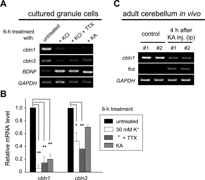
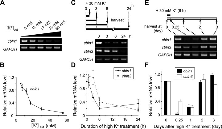
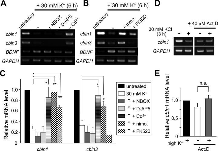
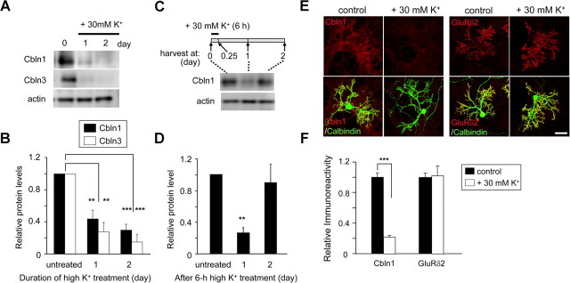
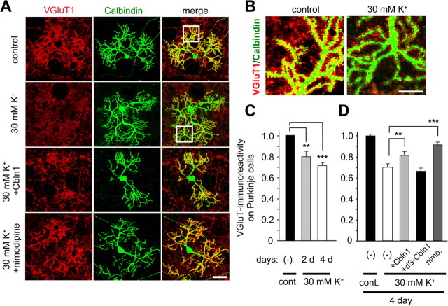
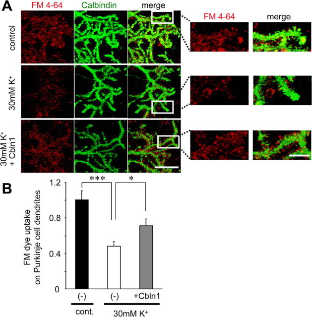
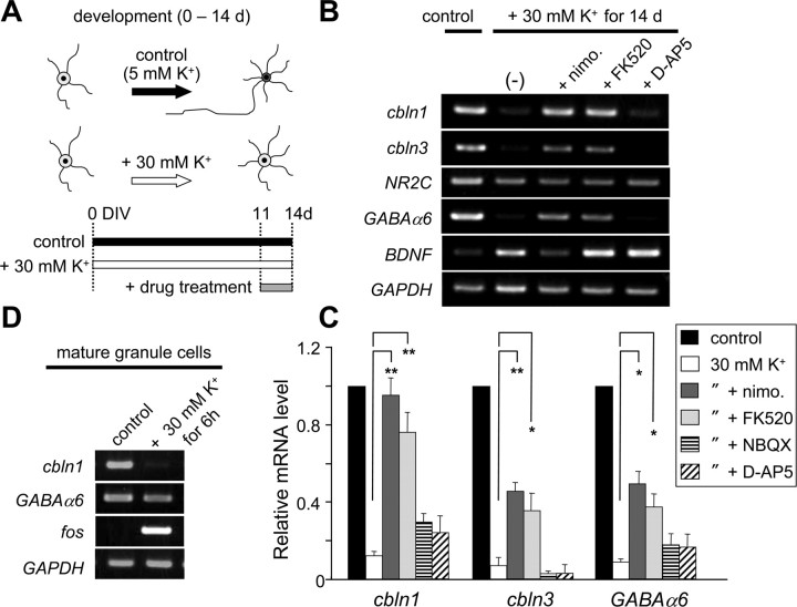
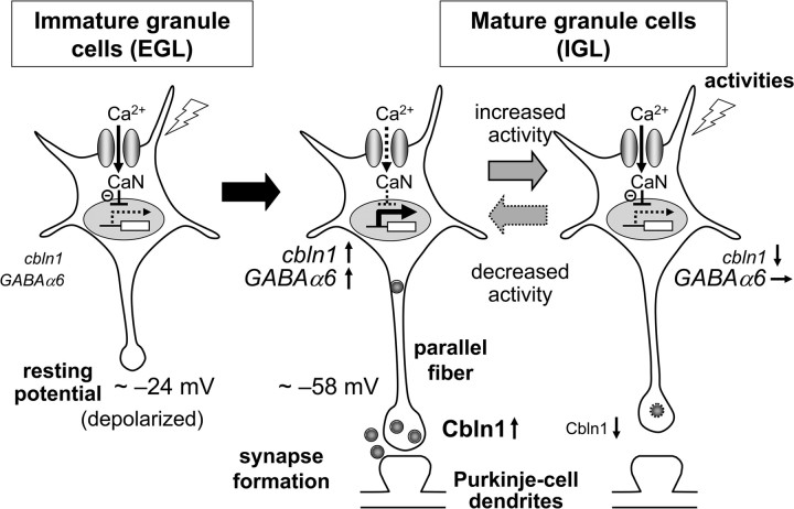
Cbln1, which belongs to the C1q/tumor necrosis factor superfamily, is released from cerebellar granule cells and plays a crucial role in forming and maintaining excitatory synapses between parallel fibers (PFs; axons of granule cells) and Purkinje cells not only during development but also in the adult cerebellum. Although neuronal activity is known to cause morphological changes at synapses, how Cbln1 signaling is affected by neuronal activity remains unclear. Here, we show that chronic stimulation of neuronal activity by elevating extracellular K(+) levels or by adding kainate decreased the expression of cbln1 mRNA within several hours in mature granule cells in a manner dependent on L-type voltage-dependent Ca(2+) channels and calcineurin. Chronic activity also reduced Cbln1 protein levels within a few days, during which time the number of excitatory synapses on Purkinje cell dendrites was reduced; this activity-induced reduction of synapses was prevented by the addition of exogenous Cbln1 to the culture medium. Therefore, the activity-dependent downregulation of cbln1 may serve as a new presynaptic mechanism by which PF-Purkinje cell synapses adapt to chronically elevated activity, thereby maintaining homeostasis. In addition, the expression of cbln1 mRNA was prevented when immature granule cells were maintained in high-K(+) medium. Since immature granule cells are chronically depolarized before migrating to the internal granule layer, this depolarization-dependent regulation of cbln1 mRNA expression may also serve as a developmental switch to facilitate PF synapse formation in mature granule cells in the internal granule layer.


Figures

Figure 1.
Figure 1.
Downregulation of cbln1 mRNA by increased neuronal activities in vitro and in vivo. A, Activity-dependent changes in expression of cbln1 and cbln3 mRNAs in mature granule cells. Granule cells maintained in 5 mm K+ for 2 weeks were treated with media containing high K+ (KCl; 30 mm), high K+ plus TTX (2 μm), or kainate (KA; 50 μm) for 6 h. Expression levels of the mRNAs for cbln1, cbln3, BDNF, and GAPDH were determined using semiquantitative PCR as described in Materials and Methods. B, Quantitative analysis of the expression levels of cbln1 and cbln3 mRNAs. The band intensities of cbln1 or cbln3 RT-PCR products were normalized to those of GAPDH. The value of untreated granule cells was arbitrarily defined as 1.0. n = 3 independent experiments. *p < 0.05, **p < 0.01. C, Activity-dependent reduction in expression of cbln1 mRNA in mature cerebellum in vivo. Four hours after the intraperitoneal injection of kainate (20 μg/g), the expression levels of cbln1, c-fos, and GAPDH mRNAs were determined by semiquantitative RT-PCR analysis in two 8-week-old C57BL/6J mice.
Figure 2.
Figure 2.
Depolarization conditions required to repress cbln1 mRNA expression. Cerebellar granule cells were maintained in 5 mm K+ for 2 weeks and were treated with high K+ under various conditions. A, B, Effects of extracellular K+ concentrations. Granule cells were treated with media containing various concentrations of extracellular K+ for 6 h. Expression levels of mRNAs for cbln1 and GAPDH were determined using semiquantitative PCR. The band intensities of the cbln1 transcripts were normalized to those of GAPDH, and the value of untreated (5 mm K+) granule cells was arbitrarily defined as 1.0 (B). n = 4 independent experiments. C, D, Effects of duration of high-K+ treatment. Granule cells were treated with 30 mm K+ for 1, 3, 6, or 24 h. The expression levels of mRNAs for cbln1, cbln3, and GAPDH were determined at the end of each treatment using semiquantitative PCR. The band intensities of the cbln1 and cbln3 transcripts were normalized to those of GAPDH, and the value of untreated (0 h) granule cells was arbitrarily defined as 1.0 (D). n = 3 independent experiments. E, F, Effects of elapsed time after high-K+ treatment. Granule cells were treated with 30 mm K+ for 6 h and the cbln1, cbln3, and GAPDH expression levels were analyzed at 0 and 6 h (at the end of the treatment) and 1, 2, or 3 d after treatment. The band intensities of the cbln1 and cbln3 transcripts were normalized to those of GAPDH, and the value of untreated (0 h) granule cells was arbitrarily defined as 1.0 (F). n = 3 independent experiments.
Figure 3.
Figure 3.
Effects of various inhibitors on high-K+-induced repression of cbln1 and cbln3 mRNAs. Cerebellar granule cells maintained in 5 mm K+ for 2 weeks were treated with high K+ for 6 h (A–C) or 3 h (D, E) in the presence of various inhibitors. A, Effects of an AMPA receptor blocker, NBQX (25 μm), an NMDA receptor blocker, d-AP5 (100 μm), and a general Ca2+ channel inhibitor, Cd2+ (10 μm). B, Effects of an l-type voltage-gated Ca2+ channel blocker, nimodipine (nimo.; 50 μm), and a calcineurin inhibitor, FK520 (0.5 μg/ml). C, Quantitative analysis of the effects of inhibitors on the high-K+-induced repression of the expression of cbln1 and cbln3 mRNAs. The band intensities of cbln1 or cbln3 RT-PCR products were normalized to those of GAPDH. The value of untreated granule cells was arbitrarily defined as 1.0. n = 4–10 independent experiments. *p < 0.05, **p < 0.01. D, Effects of a transcription blocker, actinomycin D (Act. D; 40 μm). E, Quantitative analysis of the effects of actinomycin D on the high-K+-induced repression of the expression of cbln1 mRNA. The band intensities of cbln1 RT-PCR products were normalized to those of GAPDH. The value of granule cells treated with high K+ alone was arbitrarily defined as 1.0. n = 4 independent experiments. n.s., No significant difference.
Figure 4.
Figure 4.
Increased neuronal activity reduced the expression of Cbln1 proteins in granule cells. A, High-K+-induced reduction of Cbln1 and Cbln3 proteins in granule cells. Mature granule cells maintained in 5 mm K+ for 2 weeks were treated with 30 mm K+ for 1 or 2 d, and the cell lysates were subjected to immunoblot analysis using specific antibodies against Cbln1, Cbln3, and actin. B, Quantitative analysis of the high-K+-induced reduction of Cbln1 and Cbln3 proteins. Band intensities of Cbln1 or Cbln3 were normalized with those of actin, and the value of untreated (0 d) granule cells was arbitrarily defined as 1.0. n = 3–4 independent experiments. **p < 0.01, ***p < 0.001 (compared with each untreated group). C, Reversibility of high-K+-induced reduction of Cbln1. Mature granule cells maintained in 5 mm K+ for 2 weeks were treated with 30 mm K+ for 6 h. The cell lysates were harvested at 0 d (untreated) or 1 or 2 d after treatment and subjected to immunoblot analysis using specific antibodies against Cbln1 and actin. D, Quantitative analysis of data shown in C. Band intensities were normalized to those of actin, and the value of untreated granule cells was arbitrarily defined as 1.0. n = 8 (in 3 independent experiments). E, Immunocytochemical analysis of endogenous Cbln1 and GluRδ2 in mixed cerebellar cultures after high-K+ treatment. Mixed cerebellar cultures maintained in normal K+ (5 mm) for 18–20 d were treated with media containing normal K+ (control) or high K+ (30 mm) in the presence of TTX (2 μm) for 4 d. Cells were fixed and immunostained using an anti-Cbln1 antibody (red; left), an anti-GluRδ2 antibody (red; right), or an anti-calbindin antibody (green). Scale bar, 50 μm. F, Quantitative analysis of the changes in Cbln1 and GluRδ2 proteins in mixed cerebellar cultures. The fluorescence intensity of Cbln1 or GluRδ2 immunoreactivities was measured on the dendrites of Purkinje cell dendrites, which were identified by calbindin immunoreactivity. The mean fluorescence intensity of Cbln1 or GluRδ2 was normalized to that of calbindin, and the value of control cultures was arbitrarily defined as 1.0. n = 4–5 independent experiments. ***p < 0.001.
Figure 5.
Figure 5.
Increased neuronal activity reduced VGluT1 immunoreactivity on Purkinje cells. A, Mixed cerebellar cultures maintained in normal K+ (5 mm) for 18–20 d were treated with media containing normal K+ (control), high K+ (30 mm), high K+ plus exogenous Cbln1 (2 μg/ml), or high K+ plus nimodipine (50 μm) for 4 d. Cells were fixed and immunostained for VGluT (red) and calbindin (green), markers for glutamatergic presynaptic terminals and Purkinje cells, respectively. The right column contains the merged images of VGluT1 and calbindin immunoreactivities. B, Enlarged images of the Purkinje cell dendrites shown in the white box in A. C, Quantitative analysis of the presynaptic marker VGluT1 on Purkinje cells after control (5 mm; cont.) or high-K+ (30 mm) treatment for 2 and 4 d. After subtracting the background level, the mean intensity of VGluT1 within the calbindin-positive Purkinje cell dendrites was normalized to that of calbindin. The value of the control cultures was arbitrarily defined as 1.0. n = 4–5 independent experiments. **p < 0.001, ***p < 0.0001. D, Quantitative analysis of the presynaptic marker VGluT1 on Purkinje cells after control (5 mm; cont.) or high-K+ (30 mm) treatment together with none (−), exogenous Cbln1 (2 μg/ml), mutant Cbln1 (dS–Cbln1, 2 μg/ml), or nimodipine (nimo.; 50 μm) for 4 d. Exogenous Cbln1 or nimodipine, but not dS–Cbln1, significantly rescued the reduced VGluT1 immunoreactivity seen on high-K+-treated Purkinje cell dendrites. n = 4–5 independent experiments. **p < 0.001, ***p < 0.0001. Scale bars: A, 50 μm; B, 20 μm.
Figure 6.
Figure 6.
Increased neuronal activity reduced functional presynaptic terminals on Purkinje cells. A, High-K+-induced reduction in presynaptic terminals labeled with FM4-64. Mixed cerebellar cultures maintained in normal K+ (5 mm) for 18–20 d were treated with media containing normal K+ (control), high K+ (30 mm), and high K+ plus exogenous Cbln1 (2 μg/ml) for 3 d. The functional presynaptic terminals were labeled with FM4-64 (red) as described in Materials and Methods. Cells were subsequently fixed and immunostained for calbindin (green), a marker for Purkinje cells. The right panels indicate enlarged images of the Purkinje cell dendrites shown in white boxes in left panels. Scale bars: left, 20 μm; right, 5 μm. B, Quantitative analysis of the presynaptic marker FM4-64 on Purkinje cells after control (5 mm; cont.) or high-K+ (30 mm) treatment together with none (−) or exogenous Cbln1 (2 μg/ml) for 3 d. Exogenous Cbln1 significantly rescued the reduced presynaptic terminals labeled with FM4-64 seen on high-K+-treated Purkinje cell dendrites. n = 18 neurons each from three independent experiments. *p < 0.05, ***p < 0.001.
Figure 7.
Figure 7.
Depolarization blocked the expression of cbln1 and cbln3 mRNA in developing granule cells. A, Schematic diagram indicating the timing of the drug treatment. Cerebellar granule cells were maintained in normal K+ (5 mm; control) or high-K+ (30 mm) media from 0 DIV through to 14 DIV. Nimodipine (50 μm), FK520 (0.5 μg/ml), or d-AP5 (100 μm) was added for the last 3 d. B, Effects of high K+ and various blockers on the developmental expression of various genes. Expression levels of mRNAs for cbln1, cbln3, NR2C, GABAα6, BDNF, and GAPDH were determined using semiquantitative PCR. C, Quantitative analysis of data shown in B. The band intensities of cbln1, cbln3, and GABAα6 transcripts were normalized to those of GAPDH, and the value of the control granule cells maintained in 5 mm K+ was arbitrarily defined as 1.0. n = 4–5 independent experiments. *p < 0.05, **p < 0.01. nimo, Nimodipine. D, Insensitivity of GABAα6 expression to high-K+ treatment in mature granule cells. The expression of GABAα6 was not downregulated by 30 mm K+ treatment for 6 h in mature granule cells maintained in normal K+ for 2 weeks. The same treatment downregulated cbln1 expression and upregulated c-fos expression. The result represents n = 3 independent experiments.
Figure 8.
Figure 8.
A model for the activity-dependent regulation of cbln1. In immature granule cells in the EGL, the resting membrane potentials are depolarized because of high extracellular K+ concentrations. Thus, Ca2+ influx through l-VGCC activates CaN and inhibits the expression of cbln1 mRNA. In mature granule cells in the IGL, the resting membrane potentials are repolarized, leading to the removal of the CaN-dependent repression of cbln1 mRNA expression. Together with other developmentally upregulated genes, such as GABAα6, increased cbln1 may mediate synaptogenesis between parallel fibers and Purkinje cell dendrites. When neuronal activities are chronically increased in mature granule cells, Ca2+ influx through l-VGCC again activates CaN and downregulates the expression of cbln1 mRNA, but not GABAα6 mRNA, leading to the loss of parallel fiber–Purkinje cell synapses. This activity-dependent downregulation of cbln1 may serve as a presynaptic mechanism by which PF–Purkinje cell synapses adapt to chronically elevated activity levels, thereby maintaining homeostasis.

Similar articles

Cited by

References

    1. Bading H, Ginty DD, Greenberg ME. Regulation of gene expression in hippocampal neurons by distinct calcium signaling pathways. Science. 1993;260:181–186. - PubMed
    1. Bao D, Pang Z, Morgan JI. The structure and proteolytic processing of Cbln1 complexes. J Neurochem. 2005;95:618–629. - PubMed
    1. Bessho Y, Nakanishi S, Nawa H. Glutamate receptor agonists enhance the expression of BDNF mRNA in cultured cerebellar granule cells. Brain Res Mol Brain Res. 1993;18:201–208. - PubMed
    1. Bessho Y, Nawa H, Nakanishi S. Selective up-regulation of an NMDA receptor subunit mRNA in cultured cerebellar granule cells by K(+)-induced depolarization and NMDA treatment. Neuron. 1994;12:87–95. - PubMed
    1. Black JE, Isaacs KR, Anderson BJ, Alcantara AA, Greenough WT. Learning causes synaptogenesis, whereas motor activity causes angiogenesis, in cerebellar cortex of adult rats. Proc Natl Acad Sci U S A. 1990;87:5568–5572. - PMC - PubMed

Publication types

MeSH terms

LinkOut - more resources