Comparative Study
. 2009 Apr 29;29(17):5558-72.
doi: 10.1523/JNEUROSCI.0520-09.2009.

The translational repressors Nanos and Pumilio have divergent effects on presynaptic terminal growth and postsynaptic glutamate receptor subunit composition

Affiliations
Comparative Study

The translational repressors Nanos and Pumilio have divergent effects on presynaptic terminal growth and postsynaptic glutamate receptor subunit composition

Kaushiki P Menon et al. J Neurosci. .

Abstract

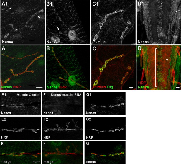
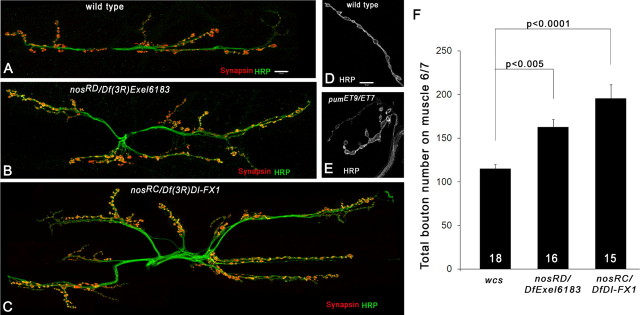
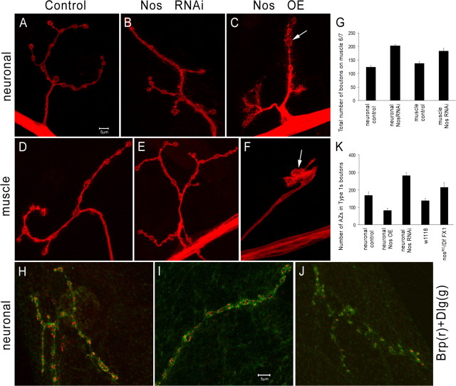
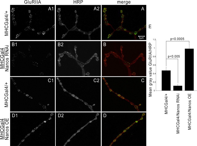
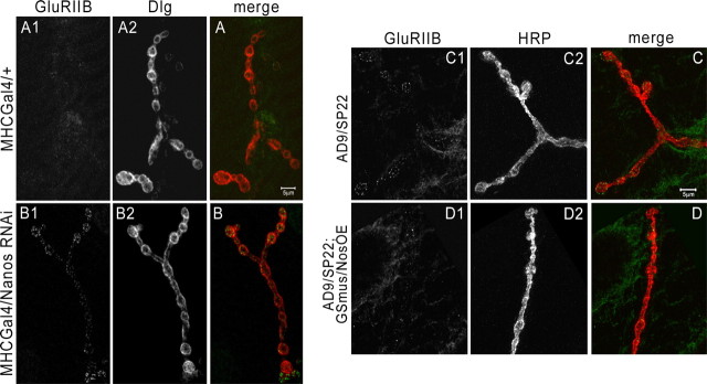
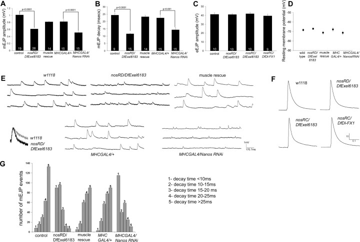
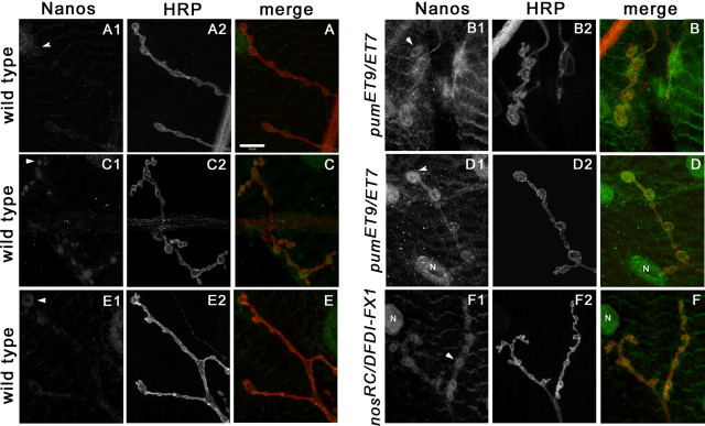
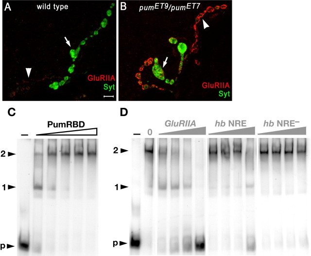
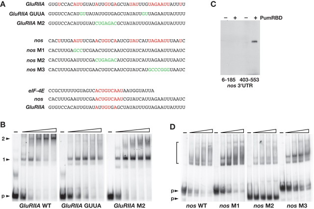
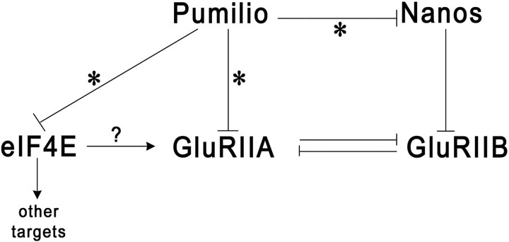
Pumilio (Pum) is a translational repressor that binds selectively to target mRNAs and recruits Nanos (Nos) as a corepressor. In the larval neuromuscular system, Pum represses expression of the translation factor eIF-4E and the glutamate receptor subunit GluRIIA. Here, we show that Nos, like Pum, is expressed at the neuromuscular junction (NMJ) and in neuronal cell bodies. Surprisingly, however, Nos and Pum have divergent functions on both the presynaptic and postsynaptic sides of the NMJ. In nos mutant and nos RNA interference larvae, the number of NMJ boutons is increased, whereas loss of Pum reduces the bouton number. On the postsynaptic side, Nos acts in opposition to Pum in regulating the subunit composition of the glutamate receptor. NMJ active zones are associated with GluRIIA- and GluRIIB-containing receptor clusters. Loss of Nos causes downregulation of GluRIIA and increases the levels of GluRIIB. Consistent with this finding, the electrophysiological properties of NMJs lacking postsynaptic Nos suggest that they use primarily GluRIIB-containing receptors. Nos can regulate GluRIIB in the absence of GluRIIA, suggesting that the effects of Nos on GluRIIB levels are at least partially independent of synaptic competition between GluRIIA and GluRIIB. Nos is a target for Pum repression, and Pum binds selectively to the 3' untranslated regions of the nos and GluRIIA mRNAs. Our results suggest a model in which regulatory interplay among Pum, Nos, GluRIIA, and GluRIIB could cause a small change in Pum activity to be amplified into a large shift in the balance between GluRIIA and GluRIIB synapses.


Figures

Figure 1.
Figure 1.
Nos is localized to the larval NMJ. A–C, Muscle 4 NMJs of A2 segments stained with mouse Nos antibody (A1, green in A), rat Nos antibody (B1, green in B), and Pum antibody (C1, red in C). Double labeling with anti-HRP in red (A, B) and with anti-Dlg in green (C) is shown. Type 1b boutons are indicated by arrows (A1, B1), and type 1s boutons are indicated by the arrowhead (A1). D, Larval VNC staining with mouse Nos antibody (D1, green in D) and anti-HRP (red in D). The asterisk indicates axons, and the bracket shows medial neurons that stain with Nos antibody and anti-HRP. E, F, Muscle 6/7 NMJs of A3 segments stained with mouse Nos antibody (E1, F1) and anti-HRP (E2, F2). E and F are merged images. The muscle control NMJ (E) is from C57-GAL4 crossed to w1118. The muscle Nos RNAi NMJ (F) is from C57-GAL4 crossed to Nos RNAi. Note the reduction in staining in F1 compared with E1; the anti-HRP signal is the same in E2 and F2. All panels are confocal z-series projections, except for D1, D, G1, and G, which are single sections. G, A cross section of a 6/7 A2 NMJ labeled with mouse Nos antibody (G1, green in G) and anti-HRP (G2, red in G). Scale bars: A, D, E, 10 μm; C, 5 μm.
Figure 2.
Figure 2.
Nos mutant NMJs have an increased number of synaptic boutons. A–C, Muscle 6/7 NMJs of A2 segments labeled with synapsin (red) and anti-HRP (green) antibodies in wild-type (WCS) and two nos mutant transheterozygotes, nosRD/Df(3R)Exel6183 and nosRC/Df(3R)Dl-FX1. D, E, Muscle 4 NMJs of A3 segments labeled with anti-HRP antibody in wild type (w1118) and pumET9/pumET7. Note the large boutons and the fused bouton phenotype in E. F, A graph of the total bouton number at 6/7 A2 NMJs for three genotypes: WCS, nosRD/Df(3R)Exel6183, and nosRC/Df(3R)Dl-FX1. The differences between WCS and nos mutants are highly significant (p < 0.005 and p < 0.0001, Student's t test). Scale bars: A, D, 10 μm.
Figure 3.
Figure 3.
Perturbation of Nos alters bouton and AZ numbers. A–F, Muscle 4 NMJs of A2 segments labeled with anti-HRP (red) antibody for neuronal (A–C) and muscle (D–F) Nos RNAi and Nos overexpression. A–C, Neuronal control (C155-GAL4 crossed to w1118; A), C155-GAL4 crossed to Nos RNAi (B), and OK6-GAL4 crossed to a UAS-Nanos line (C, Nos OE). Neuronal drivers C155-GAL4 and OK6-GAL4 produced similar phenotypes when used to overexpress Nos. The arrow in C shows an irregularly shaped type 1b bouton. D–F, Muscle control (24B-GAL4 crossed to w1118; D), muscle knockdown of Nos (24B-GAL4 crossed to a Nos RNAi line; E), and muscle overexpression of Nos (Nos OE; 24B-GAL4 crossed to a UAS-Nanos line; F). Note the abnormal structure of the entire muscle 4 NMJ when Nos is overexpressed in muscles (F, arrow). G, A graph of the total bouton number at muscle 6/7 clefts in neuronal control (n = 11), neuronal Nos RNAi (n = 12), muscle control (n = 15), and muscle Nos RNAi (n = 12) larvae. The difference in bouton number between NMJs of neuronal control and neuronal Nos RNAi larvae and between muscle control and muscle Nos RNAi are highly significant (Student's t test; p < 0.0001 and p < 0.001, respectively). H–J, Type 1s boutons of muscle 4 NMJs in A2 segments labeled for the AZ marker Brp (red) and Dlg (green) are shown for neuronal control (H), neuronal Nos RNAi (I), and neuronal Nos overexpression (J). K, The total number of AZs in type 1s boutons of muscle 4 NMJs in A2 segments were quantitated in five genotypes: neuronal control (n = 9), neuronal Nos overexpression (Nos OE; n = 7), neuronal Nos RNAi (n = 8), wild type (w1118; n = 5), and nosRC/Df(3R)Dl-FX1 (n = 7). The differences between control, neuronal OE (Student's t test, p < 0.005), and neuronal RNAi (p < 0.0005) lines are highly significant. Scale bars: A, I, 5 μm.
Figure 4.
Figure 4.
Knockdown of postsynaptic Nos causes downregulation of GluRIIA, and Nos overexpression upregulates GluRIIA. A–D, Muscle 4 NMJs in A2 segments labeled with anti-GluRIIA and anti-HRP. A, C, Control NMJs (MHC-GAL4 crossed to w1118). B, An NMJ from Nos RNAi crossed to MHC-GAL4. D, An NMJ from UAS-Nos crossed to MHC-GAL4 (Nanos OE). A, B, GluRIIA levels are decreased when Nos is knocked down in muscles (compare A1, B1; green in A and B). Anti-HRP (neuronal marker) levels are similar in both control and Nos RNAi NMJs (A2, B2; red in A and B). C, D, GluRIIA levels are increased when Nos is overexpressed in muscles (compare C1, D1; green in C and D). Anti-HRP labeling is similar in control and overexpression larvae (C2, D2; red in C and D). Two control NMJs are shown in this figure because the larvae for the RNAi or the overexpression crosses were processed and imaged at the same time along with their corresponding control animals in the same tube. Scale bar: A, 5 μm. E, Quantitation of GluRIIA at muscle 4 NMJs in control, Nos RNAi, and Nos overexpression (Nos OE). GluRIIA levels in distal boutons were quantitated and normalized against anti-HRP. This is represented as mean gray value of GluRIIA/HRP per hemisegment (see Materials and Methods). The values for MHC-GAL4/Nos RNAi (0.11 ± 0.03, n = 16) and MHC-GAL4/Nos OE (0.98 ± 0.11, n = 16) were significantly different from control MHC-GAL4/+ (0.47 ± 0.08, n = 35) (Student's t test; p < 0.005 and p < 0.0005, respectively).
Figure 5.
Figure 5.
Knockdown of Nos in muscles upregulates GluRIIB, and Nos overexpression in muscles in a GluRIIA mutant downregulates GluRIIB. A, B, Muscle 4 NMJs in A2 segments labeled with anti-GluRIIB and anti-Dlg. MHC-GAL4/+ (A) is a control NMJ (MHC-GAL4 muscle driver crossed to w1118), and MHC-GAL4/Nos RNAi (B) is a Nos RNAi line crossed to MHC-GAL4. GluRIIB levels are increased when Nos is knocked down in muscles (compare A1, B1; green in A and B). Dlg labeling is the same in control and Nos RNAi NMJs (A2, B2; red in A and B). GluRIIB labeling from distal boutons at muscle 4 NMJs was quantitated (see Materials and Methods), and these values were normalized against Dlg intensity. The mean gray value of GluRIIB/Dlg per hemisegment for MHC-GAL4/Nos RNAi was 0.41 ± 0.1 (n = 18), significantly different from control MHC-GAL4/+ (0.14 ± 0.08; n = 18; Student's t test, p < 0.02). C, D, Muscle 4 NMJs in A2 segments labeled with anti-GluRIIB and anti-HRP. C, Control NMJ, AD9/SP22, is a transheterozygote of a GluRIIA null mutant GluRIIAAD9 over a deficiency GluRIIASP22 that removes both GluRIIA and GluRIIB. D, AD9/SP22;GSmus/NosOE is an AD9/SP22 animal in which Nos is overexpressed from the MHC-GeneSwitch-GAL4 driver. Note that GluRIIB levels are decreased at the boutons of the NMJ in animals where Nos is overexpressed in a GluRIIA mutant (compare D1, C1; green in C and D). The magnitude of the decrease is approximately threefold (see Results). HRP labeling is the same in both control and experimental NMJs (C2, D2; red in C and D). Scale bars: A, C, 5 μm.
Figure 6.
Figure 6.
The amplitudes of mEJP events are decreased in nanos mutants. For electrophysiological analysis of the muscle 6/7 NMJ, recordings were done from muscle 6 in A2 and A3 segments. The number of animals that were analyzed for each genotype is indicated at the bottom of the bars. A, A graph of the amplitudes of mEJPs in various genotypes. Controls were two wild-type genotypes, w1118 (n = 10) and WCS (n = 9), and a driver crossed to w1118 control (n = 15) for the RNAi animals. Amplitudes were reduced in mutants and muscle RNAi larvae. B, A graph of the decay time (time constant) of mEJPs in the same genotypes. Decay was faster in mutants and muscle RNAi larvae. C, A graph of eEJPs in various genotypes. There were no significant differences among genotypes. D, A graph of resting membrane potential in the five genotypes in A and B. All the animals that were used for recordings had resting potentials that were more negative than −60 mV. This indicates that all preparations were equally healthy. E, Three representative traces of spontaneous events for the five genotypes in A and B. To show the faster decay in the mutant compared with the control, one spontaneous event from a control (w1118, gray) trace and one from a nanos transheterozygote [nosRD/Df(3R)Exel6183, black] trace that had the same mEJP amplitudes are shown. F, Representative traces of evoked responses from the three genotypes in C. Each trace is an average of 10 sequential eEJPs from one animal. G, A histogram showing the distribution of decay times of mEJP events in the different genotypes. The first 50 mEJP events from traces of five animals (a total of 250 events per genotype) were analyzed and put in bins corresponding to decay times of <10, 10–15, 15–20, 20–25, and >25 ms. The mutant and muscle Nos RNAi animals have a much larger number of events with fast decay times compared with controls and muscle rescue animals.
Figure 7.
Figure 7.
Nos protein expression at the NMJ is increased in pumilio and nanos mutants. Muscle 4 NMJs labeled with rat Nos antibody (A, B, E, F) or mouse Nos antibody (C, D) and double stained with anti-HRP. NMJs are from wild-type (w1118; A, C, E), pum mutants (pumET7/pumET9; B, D), and a nos mutant [nosRC/Df(3R)Dl-FX1; F]. A–D, Both Nos antibodies reveal a large increase in staining at the postsynaptic side of the NMJ and on the muscle surface in pum mutants [compare A1, B1 (green in A and B for the rat Nos antibody); C2, D2 (green in C and D for the mouse Nos antibody)]. Segment A3 (A, B) and segment A2 (C, D) are shown. Scale bar: A, 10 μm. E, F, The levels of Nos-immunoreactive protein at the A3 NMJ and on the muscle surface are increased in this nos mutant (compare E1, F1; green in E and F), as well as in two other transheterozygous nos genotypes (data not shown). Anti-HRP labeling is similar in all genotypes. The different appearance of the NMJs in B and D reflects the pum phenotype. Arrowheads indicate type 1b boutons, and N indicates a nucleus.
Figure 8.
Figure 8.
Pum binds selectively to the GluRIIA 3′UTR. A, B, Upregulation of GluRIIA in pum mutants (image from Menon et al., 2004). Muscle 12 NMJs are shown, labeled with anti-GluRIIA (red) and anti-Synaptotagmin (Syt; green), which strongly labels 1b boutons (arrows) and weakly labels 1s boutons (arrowheads). Note the dramatic increase in GluRIIA staining in the mutant, especially around type 1s boutons. C, Gel mobility shift assay (EMSA) for interaction of PumRBD with a 32P-labeled GluRIIA 3′UTR RNA probe. Binding reactions contained the same amount of RNA probe and either 0 (−) or an increasing concentration of PumRBD protein (ramp above lanes). D, Binding of PumRBD to the 32P-labeled GluRIIA 3′UTR RNA probe was challenged by an increasing (300-, 500-, 1000-, and 5000-fold) molar excess of an unlabeled competitor GluRIIA 3′UTR RNA, hb NRE RNA, or hb NRE RNA. Binding reactions contained either no (−) or a constant amount of PumRBD. Complexes 1 and 2 and unbound probe (p) are indicated. Note that the lane with the highest amount of hb competitor (right lane in middle panel) has probe and complex bands of the same intensity as the second lane from the right in the left panel, which has a fivefold lower amount of GluRIIA competitor. This shows that the GluRIIA sequence competes more effectively for binding than does the hb NRE.
Figure 9.
Figure 9.
Pum recognizes conserved noncanonical binding sites in the nanos and GluRIIA 3′UTRs. A, The sequences of the GluRIIA and nos 405–453 fragments bound by Pum, with nucleotides conserved between the two indicated in red. The sequences of the GluRIIA GUUA and M2 mutants and the nos M1, M2, and M3 mutants are shown below the respective wild-type sequences, with altered nucleotides in green. The lower alignment shows similarity among the Pum-binding regions of the eIF-4E, nos, and GluRIIA 3′UTRs. Note that there is a 9 nt sequence spanning the M2 motif that is identical between eIF-4E and nos. B, Gel mobility shift assays of PumRBD binding to 32P-labeled GluRIIA wild-type, GUUA, or M2 mutant RNA probes. Binding reactions contained the same amount of RNA probe and either 0 (−) or an increasing concentration of PumRBD protein (ramp above lanes). C, UV-crosslinking of the PumRBD to 32P-labeled nos RNA probes. Pum binds to a fragment encompassing nos 3′UTR nucleotides 405–453, but not to a fragment containing nucleotides 6–185. −, Reaction without PumRBD; +, reaction containing PumRBD. D, Gel mobility shift assays of PumRBD binding to 32P-labeled nos wild-type, M1, M2, and M3 mutant RNA probes. Binding reactions contained the same amount of RNA probe and either 0 (−) or an increasing concentration of PumRBD protein. The Pum-nos RNA complex migrates as a smear of several bands (bracket); the overall intensity of this smear is decreased in the M2 lanes but not for the other mutants. Note also that the unbound M2 mutant probe is not depleted even at the highest concentration of PumRBD, whereas the other probes are. The reason for the differing mobility of the M2 probe is not known but presumably reflects an inherent sequence and/or structural difference. WT, Wild type.
Figure 10.
Figure 10.
A diagram of the regulatory interactions among Pum, Nos, GluRIIA, GluRIIB, and eIF-4E. Pum represses eIF-4E, Nos, and GluRIIA and directly binds to all three target mRNAs (*). eIF-4E is often limiting for translation, so its elevation in pum mutants may help stimulate translation of GluRIIA and other postsynaptic mRNAs. Others have shown that GluRIIA and GluRIIB have mutually repressive interactions (see Discussion for references). This diagram predicts that the regulatory interplay among these factors should act as an amplifier, converting a small decrease in Pum activity (possibly in response to changes in environmental conditions) into a large shift in the GluRIIA/GluRIIB ratio.

Similar articles

Cited by

References

    1. Bergsten SE, Huang T, Chatterjee S, Gavis ER. Recognition and long-range interactions of a minimal nanos RNA localization signal element. Development. 2001;128:427–435. - PubMed
    1. Bernstein D, Hook B, Hajarnavis A, Opperman L, Wickens M. Binding specificity and mRNA targets of a C. elegans PUF protein, FBF-1. RNA. 2005;11:447–458. - PMC - PubMed
    1. Brand AH, Perrimon N. Targeted gene expression as a means of altering cell fates and generating dominant phenotypes. Development. 1993;118:401–415. - PubMed
    1. Brechbiel JL, Gavis ER. Spatial regulation of nanos is required for its function in dendrite morphogenesis. Curr Biol. 2008;18:745–750. - PMC - PubMed
    1. Chen G, Li W, Zhang QS, Regulski M, Sinha N, Barditch J, Tully T, Krainer AR, Zhang MQ, Dubnau J. Identification of synaptic targets of Drosophila pumilio. PLoS Comput Biol. 2008;4:e1000026. - PMC - PubMed

Publication types

MeSH terms