Comparative Study
. 2009 Apr 29;29(17):5690-700.
doi: 10.1523/JNEUROSCI.0265-09.2009.

SRC family kinases are required for limb trajectory selection by spinal motor axons

Affiliations
Comparative Study

SRC family kinases are required for limb trajectory selection by spinal motor axons

Tzu-Jen Kao et al. J Neurosci. .

Abstract

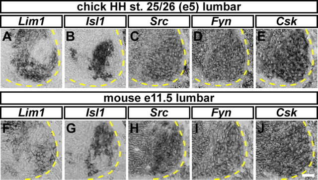
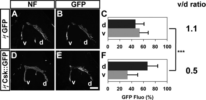
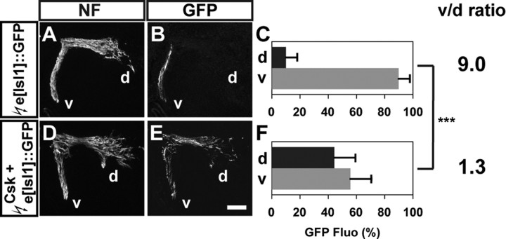
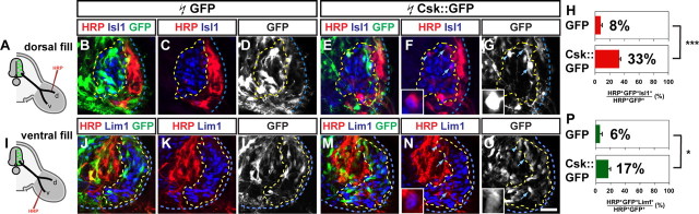
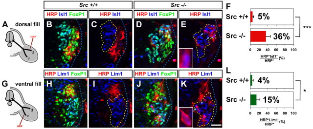
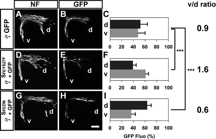
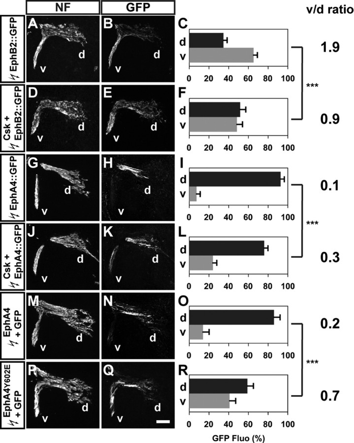
Signal relay by guidance receptors at the axonal growth cone is a process essential for the assembly of a functional nervous system. We investigated the in vivo function of Src family kinases (SFKs) as growth cone guidance signaling intermediates in the context of spinal lateral motor column (LMC) motor axon projection toward the ventral or dorsal limb mesenchyme. Using in situ mRNA detection we determined that Src and Fyn are expressed in LMC motor neurons of chick and mouse embryos at the time of limb trajectory selection. Inhibition of SFK activity by C-terminal Src kinase (Csk) overexpression in chick LMC axons using in ovo electroporation resulted in LMC axons selecting the inappropriate dorsoventral trajectory within the limb mesenchyme, with medial LMC axon projecting into the dorsal and ventral limb nerve with apparently random incidence. We also detected LMC axon trajectory choice errors in Src mutant mice demonstrating a nonredundant role for Src in motor axon guidance in agreement with gain and loss of Src function in chick LMC neurons which led to the redirection of LMC axons. Finally, Csk-mediated SFK inhibition attenuated the retargeting of LMC axons caused by EphA or EphB over-expression, implying the participation of SFKs in Eph-mediated LMC motor axon guidance. In summary, our findings demonstrate that SFKs are essential for motor axon guidance and suggest that they play an important role in relaying ephrin:Eph signals that mediate the selection of motor axon trajectory in the limb.


Figures

Figure 1.
Figure 1.
Expression of Src, Fyn, and Csk in chick and mouse LMC motor neurons. A–J, Detection of mRNA in consecutive sections of the spinal cord. All chick sections (A–E) are HH st. 25/26 lumbar spinal cord; all mouse sections (F–J) are e11.5 lumbar spinal cord. A, B, Detection of Lim1 and Isl1 mRNA in the chick spinal cord. Lim1 (A) and Isl1 (B) expression highlights lateral and medial LMC motor neurons, respectively. C, D, Detection of Src (C) and Fyn (D) mRNA, in both medial and lateral chick LMC neurons. E, Detection of Csk mRNA in LMC in both medial and lateral chick LMC neurons. F, G, Detection of Lim1 (F) and Isl1 (G) mRNA in mouse LMC motor neurons. H–J, Detection of Src (H), Fyn (I), and Csk (J) mRNA in medial and lateral mouse LMC neurons. Scale bar, 25 μm (A–J).
Figure 2.
Figure 2.
SFK inhibition by Csk overexpression redirects LMC motor axons into the dorsal limb nerve. A, D, Neurofilament detection in the limb nerves of chick HH st. 28/29 embryos electroporated with GFP (A) or Csk::GFP (D) expression plasmids. B, E, GFP detection in the limb nerves of chick HH st. 28/29 embryos electroporated with GFP (B) or Csk::GFP (E) expression plasmids. C, F, Quantification of GFP+ axons within dorsal and ventral limb nerves expressed as a percentage of total GFP+ axons [GFP Fluo (%)]. In GFP electroporated embryos, 47 ± 14% of GFP+ axons were detected in the dorsal limb nerves, and 53 ± 14% of GFP+ axons were detected in the ventral limb nerves, resulting in a v/d ratio [(average ventral GFP Fluo %)/(average dorsal GFP Fluo %)] of 1.1 (C). In Csk::GFP electroporated embryos, 68 ± 17% of GFP+ axons were detected in the dorsal limb nerves and 32 ± 17% of GFP+ axons were detected in the ventral limb nerves, resulting in a v/d ratio of 0.5 (F). Number of embryos quantified: C, n = 10; F, n = 12. Proportions of GFP+ axons in Csk::GFP expressing embryos and those in GFP expressing embryos are significantly different (p < 0.001) using Student's unpaired t test. v/d ratio = (average ventral GFP Fluo %)/(average dorsal GFP Fluo %); NF, neurofilament; d, dorsal; v, ventral. Error bars indicate SD. ***p < 0.001; all values are expressed as mean ± SD. Scale bar: (A, B, D, E), 200 μm.
Figure 3.
Figure 3.
SFK inhibition by Csk overexpression apparently randomizes medial LMC motor axon projections. A, D, Neurofilament detection in the limb nerves of chick HH st. 28/29 embryos electroporated with the medial LMC marker, e[Isl1]::GFP (A), or Csk and e[Isl1]::GFP (D) expression plasmids. B, E, GFP detection in the limb nerves of chick HH st. 28/29 embryos electroporated with e[Isl1]::GFP (B) or Csk and e[Isl1]::GFP (E) expression plasmids. C, F, Quantification of GFP+ axons within dorsal and ventral limb nerves expressed as the percentage of total GFP+ axons [GFP Fluo (%)]. In e[Isl1]::GFP electroporated embryos, 10 ± 8% of GFP+ axons were detected in the dorsal limb nerves, and 90 ± 8% of GFP+ axons were detected in the ventral limb nerves (v/d ratio of 9.0; C). In Csk and e[Isl1]::GFP coelectroporated embryos, 44 ± 15% of GFP+ axons were detected in the dorsal limb nerves and 56 ± 15% of GFP+ axons were detected in the ventral limb nerves, resulting (v/d ratio of 1.3; F). Number of embryos quantified: (C) n = 6, (F) n = 6. Proportions of GFP+ axons in Csk and e[Isl1]::GFP expressing embryos and those in e[Isl1]::GFP expressing embryos are significantly different (p < 0.001) using Student's unpaired t test. v/d ratio = (average ventral GFP Fluo %)/(average dorsal GFP Fluo %); NF, neurofilament; d, dorsal; v, ventral. Error bars indicate SD. ***p < 0.001; all values are expressed as mean ± SD. Scale bar: (A, B, D, E), 200 μm.
Figure 4.
Figure 4.
SFK function is required for the fidelity of LMC motor axon limb trajectories. A, I, Diagram depicting retrograde labeling of LMC neurons by HRP injections into dorsal (A) or ventral (I) hindlimb shank muscles of chick HH st. 28/29 embryos overexpressing Csk::GFP or GFP. B–G, Detection of HRP (red), Isl1 (blue), and GFP (green) in the LMC region of GFP (B–D) or Csk::GFP (E–G) electroporated embryos injected with HRP into dorsal shank muscles. Examples of electroporated medial LMC motor neurons labeled with HRP are indicated by arrows (F, G) and arrowheads (F, G). Examples indicated by arrowheads are shown at a higher magnification (insets of F, G). H, Proportions (%) of electroporated medial LMC motor neurons labeled with HRP in embryos injected with HRP into dorsal shank muscles. In dorsally filled GFP expressing embryos, 8 ± 2% of HRP+ GFP+ LMC neurons were Isl1+. In dorsally filled Csk::GFP expressing embryos, 33 ± 2% of HRP+ GFP+ LMC neurons were Isl1+. Number of embryos analyzed: n = 4 for both GFP and Csk::GFP expressing embryos. J–O, Detection of HRP (red), Lim1 (blue), and GFP (green) in the LMC region of GFP (J–L) or Csk::GFP (M–O) electroporated embryos injected with HRP into ventral shank muscles. Examples of electroporated lateral LMC motor neuron labeled with HRP are indicated by arrows (N, O) and arrowheads (N, O). Examples indicated by arrowheads are shown at a higher magnification (insets of N, O). P, Proportions (%) of electroporated lateral LMC motor neurons labeled with HRP in embryos injected with HRP into ventral shank muscles. In ventrally filled GFP expressing embryos, 6 ± 5% of HRP+ GFP+ LMC neurons were Lim1+. In ventrally filled Csk::GFP expressing embryos, 17 ± 3% of HRP+ GFP+ LMC neurons were Lim1+. Number of embryos quantified: n = 4 for GFP and Csk::GFP expressing embryos. Proportions of HRP+ GFP+ medial LMC neurons in dorsally filled Csk::GFP expressing embryos and those in dorsally filled GFP expressing embryos are significantly different (p < 0.001) using Student's unpaired t test. Proportions of HRP+ GFP+ lateral LMC neurons in ventrally filled Csk::GFP expressing embryos and those in ventrally filled GFP expressing embryos are significantly different (p < 0.05) using Student's unpaired t test. Error bars indicate SD. ***p < 0.001; *p < 0.05. All values are expressed as mean ± SD. Scale bars: (B–G, J–O), 25 μm; or 7 μm (insets of F, G, N, O).
Figure 5.
Figure 5.
Src is required for the fidelity of LMC motor axon trajectory selection. A, G, Diagram depicting retrograde labeling of LMC neurons by HRP injections into dorsal (A) or ventral (G) limb muscles of mouse e12.5 embryos. B–E, Detection of HRP (red), Isl1 (blue), and FoxP1 (green), which marks all LMC motor neurons, in the LMC region of Src+/+ (B, C) or Src−/− (D, E) embryos injected with HRP into dorsal forelimb muscles. Examples of medial LMC motor neurons labeled with HRP are indicated by arrows and arrowheads (E). Examples indicated by arrowheads are shown at a higher magnification (inset of E). F, Proportions (%) of medial LMC motor neurons labeled with HRP in embryos injected with HRP into dorsal forelimb muscles. In dorsally filled Src+/+ embryos, 5 ± 3% of HRP+ LMC neurons were Isl1+. In dorsally filled Src−/− embryos, 36 ± 14% of HRP+ LMC neurons were Isl1+. Number of embryos quantified: n = 5. H–K, Detection of HRP (red), Lim1 (blue), and FoxP1 (green) in the LMC region of Src+/+ (H, I) or Src−/− (J, K) embryos injected with HRP into ventral forelimb muscles. Examples of lateral LMC motor neurons labeled are indicated by arrows and arrowheads (K). Examples indicated by arrowheads are shown at a higher magnification (inset of K). L, Proportions (%) of lateral LMC motor neurons filled with HRP in embryos injected with HRP into ventral forelimb muscles. In ventrally filled Src+/+ embryos, 4 ± 3% of HRP+ LMC neurons were Lim1+. In ventrally filled Src−/− embryos, 15 ± 11% of HRP+ LMC neurons were Lim1+. Number of embryos quantified: n = 4. Proportions of HRP+ medial LMC neurons in dorsally filled Src−/− embryos and those in dorsally filled Src+/+ embryos are significantly different (p < 0.001) using Student's unpaired t test. Proportions of HRP+ lateral LMC neurons in ventrally filled Src−/− embryos and those in ventrally filled Src+/+ embryos are significantly different (p < 0.05) using Student's unpaired t test. Error bars = SD; ***p < 0.001; * = p < 0.05; all values expressed as mean ± SD. Scale bars: (B–E, HK), 25 μm or (insets of E, K), 7 μm.
Figure 6.
Figure 6.
Overexpression of constitutively active form (SrcY527F) or dominant negative form (SrcDN) of Src induces LMC axon trajectory choice errors. A, D, G, Neurofilament detection in the limb nerves of chick HH st. 28/29 GFP (A), SrcY527F and GFP (D), or SrcDN and GFP (G) expressing embryos. B, E, H, GFP detection in the limb nerves of HH st. 28/29 GFP (B), SrcY527F and GFP (E), or SrcDN and GFP (H) expressing embryos. C, F, I, Quantification of GFP+ axons within dorsal and ventral limb nerves expressed as the percentage of total GFP+ axons [GFP Fluo (%)]. In GFP electroporated embryos, 52 ± 11% of GFP+ axons were detected in the dorsal limb nerves, and 48 ± 11% of GFP+ axons were detected in the ventral limb nerves (v/d ratio of 0.9; C). In SrcY527F and GFP coelectroporated embryos, 39 ± 5% of GFP+ axons were detected in the dorsal limb nerves and 61 ± 5% of GFP+ axons were detected in the ventral limb nerves (v/d ratio of 1.6; F). In SrcDN and GFP coelectroporated embryos, 64 ± 7% of GFP+ axons were detected in the dorsal limb nerves and 36 ± 7% of GFP+ axons were detected in the ventral limb nerves (v/d ratio of 0.6; I). Number of embryos quantified: C, n = 5; F, n = 6; I, n = 5. Proportions of GFP+ axons in SrcY527F and GFP expressing embryos and those in GFP expressing embryos are significantly different (p < 0.001) using Student's unpaired t test. Proportions of GFP+ axons in SrcDN and GFP expressing embryos and those in GFP expressing embryos are significantly different (p < 0.001) using Student's unpaired t test. v/d ratio = (average ventral GFP Fluo %)/(average dorsal GFP Fluo %); NF, neurofilament; d, dorsal; v, ventral; error bars = SD. ***p < 0.001; all values expressed as mean ± SD. Scale bar: (A, B, D, E, G, H), 100 μm.
Figure 7.
Figure 7.
SFK inhibition by Csk attenuates EphB2- and EphA4-induced LMC motor axon redirection. A, D, G, J, M, P, Neurofilament detection in the limb nerves of chick HH st. 28/29 EphB2::GFP (A), Csk and EphB2::GFP (D), EphA4::GFP (G), Csk and EphA4::GFP (J), EphA4 and GFP (M), or EphA4Y602E and GFP (P) expressing embryos. B, E, H, K, N, Q, GFP detection in the limb nerves of HH st. 28/29 EphB2::GFP (B), Csk and EphB2::GFP (E), EphA4::GFP (H), Csk and EphA4::GFP (K), EphA4 and GFP (N), or EphA4Y602E and GFP (Q) expressing embryos. C, F, I, L, O, R, Quantification of GFP+ axons within dorsal and ventral limb nerves expressed as the percentage of total GFP+ axons [GFP Fluo (%)]. In EphB2::GFP electroporated embryos, 35 ± 4% of GFP+ axons were detected in the dorsal limb nerves, and 65 ± 4% of GFP+ axons were detected in the ventral limb nerves (v/d ratio of 1.9; C). In Csk and EphB2::GFP coelectroporated embryos, 52 ± 6% of GFP+ axons were detected in the dorsal limb nerves, and 48 ± 6% of GFP+ axons were detected in the ventral limb nerves (v/d ratio of 0.9; F). In EphA4::GFP electroporated embryos, 91 ± 4% of GFP+ axons were detected in the dorsal limb nerves, and 9 ± 4% of GFP+ axons were detected in the ventral limb nerves (v/d ratio of 0.1; I). In Csk and EphA4::GFP coelectroporated embryos, 76 ± 4% of GFP+ axons were detected in the dorsal limb nerves, and 24 ± 4% of GFP+ axons were detected in the ventral limb nerves (v/d ratio of 0.3; L). In EphA4 and GFP coelectroporated embryos, 85 ± 6% of GFP+ axons were detected in the dorsal limb nerves, and 15 ± 6% of GFP+ axons were detected in the ventral limb nerves (v/d ratio of 0.2; O). In EphA4Y602E and GFP coelectroporated embryos, 59 ± 6% of GFP+ axons were detected in the dorsal limb nerves, and 41 ± 6% of GFP+ axons were detected in the ventral limb nerves (v/d ratio of 0.7; R). Number of embryos quantified: C, n = 8; F, n = 9; I, n = 8; L, n = 7; O, n = 5; R, n = 4. Proportions of GFP+ axons in Csk and EphB2::GFP expressing embryos and those in EphB2::GFP expressing embryos are significantly different (p < 0.001) using Student's unpaired t test. Proportions of GFP+ axons in Csk and EphA4::GFP expressing embryos and those in EphA4::GFP expressing embryos are significantly different (p < 0.001) using Student's unpaired t test. Proportions of GFP+ axons in EphA4Y602E and GFP expressing embryos and those in EphA4 and GFP expressing embryos are significantly different (p < 0.001) using Student's unpaired t test. v/d ratio = (average ventral GFP Fluo %)/(average dorsal GFP Fluo %); NF, neurofilament; d, dorsal; v, ventral; error bars = SD. ***p < 0.001; all values expressed as mean ± SD. Scale bar, 100 μm in all images.

Similar articles

Cited by

References

    1. Beg AA, Sommer JE, Martin JH, Scheiffele P. α2-Chimaerin is an essential EphA4 effector in the assembly of neuronal locomotor circuits. Neuron. 2007;55:768–778. - PubMed
    1. Bisson N, Poitras L, Mikryukov A, Tremblay M, Moss T. EphA4 signaling regulates blastomere adhesion in the Xenopus embryo by recruiting Pak1 to suppress Cdc42 function. Mol Biol Cell. 2007;18:1030–1043. - PMC - PubMed
    1. Bladt F, Aippersbach E, Gelkop S, Strasser GA, Nash P, Tafuri A, Gertler FB, Pawson T. The murine Nck SH2/SH3 adaptors are important for the development of mesoderm-derived embryonic structures and for regulating the cellular actin network. Mol Cell Biol. 2003;23:4586–4597. - PMC - PubMed
    1. Cowan CW, Shao YR, Sahin M, Shamah SM, Lin MZ, Greer PL, Gao S, Griffith EC, Brugge JS, Greenberg ME. Vav family GEFs link activated Ephs to endocytosis and axon guidance. Neuron. 2005;46:205–217. - PubMed
    1. Cursi S, Rufini A, Stagni V, Condò I, Matafora V, Bachi A, Bonifazi AP, Coppola L, Superti-Furga G, Testi R, Barilà D. Src kinase phosphorylates Caspase-8 on Tyr380: a novel mechanism of apoptosis suppression. EMBO J. 2006;25:1895–1905. - PMC - PubMed

Publication types

MeSH terms

LinkOut - more resources