. 2009 Jul;110(1):212-23.
doi: 10.1093/toxsci/kfp084. Epub 2009 Apr 29.

Signal transducer and activator of transcription 1 (STAT1) is essential for chromium silencing of gene induction in human airway epithelial cells

Affiliations

Signal transducer and activator of transcription 1 (STAT1) is essential for chromium silencing of gene induction in human airway epithelial cells

Antonia A Nemec et al. Toxicol Sci. 2009 Jul.

Abstract

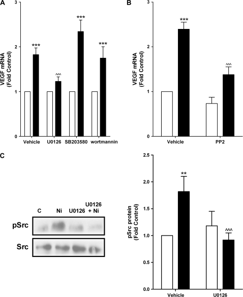
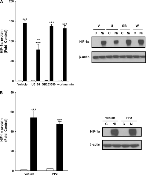
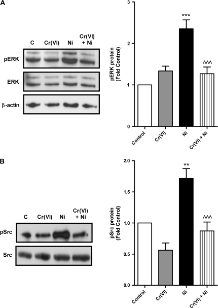
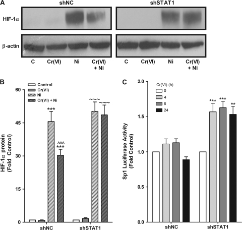
Hexavalent chromium (Cr(VI)) promotes lung injury and pulmonary diseases through poorly defined mechanisms that may involve the silencing of inducible protective genes. The current study investigated the hypothesis that Cr(VI) actively signals through a signal transducer and activator of transcription 1 (STAT1)-dependent pathway to silence nickel (Ni)-induced expression of vascular endothelial cell growth factor A (VEGFA), an important mediator of lung injury and repair. In human bronchial airway epithelial (BEAS-2B) cells, Ni-induced VEGFA transcription by stimulating an extracellular regulated kinase (ERK) signaling cascade that involved Src kinase-activated Sp1 transactivation, as well as increased hypoxia-inducible factor-1 alpha (HIF-1 alpha) stabilization and DNA binding. Ni-stimulated ERK, Src, and HIF-1 alpha activities, as well as Ni-induced VEGFA transcript levels were inhibited in Cr(VI)-exposed cells. We previously demonstrated that Cr(VI) stimulates STAT1 to suppress VEGFA expression. In BEAS-2B cells stably expressing STAT1 short hairpin RNA, Cr(VI) increased VEGFA transcript levels and Sp1 transactivation. Moreover, in the absence of STAT1, Cr(VI), and Ni coexposures positively interacted to further increase VEGFA transcripts. This study demonstrates that metal-stimulated signaling cascades interact to regulate transcription and induction of adaptive or repair responses in airway cells. In addition, the data implicate STAT1 as a rate limiting mediator of Cr(VI)-stimulated gene regulation and suggest that cells lacking STAT1, such as many tumor cell lines, have opposite responses to Cr(VI) relative to normal cells.


Figures

FIG. 1.
FIG. 1.
Cr(VI) inhibits Ni-induced VEGFA mRNA and protein levels. BEAS-2B cells were exposed to 5μM Cr(VI), 200μM Ni, or Cr(VI) for 2 h prior to adding 200μM Ni for (A) 24 h and (B). 48 h. (A) VEGFA mRNA levels were measured by real-time PCR. (B) Conditioned medium was collected and VEGF protein release was measured by ELISA. Data represent mean ± SEM of fold control (n = 3). ** and *** designate p < 0.01 and p < 0.001, respectively compared with untreated cells (control); ∧∧ and ∧∧∧ designate p < 0.01 and p < 0.001, respectively compared with cells treated with Ni alone.
FIG. 2.
FIG. 2.
ERK mediates Ni-induced VEGFA mRNA levels. BEAS-2B cells were pretreated with (A) 10μM U0126, 20μM SB203580, or 1μM wortmannin or (B) 10μM PP2 prior to adding vehicle (white bars) or 200μM Ni (black bars) for 24 h. VEGFA mRNA levels were measured by real-time PCR. Data represent mean ± SEM of fold control (n = 3). (C) BEAS-2B cells were pretreated with 10μM U0126 prior to adding vehicle (white bars) or 200μM Ni (black bars) for 30 min. Total Src was immunoprecipitated from whole cell lysates and immunoblotted for pSrc. ImageJ software was used to quantify the intensity of the bands and data represent mean ± SEM of fold control (n = 3). ** and *** designate p < 0.001 and p < 0.001, respectively, compared with untreated cells (control). ∧∧∧ designates p < 0.001 compared with cells treated with Ni alone.
FIG. 3.
FIG. 3.
Ni-induced HIF-1α protein stabilization requires ERK. BEAS-2B cells were pretreated with (A) 10μM U0126, 20μM SB203580, or 1μM wortmannin or (B) 10μM PP2 prior to adding vehicle (white bars) or 200μM Ni (black bars) for 24 h. Total protein was isolated and HIF-1α and β-actin protein levels were determined by western analysis. ImageJ software was used to quantify the intensity of the bands. Data represent mean ± SEM of fold control (n = 3). ***Designates p < 0.001 compared with untreated cells (control); ∧∧∧ designates p < 0.001 compared with cells treated with Ni alone.
FIG. 4.
FIG. 4.
Cr(VI) abrogates Ni-stimulated ERK signaling. (A) BEAS-2B cells were exposed to 5μM Cr(VI), 200μM Ni, or Cr(VI) for 2h prior to adding 200μM Ni for 5 min. Total protein was isolated and phosphorylated ERK (pERK), total ERK, and β-actin protein levels were determined by western analysis. (B) BEAS-2B cells were exposed to 5μM Cr(VI), 200μM Ni, or Cr(VI) for 2 h prior to adding 200μM Ni for 30 min. Total Src was immunoprecipitated from whole cell lysates and immunoblotted for pSrc. ImageJ software was used to quantify the intensity of the bands and data represent mean ± SEM of fold control (n = 3). ** and *** designate p < 0.01 and p < 0.001, respectively compared with untreated cells (control); ∧∧∧ designates p < 0.001 compared with cells treated with Ni alone.
FIG. 5.
FIG. 5.
Cr(VI) partially inhibits Ni-induced HIF-1α stabilization and HRE transactivation. (A) BEAS-2B cells were exposed to 5μM Cr(VI), 200μM Ni, or Cr(VI) for 2 h prior to adding 200μM Ni for 24 h. Total protein was isolated and HIF-1α and β-actin protein levels were determined by western analysis. ImageJ software was used to quantify the intensity of the bands. (B) BEAS-2B cells were transiently transfected with HRE-luc and eGFP. After 24 h, cells were exposed to 5μM Cr(VI), 200μM Ni, or Cr(VI) for 2 h prior to adding 200μM Ni for 8 h. Relative luciferase activity of HRE-luc was normalized to eGFP. Data represent mean ± SEM of fold control (n = 3). ** and *** designate p < 0.01 and p < 0.001, respectively, compared with untreated cells (control); ∧∧ and ∧∧∧ designate p < 0.01 and p < 0.001, respectively, compared with cells treated with Ni alone.
FIG. 6.
FIG. 6.
STAT1 is required for Cr(VI) suppression of VEGFA induction. (A) Total protein was isolated from BEAS-2B cell lines stably expressing either scrambled (shNC) or STAT1 (shSTAT1) shRNA and total STAT1 and β-actin protein levels were determined by western analysis. (B) shNC and shSTAT1 cells were exposed to 5μM Cr(VI), 200μM Ni, or Cr(VI) for 2 h prior to adding 200μM Ni for 24 h. (C) shNC and shSTAT1 cells were exposed to 5μM Cr(VI) for the indicated times. (D) Parental BEAS-2B cells were pretreated with 10 μg/ml of cycloheximide for 5 min prior to exposure to 5μM Cr(VI) for the indicated times. Total RNA was isolated and VEGFA mRNA levels were measured by real-time PCR. Data represent mean ± SEM of fold control (n = 3). * and ∼ designate p < 0.05, *** and ∼∼∼ designate p < 0.001 compared with respective untreated cells (control); ## and ∧∧∧ designate p < 0.01 and p < 0.001, respectively, compared with respective cells treated with Ni alone.
FIG. 7.
FIG. 7.
STAT1 represses HIF-1α protein stabilization and Sp1 transactivation. (A) BEAS-2B cell lines stably expressing either scrambled (shNC) or STAT1 (shSTAT1) shRNA were exposed to 5μM Cr(VI), 200μM Ni, or Cr(VI) for 2 h prior to adding 200μM Ni for 24 h. Total protein was isolated and HIF-1α and β-actin protein levels were determined by Western analysis and a representative blot from a single experiment is shown. (B) Density of the protein bands from three separate experiments were quantified with ImageJ software and presented as mean ± SEM of fold control (n = 3). *** and ∼∼∼ designate p < 0.001 compared with the respective untreated cells (control). ∧∧∧ designates p < 0.001 compared with cells treated with Ni alone. (C) shNC or shSTAT1 cells were transiently transfected with Sp1-luc and eGFP and exposed to 5μM Cr(VI) for the indicated times. Relative luciferase activity of Sp1-luc was normalized to eGFP. Data represent mean ± SEM of fold control (n = 3). ** and *** designate p < 0.01 and p < 0.001, respectively, compared with untreated cells (control).

References

    1. Acarregui MJ, Penisten ST, Goss KL, Ramirez K, Snyder JM. Vascular endothelial growth factor gene expression in human fetal lung in vitro. Am. J. Respir. Cell Mol. Biol. 1999;20(1):14–23. - PubMed
    1. Andrew AS, Klei LR, Barchowsky A. Nickel requires hypoxia-inducible factor-1α, not redox signaling to induce plasminogen activator inhibitor-1. Am. J. Physiol. Lung Cell Mol. Physiol. 2001;281:L607–L615. - PubMed
    1. Antonini JM, Stone S, Roberts JR, Chen B, Schwegler-Berry D, Afshari AA, Frazer DG. Effect of short-term stainless steel welding fume inhalation exposure on lung inflammation, injury, and defense responses in rats. Toxicol. Appl. Pharmacol. 2007;223(3):234–245. - PubMed
    1. Antonini JM, Taylor MD, Zimmer AT, Roberts JR. Pulmonary responses to welding fumes: Role of metal constituents. J. Toxicol. Environ. Health A. 2004;67(3):233–249. - PubMed
    1. Barchowsky A, Roussel RR, Krieser RJ, Mossman BT, Treadwell MD. Expression and activity of urokinase and its receptor in endothelial and pulmonary epithelial cells exposed to asbestos. Toxicol. Appl. Pharmacol. 1998;152(2):388–396. - PubMed

Publication types

MeSH terms