Review
. 2009 May;30(3):241-63.
doi: 10.1210/er.2008-0039. Epub 2009 Apr 29.

In search of adrenocortical stem and progenitor cells

Affiliations
Review

In search of adrenocortical stem and progenitor cells

Alex C Kim et al. Endocr Rev. 2009 May.

Abstract

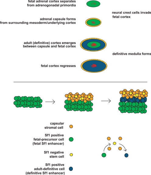
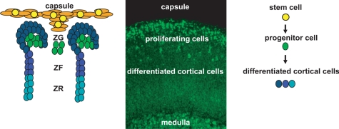
Scientists have long hypothesized the existence of tissue-specific (somatic) stem cells and have searched for their location in different organs. The theory that adrenocortical organ homeostasis is maintained by undifferentiated stem or progenitor cells can be traced back nearly a century. Similar to other organ systems, it is widely believed that these rare cells of the adrenal cortex remain relatively undifferentiated and quiescent until needed to replenish the organ, at which time they undergo proliferation and terminal differentiation. Historical studies examining cell cycle activation by label retention assays and regenerative potential by organ transplantation experiments suggested that the adrenocortical progenitors reside in the outer periphery of the adrenal gland. Over the past decade, the Hammer laboratory, building on this hypothesis and these observations, has endeavored to understand the mechanisms of adrenocortical development and organ maintenance. In this review, we summarize the current knowledge of adrenal organogenesis. We present evidence for the existence and location of adrenocortical stem/progenitor cells and their potential contribution to adrenocortical carcinomas. Data described herein come primarily from studies conducted in the Hammer laboratory with incorporation of important related studies from other investigators. Together, the work provides a framework for the emerging somatic stem cell field as it relates to the adrenal gland.


Figures

Figure 1
Figure 1
Top, Stages of adrenal gland development. Bottom, Model of capsular and definitive zone cell lineage.
Figure 2
Figure 2
Histological analysis of a regenerating mouse adrenal gland. Top, Hemotoxylin and eosin (H&E) staining of an enucleated mouse adrenal gland transplanted into the kidney capsule. Bottom, Immunohistochemistry with an anti-Sf1 antibody.
Figure 3
Figure 3
Role of Dax1 in adrenal gland physiology. Model of Sf1- and Dax1-mediated transcription.
Figure 4
Figure 4
Model of hierarchical organization of adrenocortical cells. Left, Immunohistochemistry (IHC) with anti-Sf1 reveals cortical staining. Middle, Model of Sf1 and Dax1 expression in stem/progenitor/differentiated adrenal cortex cells. Right, Immunohistochemistry with anti-Dax1 reveals membranous subcapsular expression and nuclear intracortical expression.
Figure 5
Figure 5
Comparative paradigm of nuclear receptor-mediated transcriptional feedback loop. The intracellular feedback regulation of adrenocortical steroid production parallels the regulation of bile acid synthesis in the liver.
Figure 6
Figure 6
Role of canonical Wnt signaling in adrenocortical homeostasis. Hematoxylin and eosin staining of conditional β-catenin knockout adrenals. Progressive depletion in the adrenal cortex is evident at 30 wk of age. [Reproduced with permission of Development (114)].
Figure 7
Figure 7
The role of TGFβ signaling in adrenal vs. gonadal fate determination. Histological analysis of adrenal cortex revealing follicular-like structures in the adrenal cortex of inhibin null mice (red, granulosa cell staining for anti-mullerian hormone; green, theca cell staining for LH receptor). [Reproduced with permission. Copyright 2006, The Endocrine Society (119)].
Figure 8
Figure 8
Importance of telomere stability in adrenal gland development. Top, Model of telomerase-mediated telomere elongation. Middle, Model of telomere cap complex. Bottom, Histological analysis of Acd-deficient mouse adrenals. Immunohistochemistry using anti-3β-HSD and antityrosine hydroxylase reveals aberrant adrenal development.HSD, Hydroxysteroid dehydrogenase. [The bottom panel is reproduced with permission from Oxford University Press (151)].
Figure 9
Figure 9
Cellular organization of the adrenal cortex. Left, Model of adrenocortical homeostatic growth maintenance (right, color key for model). Middle, Immunohistochemistry using anti-PCNA reveals subcapsular localization of proliferating adrenocortical cells.
Figure 10
Figure 10
Role of Pod1 in adrenocortical development. Top left, LacZ activity staining in Pod1-LacZ adrenals reveals preferential capsular staining. Bottom left, Comparative immunohistochemical analysis of heterozygous and homozygous Pod1 knockout adrenals. Staining reveals presence of Sf1-positive cells in the capsule of homozygous Pod1 knockout adrenals. Right, Low-power magnification of anti-Sf1 staining in Pod1 knockout adrenals reveals expansion of Sf1 positive cells in homozygous Pod1 knockout adrenals.
Figure 11
Figure 11
Comparative histology of transgenic Hedgehog and Wnt reporter mouse adrenals. Left, LacZ activity staining in Gli1-LacZ mouse adrenals (218) reveals preferential capsular staining. Right, LacZ activity staining reveals preferential subcapsular staining in Wnt-Gal reporter adrenals.
Figure 12
Figure 12
Summary figure.

Similar articles

Cited by

References

    1. Gottschau M 1883 Structur und embryonale Entwickelung der Nebennieren bei Saugethieren. Archiv fur Anat und Phys S:412–458
    1. Arnold J 1866 Ein Beitrag zur feineren Struktur und dem Chemismus der Nebennieren. Virchow Arch Patholog Anatomie Physiol Klin Med 35: 64–107
    1. Evelyn H-M 1927 A transitory zone in the adrenal cortex which shows age and sex relationships. Am J Anat 40:251–293
    1. Keegan CE, Hammer GD 2002 Recent insights into organogenesis of the adrenal cortex. Trends Endocrinol Metab 13:200–208 - PubMed
    1. Else T, Hammer GD 2005 Genetic analysis of adrenal absence: agenesis and aplasia. Trends Endocrinol Metab 16:458–468 - PubMed

Publication types