

Estrogen promotes germ cell and seminiferous tubule development in the baboon fetal testis
- PMID: 19403930
- PMCID: PMC2767192
- DOI: 10.1095/biolreprod.108.073494
Estrogen promotes germ cell and seminiferous tubule development in the baboon fetal testis
Abstract
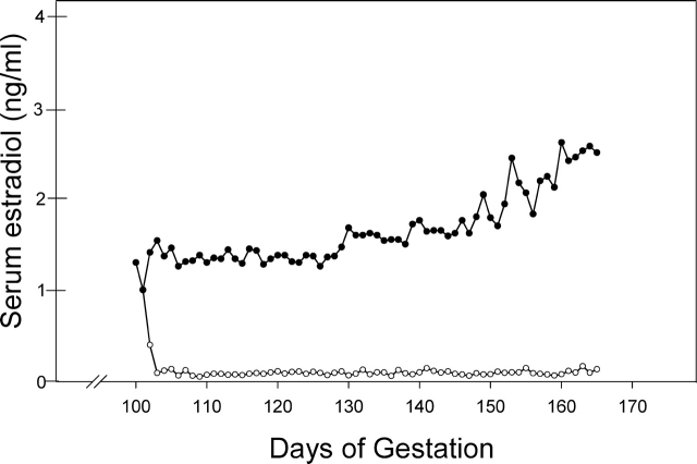
The foundation for development of the male reproduction system occurs in utero, but relatively little is known about the regulation of primate fetal testis maturation. Our laboratories have shown that estrogen regulates key aspects of the physiology of pregnancy and fetal development. Therefore, in the present study, we characterized and quantified germ cells and Sertoli cells in the fetal baboon testis in late normal gestation (i.e., Day 165; term is 184 days) and in baboons administered the aromatase inhibitor letrozole throughout the second half of gestation to assess the impact of endogenous estrogen on fetal testis development. In untreated baboons, the seminiferous cords were comprised of undifferentiated (i.e., type A) spermatogonia classified by their morphology as dark (Ad) or pale (Ap), gonocytes (precursors of type A spermatogonia), unidentified cells (UI), and Sertoli cells. In letrozole-treated baboons, serum estradiol levels were decreased by 95%. The number per milligram of fetal testis (x10(4)) of Ad spermatogonia (0.42 +/- 0.11) was 45% lower (P = 0.03), and that of gonocytes (0.58 +/- 0.06) and UI (0.45 +/- 0.12) was twofold greater (P < 0.01 and P = 0.06, respectively), than in untreated baboons. Moreover, in the seminiferous cords of estrogen-deprived baboons, the basement membrane appeared fragmented, the germ cells and Sertoli cells appeared disorganized, and vacuoles were present. We conclude that endogenous estrogen promotes fetal testis development and that the changes in the germ cell population in the estrogen-deprived baboon fetus may impair spermatogenesis and fertility in adulthood.
Figures


References
-
- Rabinovici J, Jaffe RB.Development and regulation of growth and differentiated function in human and subhuman primate fetal gonads. Endocr Rev 1990; 11: 532–557. - PubMed
-
- Aslan AR, Kogan BA, Gondos B.Testicular development. Polin RA, Fox WW, Abman SH.Fetal and Neonatal Physiology Philadelphia:W.B. Saunders;2004: 1950–1955.
-
- Cortes D, Muller J, Skakkebaek NE.Proliferation of Sertoli cells during development of the human testis assessed by stereological methods. Int J Androl 1987; 10: 589–596. - PubMed
-
- O'Shaughnessy PJ, Baker PJ, Monteiro A, Cassie S, Bhattacharya S, Fowler PA.Developmental changes in human fetal testicular cell numbers and messenger ribonucleic acid levels during the second trimester. J Clin Endocrinol Metab 2007; 92: 4792–4801. - PubMed
-
- Gaskell TL, Esnal A, Robinson LL, Anderson RA, Saunders PT.Immunohistochemical profiling of germ cells within the human fetal testis: identification of three subpopulations. Biol Reprod 2004; 71: 2012–2021. - PubMed
Publication types
MeSH terms
Substances
Grants and funding
LinkOut - more resources
Full Text Sources
