. 2009 Jun;8(6):526-33.
doi: 10.1038/nmat2444. Epub 2009 May 3.

Intravaginal gene silencing using biodegradable polymer nanoparticles densely loaded with small-interfering RNA

Affiliations

Intravaginal gene silencing using biodegradable polymer nanoparticles densely loaded with small-interfering RNA

Kim A Woodrow et al. Nat Mater. 2009 Jun.

Abstract

Vaginal instillation of small-interfering RNA (siRNA) using liposomes has led to silencing of endogenous genes in the genital tract and protection against challenge from infectious disease. Although siRNA lipoplexes are easily formulated, several of the most effective transfection agents available commercially may be toxic to the mucosal epithelia and none are able to provide controlled or sustained release. Here, we demonstrate an alternative approach using nanoparticles composed entirely of FDA-approved materials. To render these materials effective for gene silencing, we developed novel approaches to load them with high amounts of siRNA. A single dose of siRNA-loaded nanoparticles to the mouse female reproductive tract caused efficient and sustained gene silencing. Knockdown of gene expression was observed proximal (in the vaginal lumen) and distal (in the uterine horns) to the site of topical delivery. In addition, nanoparticles penetrated deep into the epithelial tissue. This is the first report demonstrating that biodegradable polymer nanoparticles are effective delivery vehicles for siRNA to the vaginal mucosa.


Figures

Figure 1
Figure 1. In vitro cytotoxicity and bioactivity of siRNA nanoparticles
Polymer PLGA nanoparticles encapsulating siRNA were evaluated for cytotoxicity using cultured (a) HepG2 hepatocytes and HeLa cervical carcinoma cells. The toxicity of free spermidine (green diamonds), spermidine-loaded PLGA nanoparticles (black squares), dsDNA/spermidine- (red circles), and siRNA/spermidine-loaded (purple stars) nanoparticles were evaluated over a range of concentrations from 0-10 mg/mL. CellTiter Blue® fluorescence (Promega) was used to measure cell viability compared to untreated cells (blue line/triangles). Neither the PLGA nanoparticles singularly or in combination with spermidine, the mimic (dsDNA), or siRNA showed cytotoxicity in the cell types and over this range of concentrations. (b) Dose-response curves comparing the bioactivity of siRNA delivered with a transfection agent (red circles) or using PLGA nanoparticles (open squares). An siRNA targeted against the luciferase gene (siLUC) was delivered to cultured HEK-293T cells stably expressing luciferase. Luciferase activity was measured with the Bright-Glo™ (Promega) reagent and separate treated wells were used to measure cell viability using CellTiter Blue®. Luciferase activity normalized to viable cell number and plotted against the amount of delivered siLUC. PLGA nanoparticles show equal or better activity compared to a commercial transfection agent (Lipofectamine RNAiMax).
Figure 2
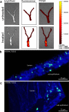
Figure 2. Fluorescent PLGA nanoparticles appear throughout the reproductive tract and penetrate deep within the vaginal tissue after topical administration
PLGA nanoparticles loaded with a fluorescent dye (coumarin-6) were vaginally instilled into the reproductive tract of female ICR mice at a dose of 750 mg per animal. The entire reproductive tract was imaged 24 h post-administration. (a) An in vivo imaging system (IVIS) was used to obtain fluorescent images of the whole tissue. Multiphoton microscopy was used to obtain deep tissue images of the vaginal tract (b) and uterine horns (c) 24 h post-treatment. Box highlights areas where nanoparticles were detected. Image dimensions are 400 mm μ 400 mm μ 75 mm. (d) Multiphoton microscopy images of the female reproductive tract in C56Bl/6 mice at 3, 5, and 7 d after a single topical treatment of fluorescent PLGA nanoparticles. Control animals were instilled with PBS and showed no fluorescence signal in the green channel used to detect nanoparticle fluorescence. Inset shows magnified areas enclosed in white box. Image dimensions are 400 mm μ 400 mm, with depths of 50-120 mm. Green, red, and blue arrows indicated x-, y-, and z-coordinates, respectively. For (b-d), Hoescht dye (blue) and coumarin-6 nanoparticles (green) were detected by excitation at 860 nm and visualized by their respective optical filters.
Figure 3
Figure 3. Release of PLGA nanoparticles densely-loaded with siRNA is sustained for several weeks
A high initial loading of siRNA (200 nmoles) was combined with spermidine and formulated into PGLA nanoparticles. (a) Under physiological pH in PBS (I = 0.2 M, pH = 7.4), cumulative release of siRNA/Spe is not significant after the initial burst release. In vitro release studies performed in acidic conditions with the same ionic strength as PBS (50 mM citrate buffer, I = 0.2 M, pH = 5.0), show linear and sustained siRNA release after 48 h. Representative SEM micrographs (inset) show that acidic conditions cause nanoparticles to exhibit pores or dimples on their surface (red arrow). (b) The siRNA released at pH 5.0 from our polymer nanoparticles is chemically intact and functional. After 24 h of controlled release at pH 5.0, siRNA was collected from the buffer, concentrated, and then analyzed by gel electrophoresis and for bioactivity in a HEK293T cell line stably expressing luciferase (293T-Luc). Released siRNA had a similar electrophoretic mobility to the stock material used as a standard (inset). The siRNA (designed to target luciferase) was also able to transfect and knockdown gene expression comparable to a positive control. Values represent the mean ± s.d.
Figure 4
Figure 4. Intravaginal delivery of siRNA using biodegradable nanoparticles causes gene silencing throughout the reproductive tract of transgenic GFP mice
A siRNA targeted against EGFP was delivered using lipoplexes (filled bar) or PLGA nanoparticles with the siEGPF pre-complexed with spermidine (diagonal bar) or protamine (hatched bar). Quantification of EGFP fluorescence per area of tissue was performed in the three different regions of the reproductive tract. Reduction in EGFP expression was seen in the vagina, cervix, and uterine horns in GFP transgenic mice 10 days after topical administration. Open bars are the cumulative negative controls (see Materials and Methods). Bars represent the mean ± s.d.
Figure 5
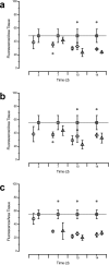
Figure 5. A single topical administration of PLGA nanoparticles loaded with siRNA causes sustained gene silencing throughout the reproductive tract
Vaginal instillation of siRNA using a commercial transfection agent or PLGA nanoparticles leads to EGFP silencing in the (a) uterine horns, (b) cervix, and (c) vaginal tract. An siRNA targeted against EGFP was formulated in LipofectamineTM RNAiMax (circles), or pre-complexed with spermidine (triangles) or protamine (diamonds) and then encapsulated into PLGA nanoparticles. Knockdown of EGFP expression was assessed by image analysis of tissue fluorescence compared to delivery of an siRNA mimic (squares). Values represent the mean ± s.d. for n = 3 per treatment group except for the negative controls where n = 18. Statistical significance was determined by a two-sample t-test with p ≤ 0.05 (*). Asterisk above the group indicates all members were statistically different compared to the control, whereas the asterisk is placed adjacent to a group when it is the only member that showed significance.
Figure 6
Figure 6. Lipid delivery of siRNA may be inflammatory and cause epithelial disruption in the vaginal tissue
Histopathology of vaginal tissue in mice instilled with PBS (a-c), siRNA polymer nanoparticles (d-e), and lipoplexes (g-i). Histologic analysis of tissue sections from the vagina and uteri in mice treated with PBS (a, b (inset of a)) or siRNA nanoparticles (d, e (inset of d) showed minimal to no inflammation by haematoxylineosin staining, exhibited equivalent vaginal epithelial thickness (b, e arrows), and showed no significant pathologic findings. In contrast, lipid-treated mice had marked vaginal epithelial hyperplasia (g, *) and frequent scattered neutrophils within the lumen (h (inset of g, arrowheads)) indicative of a mild to moderate vaginitis. Comparison of sides with anti- CD45 IHC revealed numerous CD45 positive cells within the epithelium of lipid-treated mice (i) in comparison to the nanoparticle-(f) or PBS (c) treated mice. L = vaginal lumen, E = epithelium. HE staining (a-b, d-e, g-h). DAB staining with methyl green counterstain (c, f, i). Scale bars a-b, d-e, g-h = 500 μm. Scale bar c, f, i = 100 μm.

Comment in

Similar articles

Cited by

References

    1. Grant RM, et al. Perspective - Whither or wither microbicides? Science. 2008;321:532–534. - PMC - PubMed
    1. Wilson DP, Coplan PM, Wainberg MA, Blower SM. The paradoxical effects of using antiretroviral-based microbicides to control HIV epidemics. P Natl Acad Sci USA. 2008;105:9835–9840. - PMC - PubMed
    1. Veazey RS, et al. Protection of macaques from vaginal SHIV challenge by vaginally delivered inhibitors of virus-cell fusion. Nature. 2005;438:99–102. - PubMed
    1. Richardson JL, Illum L. Penetration enhancement for polypeptides through epithelia. D. Routes of delivery case studies. 8. The vaginal route of peptide and protein drug delivery. Adv Drug Deliver Rev. 1992;8:341–366.
    1. Veazey RS, et al. Prevention of virus transmission to macaque monkeys by a vaginally applied monoclonal antibody to HIV-1 gp120. Nat Med. 2003;9:343–346. - PubMed

Publication types