. 2009;4(4):e5390.
doi: 10.1371/journal.pone.0005390. Epub 2009 Apr 30.

Muscle mitochondrial uncoupling dismantles neuromuscular junction and triggers distal degeneration of motor neurons

Affiliations

Muscle mitochondrial uncoupling dismantles neuromuscular junction and triggers distal degeneration of motor neurons

Luc Dupuis et al. PLoS One. 2009.

Abstract

Background: Amyotrophic lateral sclerosis (ALS), the most frequent adult onset motor neuron disease, is associated with hypermetabolism linked to defects in muscle mitochondrial energy metabolism such as ATP depletion and increased oxygen consumption. It remains unknown whether muscle abnormalities in energy metabolism are causally involved in the destruction of neuromuscular junction (NMJ) and subsequent motor neuron degeneration during ALS.

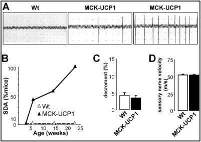
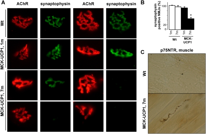
Methodology/principal findings: We studied transgenic mice with muscular overexpression of uncoupling protein 1 (UCP1), a potent mitochondrial uncoupler, as a model of muscle restricted hypermetabolism. These animals displayed age-dependent deterioration of the NMJ that correlated with progressive signs of denervation and a mild late-onset motor neuron pathology. NMJ regeneration and functional recovery were profoundly delayed following injury of the sciatic nerve and muscle mitochondrial uncoupling exacerbated the pathology of an ALS animal model.

Conclusions/significance: These findings provide the proof of principle that a muscle restricted mitochondrial defect is sufficient to generate motor neuron degeneration and suggest that therapeutic strategies targeted at muscle metabolism might prove useful for motor neuron diseases.


Conflict of interest statement

Competing Interests: The authors have declared that no competing interests exist.

Figures

Figure 1
Figure 1. Muscle phenotype of MCK-UCP1 mice.
A: body weight of male (empty diamonds) and female (empty triangles) wild-type mice, and male (filled diamonds) and female (filled triangles) MCK-UCP1 mice as a function of time. *, p<0.05 vs corresponding MCK-UCP1 (n = 7 mice per group). B: grip strength of mice as in A. *, p<0.05 vs corresponding MCK-UCP1 C: representative photomicrographs showing hematoxylin and eosin staining of gastrocnemius from wild-type mice (Wt) or MCK-UCP1 mice at 3 months (3 m) and 7 months (7 m) of age. The right panel shows the quantification of mean fiber area. *, p<0.05 (n = 4 mice per group).
Figure 2
Figure 2. MCK-UCP1 do not display muscle dystrophy.
A: serum creatine kinase (left panel) and lactate (right panel) concentrations in wild-type (Wt) or MCK-UCP1 mice at 2 months (2 m) and 7 months (7 m) of age. Non-significant differences (n = 8 mice per group). B: mRNA levels of atrogin-1 and M-cadherin in gastrocnemius from wild-type (empty circles) or MCK-UCP1 (black circles) mice at perinatal (PN), 1 month (1 m) and 7 months (7 m) of age. *, p<0.05 vs corresponding wild-type (n = 3–8 mice per group). C: representative photomicrographs showing hematoxylin and eosin staining of gastrocnemius from cardiotoxin-injured (48 hours after injury, CTX), wild-type (Wt) or MCK-UCP1 mice at 3 months (3 m) and 7 months (7 m) of age. Three mice per group were analyzed. A necrotic myofiber is indicated by an arrow. D: Evans blue dye staining as visualised by fluorescence in gastrocnemius from cardiotoxin-injured (48 hours after injury, CTX), wild-type (Wt) and MCK-UCP1 mice at 3 months (3 m) and 7 months (7 m) of age. Three mice per group were analyzed. Only a few necrotic myofibers could be detected in 7-month-old MCK-UCP1 mice (arrow).
Figure 3
Figure 3. Aging MCK-UCP1 mice show progressive NMJ deterioration.
A: representative photomicrographs showing NMJ morphology in gastrocnemius from wild-type (Wt) or MCK-UCP1 mice. Note the pretzel-shaped morphology of wild-type NMJs either at 1 month (a, b) or 7 months of age (c, d). In 1-month-old (1 m) MCK-UCP1 mice, NMJs appeared smaller and, in some rare cases, fragmented (e-h). At 7 months of age (7 m), thinner cisternae (arrow in i), ectopic AchR clusters (star in i), fragmented NMJs (j,k) and degenerated postsynaptic apparatus (l) were observed. Scale bar, 20 µm. B: quantitative morphometry of NMJs in gastrocnemius from wild-type (empty columns) or MCK-UCP1 (black columns) mice at 1 month (1 m) and 7 months (7 m) of age. *, p<0.05 vs corresponding wild type (n = 5 mice per group).
Figure 4
Figure 4. Electromyographic features of MCK-UCP1 mice.
A: representative electromyography recordings in gastrocnemius from a 7-month-old wild-type mouse (left panel), and two 7-month-old MCK-UCP1 mice showing weak (middle panel) and intense (right panel) altered electrical activity. B: fraction of mice presenting with aberrant spontaneous denervation activity in at least one muscle territory. Wild-type mice (empty triangles), MCK-UCP1 mice (filled triangles) (n = 6–10 mice per group). C: decrement observed in a repetitive stimulation test in gastrocnemius from 7-month-old wild-type (Wt) and MCK-UCP1 mice (n = 6 mice per group). D: sensory nerve velocity in 7-month-old wild-type (Wt) and MCK-UCP1 mice (n = 6 mice per group).
Figure 5
Figure 5. Aging MCK-UCP1 mice show progressive denervation and distal axonal degeneration.
A: representative photomicrographs showing AchR (red) and synaptophysin (green) staining in gastrocnemius from wild-type (Wt) or MCK-UCP1 mice at 1 month (1 m) and 7 months (7 m) of age. Scale bar, 20 µm. B: quantification of the percentage of synaptophysin positive NMJs as shown in D, *, p<0.05 vs wild-type. (n = 4 mice per group). C: representative photomicrograph showing p75NTR immunostaining in gastrocnemius from 7-month-old wild-type (Wt) or MCK-UCP1 mice. Scale bar, 20 µm.
Figure 6
Figure 6. Mild late onset motor neuron degeneration in MCK-UCP1 mice.
A: representative photomicrographs showing ventral roots from wild-type (Wt) and MCK-UCP1 mice. Note the presence of two degenerating axons in the MCK-UCP1 picture (arrows). B: distribution of the calibers of axons in the ventral roots from wild-type (empty circles) or MCK-UCP1 (filled circles) mice. Small-caliber axons represent gamma-axons, which innervate muscle spindles, whereas large-caliber axons represent alpha-axons, which innervate skeletal muscles. Note the decrease in the population of the largest caliber axons in MCK-UCP1 ventral roots. C: size distribution of axons in the ventral roots from 7-month-old wild-type (empty columns) and MCK-UCP1 (filled columns) mice. *, p<0.05 vs wild-type (n = 3 mice per genotype). D: quantification of the number of motor neurons in the lumbar spinal cord form wild-type (Wt) and MCK-UCP1 (MCK-UCP1) mice at 3 months (3 m) and 7 months (7 m) of age, after toluidine blue-staining (left panel) and ChAT immunoreactivity (right panel). *, p<0.05 vs wild-type (n = 5 mice per group). E: representative photomicrographs showing GFAP immunoreactivity in the lumbar spinal cord from 7-month-old wild-type (Wt) or MCK-UCP1 mice. Ventral horns are indicated by dashed lines. Scale bar, 20 µm.
Figure 7
Figure 7. Old MCK-UCP1 mice show increased AchR clustering in response to agrin.
representative photomicrographs showing AchR clusters in contralateral (innervated) and ipsilateral (denervated) tibialis anterior from wild-type (Wt) or MCK-UCP1 mice 8 days after sciatic nerve axotomy and 15 days after recombinant agrin injection. Scale bar, 20 µm.
Figure 8
Figure 8. MCK-UCP1 mice show delayed functional recovery after nerve crush.
A–B: Sciatic functional index (A) and grip strength (B) in 1-month-old wild-type (empty diamonds) or MCK-UCP1 mice (filled diamonds) after sciatic nerve crush, performed at day 0. Grip strength was expressed as the percentage of ipsilateral strength relative to the contralateral limb. *, p<0.05 vs corresponding wild type; #, p<0.05 vs day 0 (n = 12 mice per group). C: Mass of contralateral (empty columns) and ipsilateral (black columns) gastrocnemius from wild-type (Wt) or MCK-UCP1 mice 30 days after sciatic nerve crush. *, p<0.05 vs contralateral muscle (n = 5 mice per group). D: quantification of the percentage of synaptophysin positive NMJs as shown in E, *, p<0.05 vs wild-type. (n = 5 mice per group). E: representative photomicrographs showing AchR (red) and neurofilament/synaptophysin (green) staining in contralateral and ipsilateral gastrocnemius from wild-type (Wt) or MCK-UCP1 mice 8 days after sciatic nerve crush (e) and quantification of the experiment (d). Note the denervated endplates (stars) and the retracted nerve fibers (arrowheads). Scale bar, 20 µm.
Figure 9
Figure 9. UCP1 overexpression exacerbates motor neuron disease in mice.
A–C: Kaplan-Meier curves showing the cumulative probability of disease onset (age at the peak of body mass prior to decline, A), progression through early disease stage (when maximal body mass decreases by 10%, B), and total survival (C). The p-values, as calculated using the logrank test, are shown. D: duration of early disease stage (left) and total duration of disease (right) in SOD1(G86R) mice (empty columns) and compound SOD1(G86R)/MCK-UCP1 mice (filled columns). *, p<0.05 vs corresponding SOD1(G86R).

Similar articles

Cited by

References

    1. Gonzalez de Aguilar JL, Echaniz-Laguna A, Fergani A, Rene F, Meininger V, et al. Amyotrophic lateral sclerosis: all roads lead to Rome. J Neurochem 2007 - PubMed
    1. Boillee S, Vande Velde C, Cleveland DW. ALS: a disease of motor neurons and their nonneuronal neighbors. Neuron. 2006;52:39–59. - PubMed
    1. Boillee S, Yamanaka K, Lobsiger CS, Copeland NG, Jenkins NA, et al. Onset and progression in inherited ALS determined by motor neurons and microglia. Science. 2006;312:1389–1392. - PubMed
    1. Clement AM, Nguyen MD, Roberts EA, Garcia ML, Boillee S, et al. Wild-type nonneuronal cells extend survival of SOD1 mutant motor neurons in ALS mice. Science. 2003;302:113–117. - PubMed
    1. Desport JC, Preux PM, Magy L, Boirie Y, Vallat JM, et al. Factors correlated with hypermetabolism in patients with amyotrophic lateral sclerosis. Am J Clin Nutr. 2001;74:328–334. - PubMed

Publication types