. 2007 Sep;1(3):192-208.
doi: 10.2976/1.2773861.

How molecular motors shape the flagellar beat

How molecular motors shape the flagellar beat

Ingmar H Riedel-Kruse et al. HFSP J. 2007 Sep.

Abstract

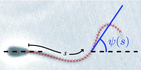
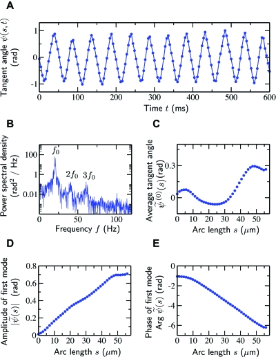
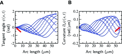
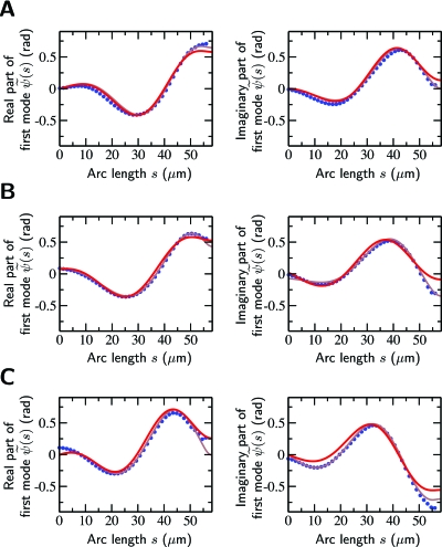
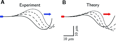
Cilia and eukaryotic flagella are slender cellular appendages whose regular beating propels cells and microorganisms through aqueous media. The beat is an oscillating pattern of propagating bends generated by dynein motor proteins. A key open question is how the activity of the motors is coordinated in space and time. To elucidate the nature of this coordination we inferred the mechanical properties of the motors by analyzing the shape of beating sperm: Steadily beating bull sperm were imaged and their shapes were measured with high precision using a Fourier averaging technique. Comparing our experimental data with wave forms calculated for different scenarios of motor coordination we found that only the scenario of interdoublet sliding regulating motor activity gives rise to satisfactory fits. We propose that the microscopic origin of such "sliding control" is the load dependent detachment rate of motors. Agreement between observed and calculated wave forms was obtained only if significant sliding between microtubules occurred at the base. This suggests a novel mechanism by which changes in basal compliance could reverse the direction of beat propagation. We conclude that the flagellar beat patterns are determined by an interplay of the basal properties of the axoneme and the mechanical feedback of dynein motors.


Figures

Figure 1
Figure 1. Snapshot of a beating bull sperm.
Superimposed on the image are red crosses tracing the contour of the flagellum as determined by the automated image analysis algorithm (see Materials and Methods section). The tangent angle ψ(s) is measured at each position defined by the arc length s. The centers of the red crosses are separated by 1.4 μm.
Figure 2
Figure 2. Schematic diagram of the axonemal architecture and its two-dimensional representation.
(A) Cross section of an axoneme with radius r as seen from the basal end. The microtubule doublets and the central pair are shown in red, the dynein motors in blue, and the radial spokes in green. The horizontal gray line indicates the plane of the beat. With this beat plane, the largest sliding displacement occurs between adjacent microtubule doublets at the top and bottom. (B) Cross section of the two-dimensional representation of the axoneme in which two flexible filaments slide relative to each other in the beat plane. The shear forces are generated by active elements which operate antagonistically between the filaments. Passive elastic elements are represented in green. The separation of the two filaments is a=2r. (C) View on the beat plane of this “two-dimensional axoneme.” Indicated are the local sliding displacement Δ(s) and the internal shear force density f(s) due to the active elements (blue) and the passive cross-linkers (green). The basal connection has a finite stiffness and friction indicated by black springs and dashpots.
Figure 3
Figure 3. Time series analysis and Fourier modes of beat patterns.
(A) Typical measured time series of the tangent angle ψ (s,t) for a given point (s=28 μm) along a flagellum whose head was clamped. (B) The corresponding power spectrum of the oscillations reveals a clear peak at a frequency f0=ω∕2π of 20 Hz containing more than 95% of the total power. (C) The time average of the tangent angle ψ˜(0)(s) at each point along the flagellum. Note that variations of ψ˜(0)(s) as a function of s lead to curved trajectories of freely swimming sperm cells. (D,E) Amplitude and phase, respectively, of the fundamental mode ψ˜(s).
Figure 4
Figure 4. The measured fundamental Fourier mode of the tangent angle and its derivative, the curvature, illustrate that two boundary conditions are satisfied.
(A) Snapshots of the measured fundamental Fourier mode of the tangent angle ψ(s,t)=ψ˜(s)eiωt+ψ˜*(s)eiωtat successive times with interval Δt=4 ms for a beat pattern satisfying the clamped head condition. (B) Same representation of the curvature, ∂sψ (s,t). The arrows point to the base (A) and tip (B) of the axoneme and indicate the expected boundary conditions (zero tangent at the base and zero curvature at the tip). The estimated error of the tangent angle is less than 10−2 rad. The estimated error of the curvature is 10−2 rad∕μm, or approximately 5% of the maximum value. Note that the last experimental data point is approximately 2 μm before the actual end of the flagellum.
Figure 5
Figure 5. Comparison of experimental and theoretical beat patterns for sliding-controlled motors.
Fit solutions to the sperm equation for the fundamental mode of the tangent angle ψ˜(s) (red line) compared to the experimental data (blue dots). (A) clamped head, (B) pivoting head, and (C) planar swimming. The frequencies of the beat patterns shown were 20, 16, and 33 Hz, respectively. The amplitude and phase of the modes were chosen to match the experimental data. The gray lines indicate improved fits obtained by allowing the dynamic stiffness χ¯ to take a different but constant value within the distal 5% of the flagellum.
Figure 6
Figure 6. Shapes of flagellar beats.
Four snapshots of flagellar shapes r(s,t) of clamped-head beat patterns which correspond to the fundamental modes of the tangent angle presented in Fig. 5A. Shown is the first quarter of the beat cycle at equally spaced time points, as well as an arrow depicting the direction of the traveling wave. (A) experiment; (B) theory.
Figure 7
Figure 7. Comparison of experimental and theoretical beat patterns for geometric clutch and curvature control mechanisms.
The fundamental mode ψ˜(s) measured from a clamped-head sperm (blue dots) is shown together with calculated beat patterns as a function of the arc length s. (A) Geometric clutch (B) curvature control. Note that the fits are worse than those for the sliding-controlled case presented in Fig. 5A.

References

    1. Afzelius, B A (1959). “Electron microscopy of the sperm tail—Results obtained with a new fixative.” J. Biophys. Biochem. Cytol. 5(2), 269. - PMC - PubMed
    1. Afzelius, B A, Dallai, R, Lanzavecchia, S, and Bellon, P L (1995). “Flagellar structure in normal human spermatozoa and in spermatozoa that lack dynein arms.” Tissue Cell 27(3), 241–247. - PubMed
    1. Bell, G I (1978). “Models for the specific adhesion of cells to cells.” Science 10.1126/science.347575 200(4342), 618–627. - DOI - PubMed
    1. Bray, D (2001). Cell Movements: From Molecules to Motility, 2nd ed., Garland, New York.
    1. Brennen, C, and Winet, H (1977). “Fluid mechanics of propulsion by cilia and flagella.” Annu. Rev. Fluid Mech. 10.1146/annurev.fl.09.010177.002011 9(1), 339–398. - DOI

LinkOut - more resources