. 2008 Apr;2(2):79-98.
doi: 10.2976/1.2889618. Epub 2008 Mar 18.

Coupling between neuronal activity and microcirculation: implications for functional brain imaging

Coupling between neuronal activity and microcirculation: implications for functional brain imaging

Ivo Vanzetta et al. HFSP J. 2008 Apr.

Abstract

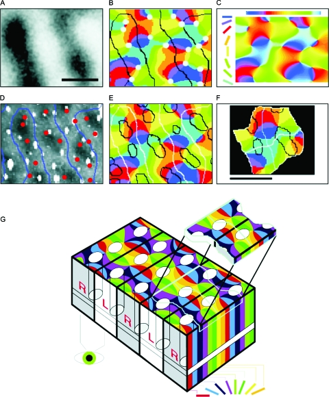
In the neocortex, neurons with similar response properties are often clustered together in column-like structures, giving rise to what has become known as functional architecture-the mapping of various stimulus feature dimensions onto the cortical sheet. At least partially, we owe this finding to the availability of several functional brain imaging techniques, both post-mortem and in-vivo, which have become available over the last two generations, revolutionizing neuroscience by yielding information about the spatial organization of active neurons in the brain. Here, we focus on how our understanding of such functional architecture is linked to the development of those functional imaging methodologies, especially to those that image neuronal activity indirectly, through metabolic or haemodynamic signals, rather than directly through measurement of electrical activity. Some of those approaches allow exploring functional architecture at higher spatial resolution than others. In particular, optical imaging of intrinsic signals reaches the striking detail of approximately 50 mum, and, together with other methodologies, it has allowed characterizing the metabolic and haemodynamic responses induced by sensory-evoked neuronal activity. Here, we review those findings about the spatio-temporal characteristics of neurovascular coupling and discuss their implications for functional brain imaging, including position emission tomography, and non-invasive neuroimaging techniques, such as funtional magnetic resonance imaging, applicable also to the human brain.


Figures

Figure 1
Figure 1. High-resolution functional optical imaging reveals the relationship between pinwheels (detection of orientated edges, shape processing), ocular dominance columns (stereo vision, depth processing), and cytochrome oxidase blobs (color vision) in the primary visual cortex of the macaque monkey.
(A) Optical map of ocular dominance from a portion of striate cortex, ∼1.5 mm×1 mm. Dark bands represent columns dominated by input from the right eye. Scale bars: 500 μm. (B) Borders of the ocular dominance columns (black contours taken from A) were overlaid onto the discrete pinwheel map (the color in the map codes for the orientation preference of each cortical location, see color of oriented bars in C). Pinwheel centers are marked with circles: ∼70% are centered on ocular dominance columns. (C) Interpolation from the eight different orientations effectively used as visual stimulus yields a map of orientation preference, which is mostly continuous except at the pinwheel centers (B). (D) Cytochrome oxidase (CO)-rich blobs are marked (dark) on the histological photograph that corresponds exactly to the cortical area that was optically imaged. (E) Overlay of maps in B and D shows the relationships between blobs (D), iso-orientation domains (B, C), and ocular dominance columns (A, B). (F) A combined view of the various maps reveals the existence of recurrent units (“hypercolumns”): here, two such fundamental modules are magnified from (E). (G) On the macroscopic scale, the sequence of hypercolumns shown in (F) can be schematically represented in the “revised ice cube model:” black lines mark the borders between columns of neurons that receive signals from different eyes. White ovals represent groups of neurons responsible for color perception (blobs). The pinwheels are formed by neurons involved in the perception of shape, with each color marking a column of neurons that respond selectively to a particular orientation in space. Note that both the blobs and the pinwheel centers lie at the center of the R or L columns. The iso-orientation lines tend to cross borders of ocular dominance columns (black lines) at right angles. The top “slice” above the “ice cube” model depicts two adjacent fundamental modules (400 μm×800 μm). Each module contains a complete set of about 60,000 neurons, processing the three features of orientation, depth, and color. (Modified from Bartfeld and Grinvald, 1992.)
Figure 2
Figure 2. Comparison of the spatio-temporal characteristics of sensory-evoked changes in oxidative metabolism and blood flow.
Left Panel: Activity maps obtained in rat whisker barrel cortex upon stimulation of two distinct whiskers (C2, top; D2, bottom, see insets for the corresponding somatotopy) from flavoprotein autofluorescence (AF) (left) and laser speckle imaging (LSI) (right). The color code depicts the percentage of signal increase from baseline. The horizontal and vertical lines facilitate the comparison across sub-panels, showing that the spatial pattern of the blood flow response was considerably less focal than that of the changes in oxidative metabolism, although their peaks of the activation roughly co-localized. Right Panel: Time course of AF and LSI responses. (A) AF (blue) and LSI (red) depict time courses from a single trial (2 s stimulation of vibrissa C2, average over area of >50% peak activation). Data points are represented by markers. Solid lines are fitted gamma curves. The shaded rectangle shows the stimulation. (B) Signal-averaged time course (20 trials). Note the excellent agreement between the single trial (A) and the average data (B). (C) Grand average (50% of the peak activation of C2 and D2 stimulations) fitted gamma curves (thick lines) ±1 standard deviation (thin lines) of the AF (blue) and LSI (red) responses. (D) Mean time course of the size of the activated area for AF (blue) and LSI (red). Shown are the areas with >25% (solid), >50% (dashed), and >75% (dotted) of the peak response. Note that the active area determined from AF is considerably smaller than that obtained from LSI. In addition, onset of the latter signal is delayed relative to the former, supporting the concept that the oxidative metabolism response is faster and more localized to neuronal activation than the blood flow response (with permission from: Weber et al., 2004).
Figure 3
Figure 3. Capillary character of blood volume (CBV) responses, shown by the morphology of high-resolution differential maps obtained by optical imaging.
(A) Image of the cortical vasculature in the primary visual cortex of an anaesthetized macaque. (B, C) Functional maps of ocular dominance columns obtained at 540 and 810 nm, two near-isosbestic wavelengths, at which the contribution of CBV changes dominates the signals. (D) Time course of the mapping signal, showing that the appearance of oximetric maps (obtained at 605 nm, blue trace) slightly precedes that of the CBV maps (obtained at 570 nm, red trace). The larger 570 nm signal was downscaled in the figure by a factor of 4 for display purposes. Inset: Zoom into the first second of the responses. (E) Image of the cortical vasculature in the primary visual cortex of an awake macaque. (F, G) Functional maps of ocular dominance columns obtained at a strongly oximetric wavelength (605 nm) and at an isosbestic wavelength (570 nm) chosen to image CBV signals. Note that (i) the patterns of the blood-volume maps are validated by the oximetric maps, known to reflect the underlying functional architecture; and (ii) the high spatial resolution of the CBV maps strongly suggests that the functional patches can only be of capillary origin. Scale bars: 1 mm. (A–C) Modified from Frostig et al., 1990; (D–G) modified from Vanzetta et al., 2004.

References

    1. Abeles, M (1991). Corticonics: neural circuits of the cerebral cortex, 1st ed., Cambridge University Press, Cambridge.
    1. Adelson, P D, Nemoto, E, Scheuer, M, Painter, M, Morgan, J, and Yonas, H (1999). “Noninvasive continuous monitoring of cerebral oxygenation periictally using near infrared spectroscopy: a preliminary report.” Epilepsia EPILAK10.1111/j.1528-1157.1999.tb02030.x 40, 1484–1489. - DOI - PubMed
    1. Akgoren, N, and Lauritzen, M (1999). “Functional recruitment of red blood cells to rat brain microcirculation accompanying increased neuronal activity in cerebellar cortex.” NeuroReport NERPEZ 10, 3257–3263. - PubMed
    1. Altman, D I, Lich, L L, and Powers, W J (1991). “Brief inhalation method to measure cerebral oxygen extraction fraction with PET: accuracy determination under pathologic conditions.” J. Nucl. Med. JNMEAQ 32, 1738–1741. - PubMed
    1. Ances, B M (2004). “Coupling of changes in cerebral blood flow with neural activity: what must initially dip must come back up.” J. Cereb. Blood Flow Metab. JCBMDN10.1097/01.WCB.0000103920.96801.12 24, 1–6. - DOI - PubMed

LinkOut - more resources