. 2009 May;60(5):1438-50.
doi: 10.1002/art.24489.

Altered mineralization of human osteoarthritic osteoblasts is attributable to abnormal type I collagen production

Affiliations

Altered mineralization of human osteoarthritic osteoblasts is attributable to abnormal type I collagen production

Denis Couchourel et al. Arthritis Rheum. 2009 May.

Abstract

Objective: Bone tissue in osteoarthritis (OA) is composed of abundant undermineralized osteoid matrix. The aim of this study was to investigate the mechanisms responsible for this abnormal matrix, using in vitro OA subchondral osteoblasts.

Methods: Primary normal and OA osteoblasts were prepared from tibial plateaus. Phenotype was determined by alkaline phosphatase activity, and osteocalcin, osteopontin, prostaglandin E2 (PGE2), and transforming growth factor beta1 (TGFbeta1) were assessed by enzyme-linked immunosorbent assay. Expression of COL1A1 and COL1A2 was determined by real-time polymerase chain reaction. The production of type I collagen was determined by the release of its C-terminal propeptide and Western blot analysis. In vitro mineralization was evaluated by alizarin red staining. Inhibition of TGFbeta1 expression was performed using a small interfering RNA technique.

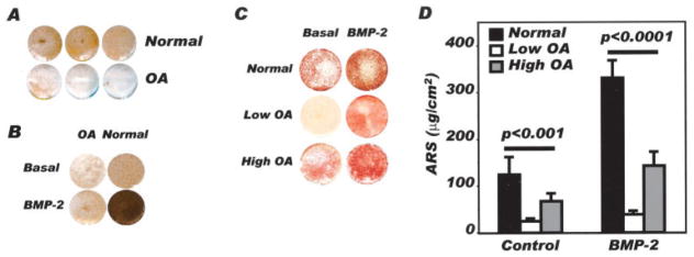
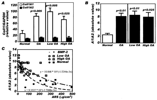
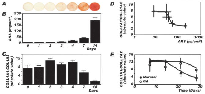
Results: Mineralization of OA osteoblasts was reduced compared with mineralization of normal osteoblasts, even in the presence of bone morphogenetic protein 2 (BMP-2). Alkaline phosphatase and osteocalcin levels were elevated in OA osteoblasts compared with normal osteoblasts, whereas osteopontin levels were similar. The COL1A1-to-COL1A2 messenger RNA ratio was 3-fold higher in OA osteoblasts compared with normal osteoblasts, and the production of collagen by OA osteoblasts was increased. Because TGFbeta1 inhibits BMP-2-dependent mineralization, and because TGFbeta1 levels are approximately 4-fold higher in OA osteoblasts than in normal osteoblasts, inhibiting TGFbeta1 levels in OA osteoblasts corrected the abnormal COL1A1-to-COL1A2 ratio and increased alizarin red staining.

Conclusion: Elevated TGFbeta1 levels in OA osteoblasts are responsible, in part, for the abnormal ratio of COL1A1 to COL1A2 and for the abnormal production of mature type I collagen. This abnormal COL1A1-to-COL1A2 ratio generates a matrix that blunts mineralization in OA osteoblasts.


Figures

Figure 1
Figure 1
Evaluation of in vitro mineralization of normal and osteoarthritis (OA) osteoblasts. Confluent normal and OA osteoblasts were incubated in BGJb medium containing 10% fetal bovine serum, 50 μg/ml ascorbic acid, and 50 μg/ml β-glycerophosphate for 30 days, in the presence or absence of 10 ng/ml bone morphogenetic protein 2 (BMP-2). Mineralization of cell cultures was evaluated by either von Kossa’s staining or alizarin red staining (ARS). A, Representative von Kossa’s staining in normal and OA osteoblast cultures (n = 3 separate individuals per group). B, Representative von Kossa’s staining in 1 normal and 1 OA osteoblast following treatment with or without 10 ng/ml BMP-2, from day 2 until day 30 (results are representative of 4 separate experiments). C, Representative alizarin red staining following treatment of normal osteoblasts, OA osteoblasts producing low levels of prostaglandin E2 (PGE2), and OA osteoblasts producing high levels of PGE2, with or without 10 ng/ml BMP-2. D, Quantification of alizarin red staining according to the method described by Gregory et al (30). Values are the mean and SEM results from 6 normal, 21 low OA, and 14 high OA preparations.
Figure 2
Figure 2
Expression of type I collagen α1 and α2 chains in normal and osteoarthritis (OA) osteoblasts by real-time polymerase chain reaction (PCR). Confluent osteoblasts were lysed in TRIzol, and RNA was extracted as described in Patients and Methods. RNA was reverse transcribed followed by PCR amplification of cDNA using specific primers. Plasmid DNAs containing the target gene sequences were used to generate the standard curves for COL1A1, COL1A2, and GAPDH. The value for each sample was calculated as the ratio of the number of molecules of the target gene:number of molecules of GAPDH. A, Expression of COL1A1 and COL1A2 under basal conditions. B, Ratio of COL1A1 to COL1A2 (A1/A2) under basal conditions in normal osteoblasts, total OA osteoblast preparations, and the subgroups of OA osteoblasts producing low levels of prostaglandin E2 (PGE2) and those producing high levels of PGE2. C, Relationship between alizarin red staining (ARS) and the COL1A1-to-COL1A2 ratio in normal osteoblasts, OA osteoblasts producing low levels of PGE2, and OA osteoblasts producing high levels of PGE2, treated or not treated with bone morphogenetic protein 2 (BMP-2). Values are the mean and SEM results from 8 normal osteoblasts, 14 total OA osteoblasts, 6 OA osteoblasts producing low levels of PGE2, and 8 OA osteoblasts producing high levels of PGE2.
Figure 3
Figure 3
Type I collagen production by normal and OA osteoblasts. Confluent osteoblasts were incubated for the last 48 hours of culture in Ham’s F-12/Dulbecco’s modified Eagle’s medium containing 0.5% bovine serum albumin. A, Culture medium was collected for the determination of collagen synthesis as the de novo release of the carboxy-terminal peptide fragment (CICP) of type I collagen, which reflects true collagen synthesis. The release of CICP was determined using a very selective enzyme-linked immunosorbent assay. Values are the mean and SD results from 9 normal and 22 OA osteoblast cultures (n = 14 OA osteoblasts producing low levels of PGE2 and 8 OA osteoblasts producing high levels of PGE2). B, Western blotting for type I collagen production by osteoblasts in 2 normal and 5 OA osteoblasts was performed. Cells lysed in radioimmunoprecipitation assay buffer and 25 μg of total protein were subjected to sodium dodecyl sulfate–polyacrylamide gel electrophoresis. Western blotting was performed with a polyclonal antibody that detects type I collagen α1 and α2 chains. Western blot analysis of actin was performed to demonstrate equivalent loading between samples. The results shown are representative of 7 normal and 10 OA osteoblast preparations (6 with low production of PGE2 and 4 with high production of PGE2). See Figure 2 for other definitions.
Figure 4
Figure 4
Relationship between mineralization and the COL1A1-to-COL1A2 ratio in SaOS-2 cells and normal and osteoarthritis (OA) osteoblasts. Confluent SaOS-2 cells were incubated for 0, 1, 2, 3, 4, 7, or 14 days in BGJb medium containing 10% fetal bovine serum, 50 μg/ml ascorbic acid, and 50 μg/ml β-glycerophosphate. Confluent normal and OA osteoblasts were incubated in the same medium in the presence of 10 ng/ml bone morphogenetic protein 2 from day 2 until day 7, day 14, day 21 or day 28 postconfluence, when cells were lysed in TRIzol for the determination of COL1A1 and COL1A2 expression. COL1A1 and COL1A2 expression was determined by real-time polymerase chain reaction, as described in Figure 2. A, Alizarin red staining (ARS) as a function of time in culture in SaOS-2 cells in the presence of mineralization medium. B, Quantification of alizarin red staining. C, Ratio of COL1A1 to COL1A2 expression. Values in B and C are the mean and SEM results from 3–7 different cell preparations per day. D, Ratio of COL1A1 to COL1A2 expression in SaOS-2 cells as a function of alizarin red staining. E, Ratio of COL1A1 to COL1A2 expression in normal and OA osteoblasts as a function of time in culture. Values in D and E are the mean ± SEM results from 3–8 normal osteoblast preparations and 8 OA osteoblast preparations.
Figure 5
Figure 5
Effect of transforming growth factor β1 (TGFβ1) on bone morphogenetic protein 2 (BMP-2)–induced alizarin red staining (ARS). A, Representative alizarin red staining for confluent normal osteoblasts (n = 5 separate experiments) incubated as described in Figure 1. B, TGFβ1 levels in normal and osteoarthritis (OA) osteoblasts as measured by selective enzyme-linked immunosorbent assay. Values are the mean and SEM results from 8 normal osteoblast preparations, 14 preparations of osteoblasts producing low levels of prostaglandin E2 (PGE2), and 14 preparations of OA osteoblasts producing high levels of PGE2. C, TGFβ1 mRNA levels, as determined by quantitative polymerase chain reaction, under basal conditions and following inhibition of TGFβ1 expression with short hairpin RNA (shRNA) plasmids. Values are the mean and SEM results from 7 OA osteoblast preparations. D, COL1A1- to -COL1A2 ratio of OA osteoblasts under basal conditions or following inhibition of TGFβ1 expression with shRNA. Values are the mean and SEM results from 7 OA osteoblast preparations. E, Top, Representative alizarin red staining of OA osteoblasts treated or not treated with TGFβ1 shRNA. Bottom, Quantification of alizarin red staining following BMP-2 treatment in OA osteoblasts treated or not treated with TGFβ1 shRNA. Values are the mean and SEM results from 6 preparations.

References

    1. Hilal G, Martel-Pelletier J, Pelletier JP, Ranger P, Lajeunesse D. Osteoblast-like cells from human subchondral osteoarthritic bone demonstrate an altered phenotype in vitro: possible role in subchondral bone sclerosis. Arthritis Rheum. 1998;41:891–9. - PubMed
    1. Mansell JP, Bailey AJ. Abnormal cancellous bone collagen metabolism in osteoarthritis. J Clin Invest. 1998;101:1596–603. - PMC - PubMed
    1. Westacott CI, Webb GR, Warnock MG, Sims JV, Elson CJ. Alteration of cartilage metabolism by cells from osteoarthritic bone. Arthritis Rheum. 1998;40:1282–91. - PubMed
    1. Radin EL, Rose RM. Role of subchondral bone in the initiation and progression of cartilage damage. Clin Orthop. 1986;213:34–40. - PubMed
    1. Li B, Aspden RM. Composition and mechanical properties of cancellous bone from the femoral head of patients with osteoporosis or osteoarthritis. J Bone Miner Res. 1997;12:641–51. - PubMed

Publication types

MeSH terms

Grants and funding