. 2009 May;60(5):1508-18.
doi: 10.1002/art.24471.

Induction of the expression of profibrotic cytokines and growth factors in normal human peripheral blood monocytes by gadolinium contrast agents

Affiliations

Induction of the expression of profibrotic cytokines and growth factors in normal human peripheral blood monocytes by gadolinium contrast agents

Peter J Wermuth et al. Arthritis Rheum. 2009 May.

Abstract

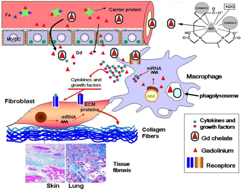
Objective: Nephrogenic systemic fibrosis (NSF) is a severe fibrosing disorder occurring in patients with renal insufficiency. The majority of patients with this disorder have documented exposure to magnetic resonance imaging contrast agents containing Gd. The purpose of this study was to examine the effects of gadolinium diethylenetriaminepentaacetic acid bismethylamide (Gd[DTPA-BMA]; Omniscan) as compared with Gd-DTPA and GdCl3 on the expression and production of cytokines and growth factors by normal human peripheral blood monocytes in vitro and to examine whether conditioned media from Gd-exposed peripheral blood monocytes could induce a profibrotic phenotype in dermal fibroblasts.

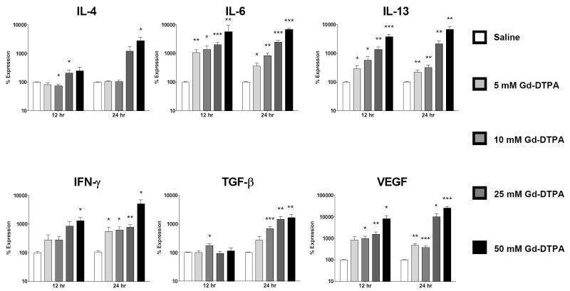
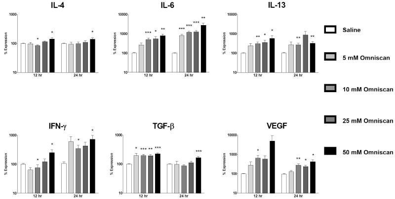
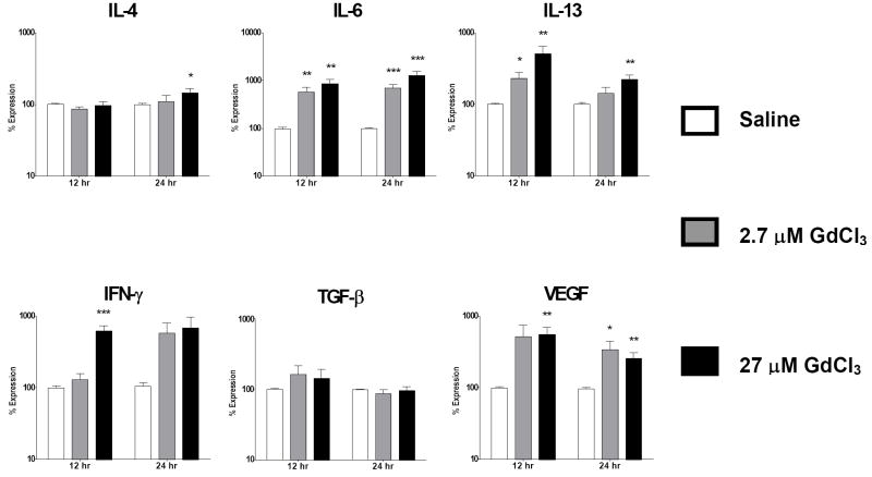
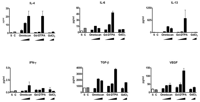
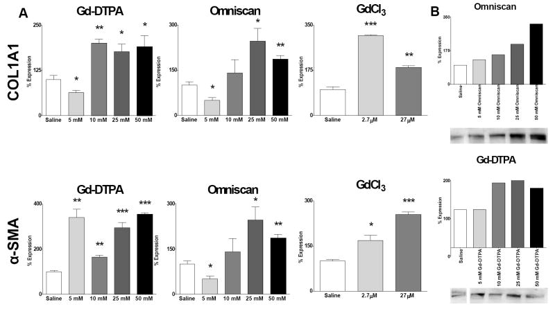
Methods: Normal human peripheral blood monocytes isolated by Ficoll-Hypaque gradient centrifugation and plastic adherence were incubated with various concentrations of Gd[DTPA-BMA], Gd-DTPA, or GdCl3. Gene expression of interleukins 4, 6, and 13, interferon-gamma, tumor necrosis factor alpha, transforming growth factor beta, connective tissue growth factor, and vascular endothelial growth factor were assessed by real-time polymerase chain reaction (PCR) analysis. Production and secretion of cytokines and growth factors by Gd compound-exposed monocytes was quantified by enzyme-linked immunosorbent assay proteome multiplex arrays. The effects of conditioned media from the Gd compound-exposed monocytes on the phenotype of normal human dermal fibroblasts were examined by real-time PCR and Western blotting.

Results: The 3 Gd-containing compounds stimulated the expression and production of numerous cytokines and growth factors by normal human peripheral blood monocytes. Conditioned media from these cells induced a profibrotic phenotype in normal human dermal fibroblasts.

Conclusion: The 3 Gd-containing compounds studied induce potent cellular responses in normal human peripheral blood monocytes, which may participate in the development of tissue fibrosis in NSF.


Figures

Figure 1
Figure 1. Upregulated cytokine/growth factor expression is induced by Gd-DTPA in normal human peripheral blood monocytes
Analysis of cytokine and growth factor expression levels by real time PCR at 12 and 24 h following exposure to Gd-DTPA. Values represent the mean (+/- standard deviation) expression levels of three replicates of five separate experiments with peripheral blood monocytes from four different normal individuals. Note that the data are presented in a semilogarithmic scale. C(t) values for cytokines were normalized with β-actin. The saline control levels were arbitrarily set at 100% expression at each time point. Values for other samples are expressed relative to the saline control. *: p<0.1; **: p < 0.01; ***: p<0.0001.
Figure 2
Figure 2. Omniscan® activates expression of profibrotic/proinflammatory cytokines in normal human peripheral blood monocytes
Analysis of cytokine and growth factor expression levels by real time PCR at 12 and 24 h following exposure to Omniscan®. Values represent the mean (+/- standard deviation) expression levels of three replicates of five separate experiments with peripheral blood monocytes from four different normal individuals. Note that the data are presented in a semilogarithmic scale. C(t) values for cytokines were normalized with β-actin. The saline control levels were arbitrarily set at 100% expression at each time point. Values for other samples are expressed relative to the saline control. *: p<0.1; **: p < 0.01; ***: p<0.0001.
Figure 3
Figure 3. GdCl3 induces upregulation of cytokine/growth factor expression in normal human peripheral blood monocytes
Analysis of cytokine and growth factor expression levels by real time PCR at 12 and 24 h following exposure to GdCl3. Values represent the mean (+/- standard deviation) expression levels of three replicates of five separate experiments with peripheral blood monocytes from four different normal individuals. Note that the data are presented in a semilogarithmic scale. C(t) values for cytokines were normalized with β-actin. The saline control levels were arbitrarily set at 100% expression at each time point. Values for other samples are expressed relative to the saline control. *: p<0.1; **: p < 0.01; ***: p<0.0001.
Figure 4
Figure 4. Amount of secreted cytokines and growth factors in cell culture supernatants from Gd-compound-exposed normal human peripheral blood monocytes
Quantitative measurement of cytokines and growth factors present in the culture media of Gd-compound-exposed cells was performed by multiplex proteome array analyses as described in Materials and Methods. Cells were incubated with either saline (S), caldiamide (C; 2500 μM), Omniscan® (5, 25, or 50 μM), Gd-DTPA (5, 25, or 50 μM), or GdCl3 (2.7 or 27 μM). Values are expressed in pg/ml and represent the mean value of values obtained from duplicate results at 3 dilutions: 1:2, 1:50 and 1:1000.
Figure 5
Figure 5. Conditioned media from Gd compound-exposed peripheral blood monocytes induce expression of COL1A1 and α-SMA transcripts and increased production of type I procollagen by normal human dermal fibroblasts
Normal dermal human fibroblasts were incubated for 24 h in standard tissue culture media containing a 1:2 dilution of culture media isolated from peripheral blood monocytes exposed to Gd-containing compounds. (A) Expression levels of COL1A1 and α-SMA transcripts determined by real time PCR. The results shown are representative of two separate experiments, each performed in triplicate. (B) Production of type I collagen assessed by Western blots and quantified employing Image J software. The saline control levels were arbitrarily set at 100%. Values for the samples are expressed as the percent increase over the saline control value. *: p<0.1; **: p < 0.01; ***: p<0.0001.
Figure 6
Figure 6. Schematic representation of proposed mechanisms of the profibrotic effects of Gd compounds
Chemical structure of gadodiamide is from (31).

References

    1. Cowper SE, Robin HS, Steinberg SM, Su LD, Supta S, LeBoit PE. Scleromyxoedema-like cutaneous disease in renal-dialysis patients. Lancet. 2000;356:1000–1001. - PubMed
    1. Cowper SE, Su LD, Bhawan J, Robin HS, LeBoit PE. Nephrogenic fibrosing dermopathy. Am J Dermatopathol. 2001;23:383–393. - PubMed
    1. Mackay-Wiggan JM, Cohen DJ, Hardy MA, Knobler EH, Grossman ME. Nephrogenic fibrosing dermopathy (scleromyxedema-like illness of renal disease) J Am Acad Dermatol. 2003;48:55–60. - PubMed
    1. Swartz RD, Crofford LJ, Phan SH, Ike RW, Su LD. Nephrogenic fibrosing dermopathy: a novel cutaneous fibrosing disorder in patients with renal failure. Am J Med. 2003;114:563–572. - PubMed
    1. Jimenez SA, Artlett CM, Sandorfi N, Derk C, Latinis K, Sawaya H, Haddad R, Shanahan JC. Dialysis-associated systemic fibrosis (nephrogenic fibrosing dermopathy): study of inflammatory cells and transforming growth factor beta1 expression in affected skin. Arthritis Rheum. 2004;50:2660–2666. - PubMed

Publication types

MeSH terms