

Mitochondrial inhibitor 3-nitroproprionic acid enhances oxidative modification of alpha-synuclein in a transgenic mouse model of multiple system atrophy
- PMID: 19405128
- PMCID: PMC2885901
- DOI: 10.1002/jnr.22089
Mitochondrial inhibitor 3-nitroproprionic acid enhances oxidative modification of alpha-synuclein in a transgenic mouse model of multiple system atrophy
Abstract
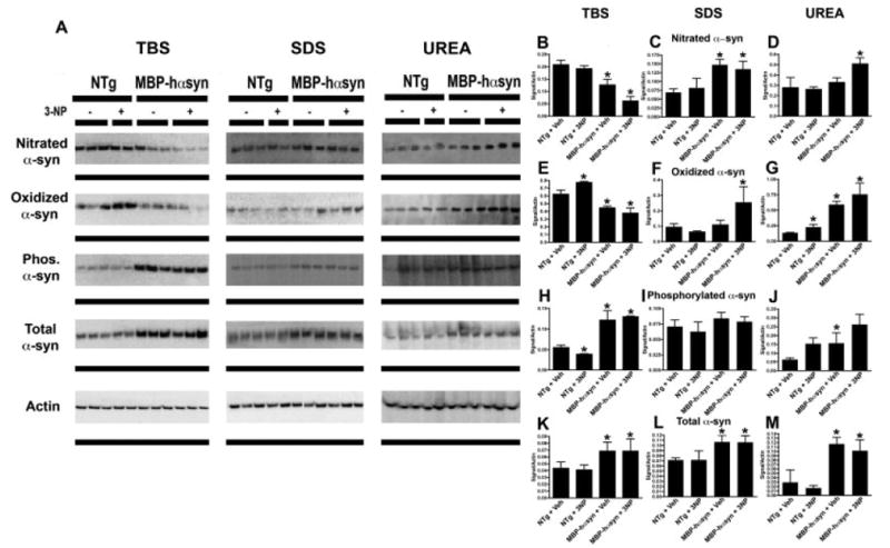
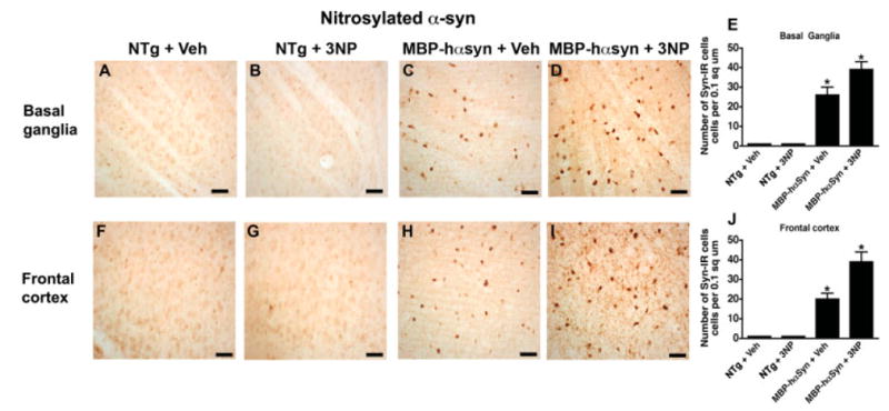
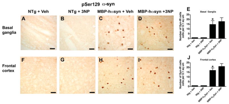
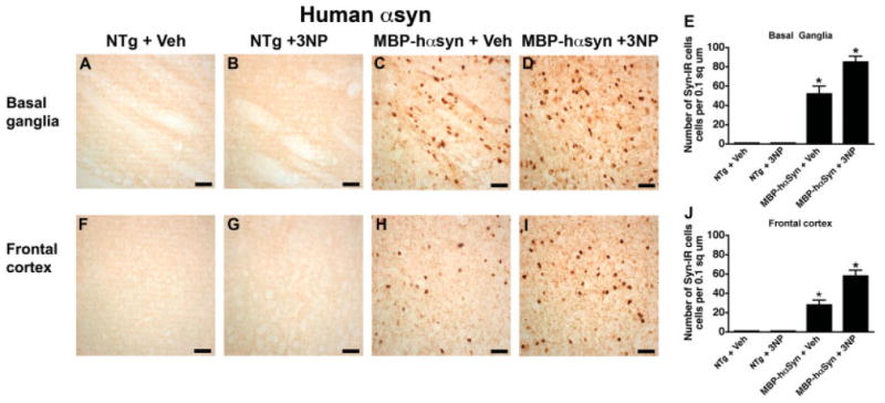
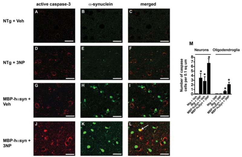
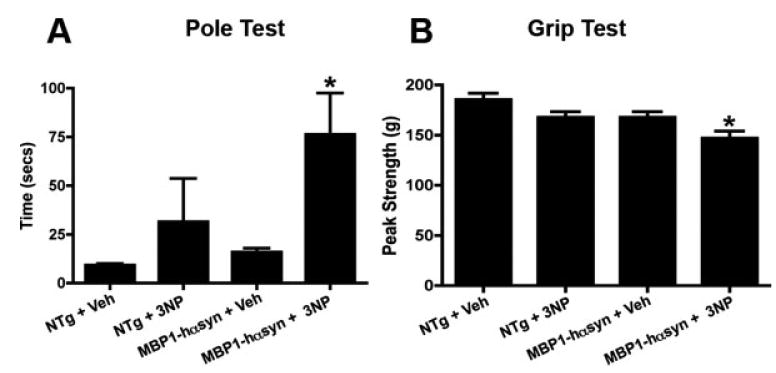
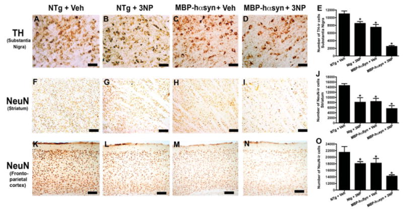
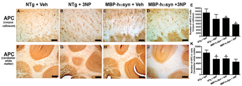
Multiple system atrophy (MSA) is a progressive neurodegenerative disease characterized by autonomic failure, parkinsonism, cerebellar ataxia, and oligodendrocytic accumulation of alpha-synuclein (alphasyn). Oxidative stress has been linked to neuronal death in MSA and the mitochondrial toxin 3-nitropropionic acid (3NP) is known to enhance the motor deficits and neurodegeneration in transgenic mice models of MSA. However, the effect of 3NP administration on alphasyn itself has not been studied. In this context, we examined the neuropathological effects of 3NP administration in alphasyn transgenic mice expressing human alphasyn (halphasyn) under the control of the myelin basic protein (MBP) promoter and the effect of this administration on posttranslational modifications of alphasyn, on levels of total alphasyn, and on its solubility. We demonstrate that 3NP administration altered levels of nitrated and oxidized alphasyn in the MBP-halphasyn tg while not affecting global levels of phosphorylated or total alphasyn. 3NP administration also exaggerated neurological deficits in the MBP-halphasyn tg mice, resulting in widespread neuronal degeneration and behavioral impairment.
Figures

Similar articles
-
Neurodegeneration in a transgenic mouse model of multiple system atrophy is associated with altered expression of oligodendroglial-derived neurotrophic factors.J Neurosci. 2010 May 5;30(18):6236-46. doi: 10.1523/JNEUROSCI.0567-10.2010. J Neurosci. 2010. PMID: 20445049 Free PMC article.
-
Alpha-synuclein deficient mice are resistant to toxin-induced multiple system atrophy.Neuroreport. 2010 Apr 21;21(6):457-62. doi: 10.1097/WNR.0b013e328338ba6b. Neuroreport. 2010. PMID: 20224454 Free PMC article.
-
Failure of Neuroprotection Despite Microglial Suppression by Delayed-Start Myeloperoxidase Inhibition in a Model of Advanced Multiple System Atrophy: Clinical Implications.Neurotox Res. 2015 Oct;28(3):185-94. doi: 10.1007/s12640-015-9547-7. Epub 2015 Jul 21. Neurotox Res. 2015. PMID: 26194617 Free PMC article.
-
Is Multiple System Atrophy a Prion-like Disorder?Int J Mol Sci. 2021 Sep 18;22(18):10093. doi: 10.3390/ijms221810093. Int J Mol Sci. 2021. PMID: 34576255 Free PMC article. Review.
-
Papp-Lantos inclusions and the pathogenesis of multiple system atrophy: an update.Acta Neuropathol. 2010 Jun;119(6):657-67. doi: 10.1007/s00401-010-0672-3. Epub 2010 Mar 23. Acta Neuropathol. 2010. PMID: 20309568 Review.
Cited by
-
Multiple system atrophy: pathogenic mechanisms and biomarkers.J Neural Transm (Vienna). 2016 Jun;123(6):555-72. doi: 10.1007/s00702-016-1545-2. Epub 2016 Apr 20. J Neural Transm (Vienna). 2016. PMID: 27098666 Review.
-
Glia and alpha-synuclein in neurodegeneration: A complex interaction.Neurobiol Dis. 2016 Jan;85:262-274. doi: 10.1016/j.nbd.2015.03.003. Epub 2015 Mar 10. Neurobiol Dis. 2016. PMID: 25766679 Free PMC article. Review.
-
Uric acid is associated with the prevalence but not disease progression of multiple system atrophy in Chinese population.J Neurol. 2013 Oct;260(10):2511-5. doi: 10.1007/s00415-013-7006-z. Epub 2013 Jun 26. J Neurol. 2013. PMID: 23801150
-
Animal models of multiple system atrophy.Clin Auton Res. 2015 Feb;25(1):9-17. doi: 10.1007/s10286-014-0266-6. Epub 2015 Jan 14. Clin Auton Res. 2015. PMID: 25585910 Free PMC article. Review.
-
Multiple system atrophy: experimental models and reality.Acta Neuropathol. 2018 Jan;135(1):33-47. doi: 10.1007/s00401-017-1772-0. Epub 2017 Oct 20. Acta Neuropathol. 2018. PMID: 29058121 Free PMC article. Review.
References
-
- Azeredo da Silveira S, Schneider BL, Cifuentes-Diaz C, Sage D, Abbas-Terki T, Iwatsubo T, Unser M, Aebischer P. Phosphorylation does not prompt, nor prevent, the formation of alpha-synuclein toxic species in a rat model of Parkinson's disease. Hum Mol Genet. 2009;18:872–887. - PubMed
-
- Beyer K. Alpha-synuclein structure, posttranslational modification and alternative splicing as aggregation enhancers. Acta Neuropathol. 2006;112:237–251. - PubMed
-
- Bieschke J, Zhang Q, Bosco DA, Lerner RA, Powers ET, Wentworth P, Jr, Kelly JW. Small molecule oxidation products trigger disease-associated protein misfolding. Acc Chem Res. 2006;39:611–619. - PubMed
-
- Chen L, Feany MB. Alpha-synuclein phosphorylation controls neurotoxicity and inclusion formation in a Drosophila model of Parkinson disease. Nat Neurosci. 2005;8:657–663. - PubMed
Publication types
MeSH terms
Substances
Grants and funding
LinkOut - more resources
Full Text Sources
Miscellaneous
