

Review
doi: 10.1146/annurev.genom.9.081307.164350.
Unraveling a multifactorial late-onset disease: from genetic susceptibility to disease mechanisms for age-related macular degeneration
Affiliations
- PMID: 19405847
- PMCID: PMC3469316
- DOI: 10.1146/annurev.genom.9.081307.164350
Item in Clipboard
Review
Unraveling a multifactorial late-onset disease: from genetic susceptibility to disease mechanisms for age-related macular degeneration
Annu Rev Genomics Hum Genet.
2009.
Abstract
Aging-associated neurodegenerative diseases significantly influence the quality of life of affected individuals. Genetic approaches, combined with genomic technology, have provided powerful insights into common late-onset diseases, such as age-related macular degeneration (AMD). Here, we discuss current findings on the genetics of AMD to highlight areas of rapid progress and new challenges. We also attempt to integrate available genetic and biochemical data with cellular pathways involved in aging to formulate an integrated model of AMD pathogenesis.
Figures
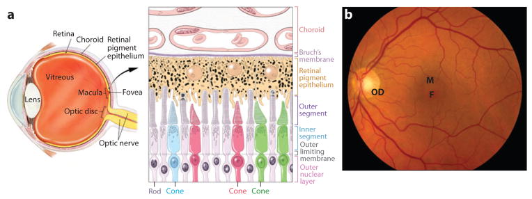
(a) Cross-section of the human eye, and schematic of the photoreceptor-retinal pigment epithelium (RPE)-choroidal layers that are affected in AMD. Cone photoreceptors are shown in red, green, or blue. RPE apical processes are intimately associated with photoreceptor outer segments. Two of the rods are shown in the active process of outer segment disc shedding. Melanosomes are shown as dark organelles in RPE cells. (b) Fundus photograph showing the retina of a normal individual. Retinal blood vessels are clearly visible. F, fovea; M, macula; OD, optic disc.
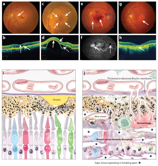
(a) Fundus photograph of the left eye of a patient with large, “soft” drusen (arrow), indicative of intermediate AMD. The patient has good vision. (b) The optical coherence tomography (OCT) image of the patient in (a) with drusen. This image has the inner retina pointing toward the top with the choroid shown at the bottom of the image, a reverse of our schematic seen in panels (i) and (j). The solid arrow points to corresponding areas of drusen that have caused an elevation of the retinal pigment epithelium, giving it a “bumpy-lumpy” appearance. This section of the OCT shows the center of fovea with the retinal layers shown as a central “foveal depression” (dashed arrow). (c) Fundus photograph of a patient with an elevated RPE detachment, which is the creamy-colored round lesion centered on the macula (outlined by the arrows). The patient still has good vision because the retina has no intraretinal or subretinal fluid. This is another form of advanced AMD that may lead to NV or GA disease. (d) The OCT image of the patient in (c) with elevated RPE detachment (long solid arrow indicates the RPE layer). There is little intraretinal fluid (short solid arrow) and subretinal fluid (dashed arrow) detected. (e) Fundus photograph of the right eye with evidence of advanced AMD, the neovascular form with evidence of subretinal hemorrhage (solid arrow), retinal hard exudates (yellow waxy lesions, dashed arrow), and large drusen. The visual acuity is 20/160. (f) Fluorescein angiogram of patient with neovascular AMD, leakage of the fluorescein dye in the late frames through the abnormal neovascular complex, resulting in hyperfluorescence (white area, solid arrow). The subretinal hemorrhage blocked the dye, resulting in an area of hypofluorescence (dashed arrow). (g) Fundus photograph of patient with the “dry” form of advanced AMD, with GA centered on the macula, a well-defined area of atrophy with the loss of the neuroretina, RPE, and the choriocapillaris. The large vessels within the atrophic area are the remaining vessels of the choroid that can be viewed readily because of the lack of the pigmented layer of the RPE (arrow). Peripheral to the GA are large drusen. The visual acuity is 20/200. (h) The OCT image of the patient in (g) with thinning of the retina and the loss of the RPE and choriocapillaris in the center of the macula (arrow). (i) Schematic of the photoreceptor-RPE-choroid region from an early AMD retina. A large drusen is indicated. (j) Schematic of the photoreceptor-RPE-choroid region from a late AMD retina. Various abnormalities are indicated.
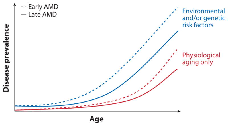
A representation of disease prevalence with age. The incidence of both early and late AMD greatly increases in all individuals at a late age (age 80 and above). We suggest that individuals with genetic susceptibility variants and those who have been exposed to environmental risk factors exhibit the disease at much earlier age (age 60 and above).
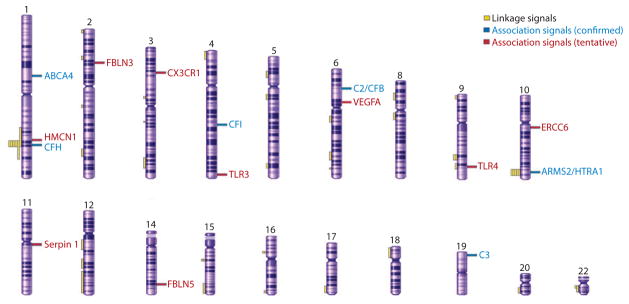
A schematic of human chromosomes showing regions of linkage identified in different studies and association signals (confirmed, and tentative requiring further validation) for AMD susceptibility loci/genes (modified from Reference 148).
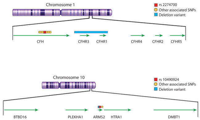
Genomic regions at chromosome 1q32 and 10q26 showing the genes and key associated single nucleotide polymorphisms (SNPs) and deletion variants.
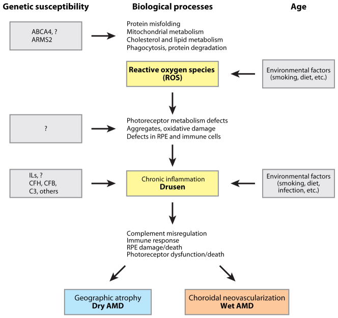
An integrated model of AMD pathogenesis. Aging is the primary driver of early cellular defects in photoreceptors and retinal pigment epithelium (RPE); these changes are exacerbated (or inhibited) by specific genetics variants. Complement and immune response pathways play critical roles in the onset and severity of disease.
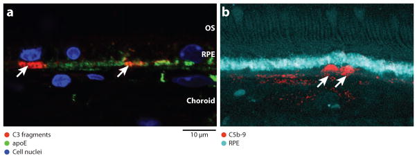
Activated complement components in sub-RPE (retinal pigment epithelium) deposits. (a) Immuno-localization of C3 fragments [C3b/iC3b/C3c (red)] and apoE (green) in basal, sub-RPE deposits (arrowheads) and Bruch’s membrane of 75-week-old APOE4-HFC mouse eye. C3 fragments are found in small, discrete patches that appear to be associated with individual RPE cells (blue is nuclear DAPI stain). (b) Immuno-localization of C5b-9 (red) in human AMD donor eye (cyan is lipofuscin autofluorescence from the RPE). C5b-9 immunoreactivity is associated with similar sub-RPE deposits (arrowheads). OS, outer segments. Images courtesy of J-D. Ding, Duke University (a) and L. Johnson, University of California, Santa Barbara (b).
References
Publication types
MeSH terms
Grants and funding
LinkOut - more resources
Full Text Sources
Other Literature Sources
Medical
Molecular Biology Databases
