

RANKL increases the level of Mcl-1 in osteoclasts and reduces bisphosphonate-induced osteoclast apoptosis in vitro
- PMID: 19405951
- PMCID: PMC2688211
- DOI: 10.1186/ar2681
RANKL increases the level of Mcl-1 in osteoclasts and reduces bisphosphonate-induced osteoclast apoptosis in vitro
Abstract
Introduction: Bisphosphonates are the most widely used class of drug for inhibiting osteoclast-mediated bone loss, but their effectiveness at preventing joint destruction in rheumatoid arthritis has generally been disappointing. We examined whether the ability of bisphosphonates to induce osteoclast apoptosis and inhibit bone resorption in vitro is influenced by the cytokine receptor activator of nuclear factor-kappa B ligand (RANKL), an important mediator of inflammation-induced bone loss.
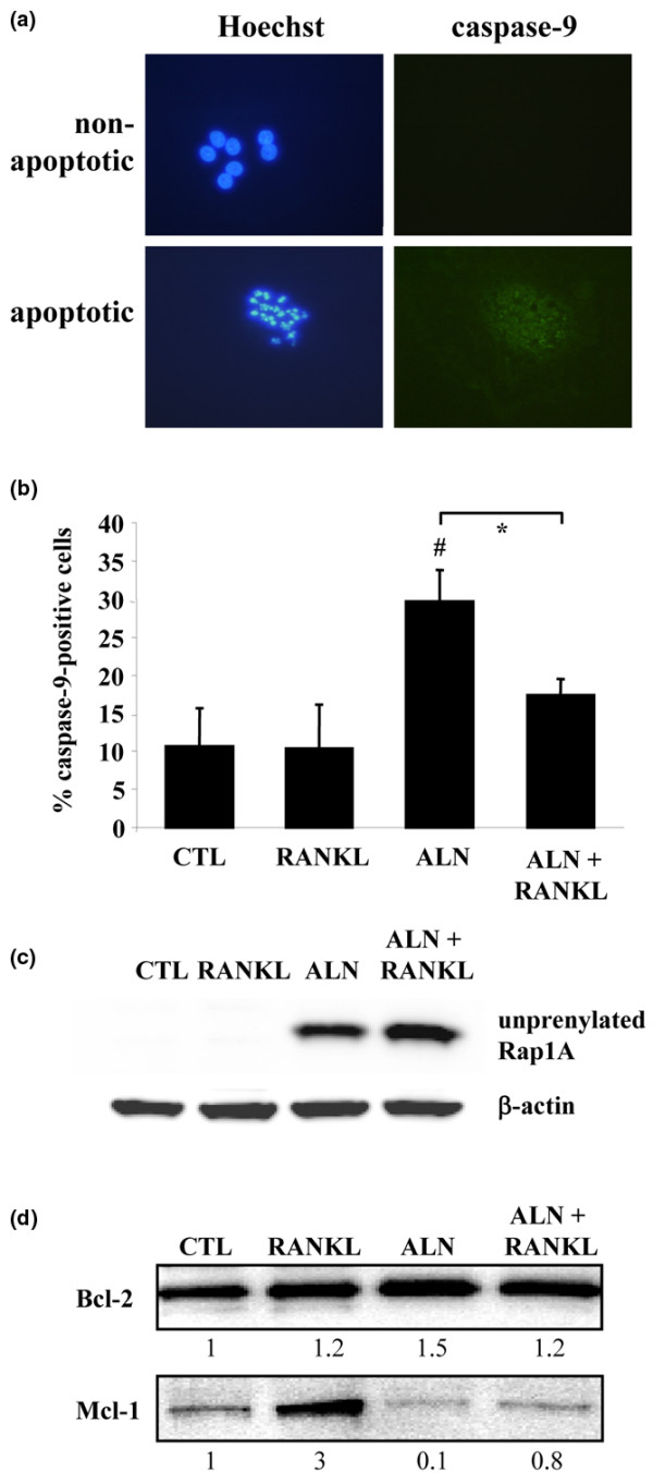
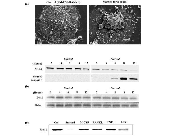
Methods: Rabbit osteoclasts were treated with the bisphosphonates clodronate or alendronate for up to 48 hours in the absence or presence of RANKL. Changes in cell morphology and induction of apoptosis were examined by scanning electron microscopy, whilst resorptive activity was determined by measuring the area of resorption cavities. Changes in the level of anti-apoptotic proteins, including Mcl-1, Bcl-2, and Bcl-x>L, were determined in rabbit osteoclasts and in cytokine-starved mouse osteoclasts by Western blotting.
Results: RANKL significantly attenuated the ability of both clodronate and alendronate to induce osteoclast apoptosis and inhibit bone resorption. Treatment of rabbit osteoclasts with RANKL was associated with an increase in the anti-apoptotic protein Mcl-1 but not Bcl-2. A role for Mcl-1 in osteoclast survival was suggested using osteoclasts generated from mouse bone marrow macrophages in the presence of RANKL + macrophage colony-stimulating factor (M-CSF) since cytokine deprivation of mouse osteoclasts caused a rapid loss of Mcl-1 (but not Bcl-2 or Bcl-xL), which preceded the biochemical and morphological changes associated with apoptosis. Loss of Mcl-1 from mouse osteoclasts could be prevented by factors known to promote osteoclast survival (RANKL, M-CSF, tumour necrosis factor-alpha [TNF-alpha], or lipopolysaccharide [LPS]).
Conclusions: RANKL protects osteoclasts from the apoptosis-inducing and anti-resorptive effects of bisphosphonates in vitro. The ability of RANKL (and other pro-inflammatory factors such as TNF-alpha and LPS) to increase the level of Mcl-1 in osteoclasts may explain the lack of effectiveness of some bisphosphonates in preventing inflammation-induced bone loss.
Figures


Similar articles
-
Antagonistic effects of different classes of bisphosphonates in osteoclasts and macrophages in vitro.J Bone Miner Res. 2003 Feb;18(2):204-12. doi: 10.1359/jbmr.2003.18.2.204. J Bone Miner Res. 2003. PMID: 12568397
-
Tumor necrosis factor prevents alendronate-induced osteoclast apoptosis in vivo by stimulating Bcl-xL expression through Ets-2.Arthritis Rheum. 2005 Sep;52(9):2708-18. doi: 10.1002/art.21236. Arthritis Rheum. 2005. PMID: 16142752
-
Activation of p38 MAPK-regulated Bcl-xL signaling increases survival against zoledronic acid-induced apoptosis in osteoclast precursors.Bone. 2014 Oct;67:166-74. doi: 10.1016/j.bone.2014.07.003. Epub 2014 Jul 9. Bone. 2014. PMID: 25016096
-
Bone Remodeling and the Role of TRAF3 in Osteoclastic Bone Resorption.Front Immunol. 2018 Sep 28;9:2263. doi: 10.3389/fimmu.2018.02263. eCollection 2018. Front Immunol. 2018. PMID: 30323820 Free PMC article. Review.
-
Mechanism of osteoclast mediated bone resorption--rationale for the design of new therapeutics.Adv Drug Deliv Rev. 2005 May 25;57(7):959-71. doi: 10.1016/j.addr.2004.12.018. Epub 2005 Apr 15. Adv Drug Deliv Rev. 2005. PMID: 15876398 Review.
Cited by
-
The Role of miR-21 in Osteoblasts-Osteoclasts Coupling In Vitro.Cells. 2020 Feb 19;9(2):479. doi: 10.3390/cells9020479. Cells. 2020. PMID: 32093031 Free PMC article.
-
A novel soft-tissue in vitro model for bisphosphonate-associated osteonecrosis.Fibrogenesis Tissue Repair. 2010 Apr 1;3:6. doi: 10.1186/1755-1536-3-6. Fibrogenesis Tissue Repair. 2010. PMID: 20359336 Free PMC article.
-
Recombinant MDA-7/IL24 Suppresses Prostate Cancer Bone Metastasis through Downregulation of the Akt/Mcl-1 Pathway.Mol Cancer Ther. 2018 Sep;17(9):1951-1960. doi: 10.1158/1535-7163.MCT-17-1002. Epub 2018 Jun 22. Mol Cancer Ther. 2018. PMID: 29934341 Free PMC article.
-
Combination of alendronate and genistein synergistically suppresses osteoclastic differentiation of RAW267.4 cells in vitro.Exp Ther Med. 2017 Aug;14(2):1769-1774. doi: 10.3892/etm.2017.4695. Epub 2017 Jun 27. Exp Ther Med. 2017. PMID: 28810648 Free PMC article.
-
A Systematic Review on the Efficacy of Bisphosphonates on Osteogenesis Imperfecta.Cureus. 2025 Jun 22;17(6):e86549. doi: 10.7759/cureus.86549. eCollection 2025 Jun. Cureus. 2025. PMID: 40698241 Free PMC article. Review.
References
-
- Rogers MJ. New insights into the molecular mechanisms of action of bisphosphonates. Curr Pharm Des. 2003;9:2643–2658. - PubMed
-
- Frith JC, Monkkonen J, Auriola S, Monkkonen H, Rogers MJ. The molecular mechanism of action of the anti-resorptive and anti-inflammatory drug clodronate: evidence for the formation in vivo of a metabolite that inhibits bone resorption and causes osteoclast and macrophage apoptosis. Arthritis Rheum. 2001;44:2201–2210. - PubMed
-
- Coxon FP, Helfrich MH, Van't Hof R, Sebti S, Ralston SH, Hamilton A, Rogers MJ. Protein geranylgeranylation is required for osteoclast formation, function, and survival: inhibition by bisphosphonates and GGTI-298. J Bone Miner Res. 2000;15:1467–1476. - PubMed
-
- Coxon FP, Thompson K, Rogers MJ. Recent advances in understanding the mechanism of action of bisphosphonates. Curr Opin Pharmacol. 2006;6:307–312. - PubMed
-
- Hughes DE, Wright KR, Uy HL, Sasaki A, Yoneda T, Roodman GD, Mundy GR, Boyce BF. Bisphosphonates promote apoptosis in murine osteoclasts in vitro and in vivo. J Bone Miner Res. 1995;10:1478–1487. - PubMed
Publication types
MeSH terms
Substances
LinkOut - more resources
Full Text Sources
Research Materials
