

Apigenin, a non-mutagenic dietary flavonoid, suppresses lupus by inhibiting autoantigen presentation for expansion of autoreactive Th1 and Th17 cells
- PMID: 19405952
- PMCID: PMC2688212
- DOI: 10.1186/ar2682
Apigenin, a non-mutagenic dietary flavonoid, suppresses lupus by inhibiting autoantigen presentation for expansion of autoreactive Th1 and Th17 cells
Abstract
Introduction: Lupus patients need alternatives to steroids and cytotoxic drugs. We recently found that apigenin, a non-mutagenic dietary flavonoid, can sensitize recurrently activated, normal human T cells to apoptosis by inhibiting nuclear factor-kappa-B (NF-kappaB)-regulated Bcl-xL, cyclooxygenase 2 (COX-2), and cellular FLICE-like inhibitory protein (c-FLIP) expression. Because sustained immune activation and hyperexpression of COX-2 and c-FLIP contribute to lupus, we treated SNF1 mice that spontaneously develop human lupus-like disease with apigenin.
Methods: SNF1 mice with established lupus-like disease were injected with 20 mg/kg of apigenin daily and then monitored for development of severe nephritis. Histopathologic changes in kidneys, IgG autoantibodies to nuclear autoantigens in serum and in cultures of splenocytes, along with nucleosome-specific T helper 1 (Th1) and Th17 responses, COX-2 expression, and apoptosis of lupus immune cells were analyzed after apigenin treatment.
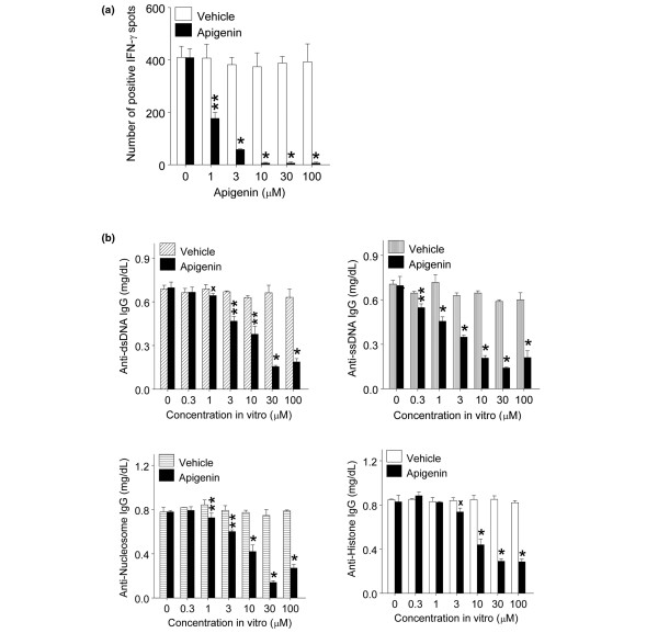
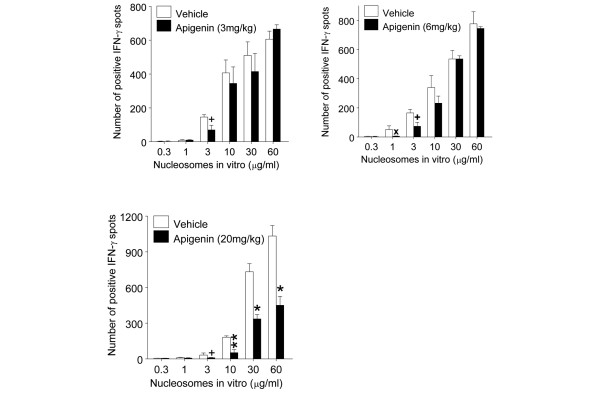
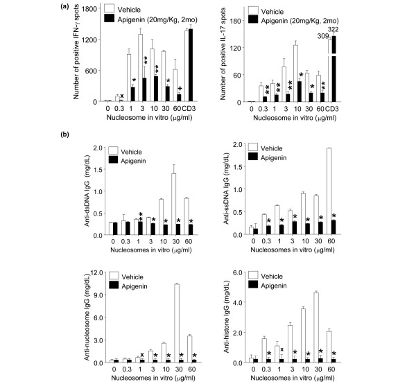
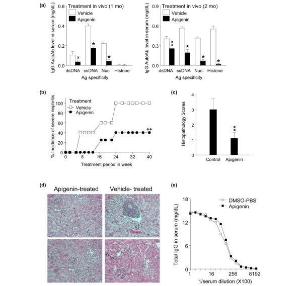
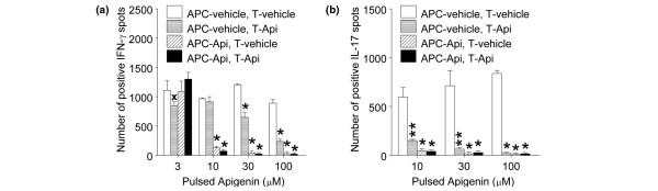
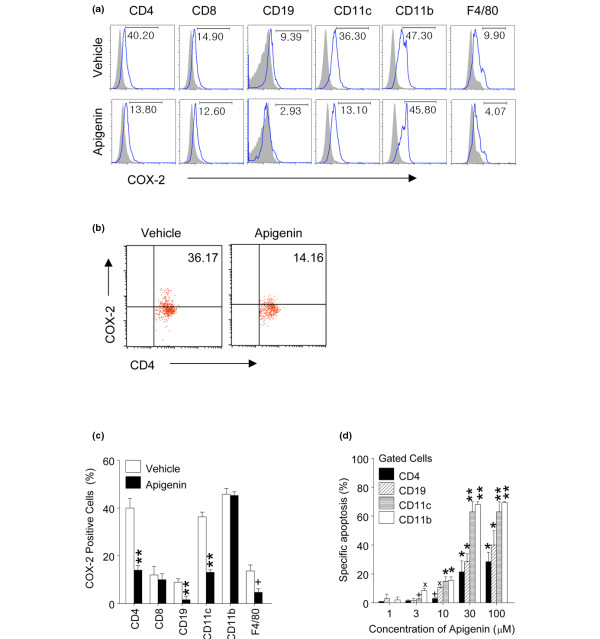
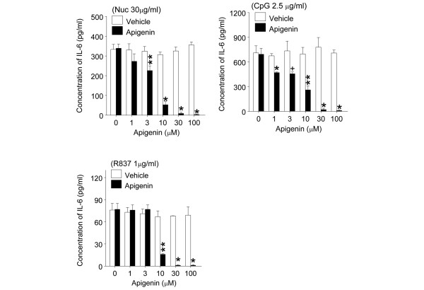
Results: Apigenin in culture suppressed responses of Th1 and Th17 cells to major lupus autoantigen (nucleosomes) up to 98% and 92%, respectively, and inhibited the ability of lupus B cells to produce IgG class-switched anti-nuclear autoantibodies helped by these Th cells in presence of nucleosomes by up to 82%. Apigenin therapy of SNF1 mice with established lupus suppressed serum levels of pathogenic autoantibodies to nuclear antigens up to 97% and markedly delayed development of severe glomerulonephritis. Apigenin downregulated COX-2 expression in lupus T cells, B cells, and antigen-presenting cells (APCs) and caused their apoptosis. Autoantigen presentation and Th17-inducing cytokine production by dendritic cells were more sensitive to the inhibitory effect of apigenin in culture, as evident at 0.3 to 3 muM, compared with concentrations (10 to 100 microM) required for inducing apoptosis.
Conclusions: Apigenin inhibits autoantigen-presenting and stimulatory functions of APCs necessary for the activation and expansion of autoreactive Th1 and Th17 cells and B cells in lupus. Apigenin also causes apoptosis of hyperactive lupus APCs and T and B cells, probably by inhibiting expression of NF-kappaB-regulated anti-apoptotic molecules, especially COX-2 and c-FLIP, which are persistently hyperexpressed by lupus immune cells. Increasing the bioavailability of dietary plant-derived COX-2 and NF-kappaB inhibitors, such as apigenin, could be valuable for suppressing inflammation in lupus and other Th17-mediated diseases like rheumatoid arthritis, Crohn disease, and psoriasis and in prevention of inflammation-based tumors overexpressing COX-2 (colon, breast).
Figures
Similar articles
-
Megakaryocyte progenitors are the main APCs inducing Th17 response to lupus autoantigens and foreign antigens.J Immunol. 2012 Jun 15;188(12):5970-80. doi: 10.4049/jimmunol.1200452. Epub 2012 May 4. J Immunol. 2012. PMID: 22561152 Free PMC article.
-
Naturally processed chromatin peptides reveal a major autoepitope that primes pathogenic T and B cells of lupus.J Immunol. 2002 Mar 1;168(5):2530-7. doi: 10.4049/jimmunol.168.5.2530. J Immunol. 2002. PMID: 11859148
-
Nucleosomal peptide epitopes for nephritis-inducing T helper cells of murine lupus.J Exp Med. 1996 Jun 1;183(6):2459-69. doi: 10.1084/jem.183.6.2459. J Exp Med. 1996. PMID: 8676066 Free PMC article.
-
Role of nucleosomes for induction and glomerular binding of autoantibodies in lupus nephritis.Curr Opin Nephrol Hypertens. 1999 May;8(3):299-306. doi: 10.1097/00041552-199905000-00005. Curr Opin Nephrol Hypertens. 1999. PMID: 10456260 Review.
-
Lupus nephritis: role of antinucleosome autoantibodies.Semin Nephrol. 2011 Jul;31(4):376-89. doi: 10.1016/j.semnephrol.2011.06.009. Semin Nephrol. 2011. PMID: 21839371 Review.
Cited by
-
Demethylzeylasteral (T-96) Treatment Ameliorates Mice Lupus Nephritis Accompanied by Inhibiting Activation of NF-κB Pathway.PLoS One. 2015 Jul 24;10(7):e0133724. doi: 10.1371/journal.pone.0133724. eCollection 2015. PLoS One. 2015. PMID: 26208003 Free PMC article.
-
The Potential Protective Effects of Polyphenols in Asbestos-Mediated Inflammation and Carcinogenesis of Mesothelium.Nutrients. 2016 May 9;8(5):275. doi: 10.3390/nu8050275. Nutrients. 2016. PMID: 27171110 Free PMC article. Review.
-
Promising Strategies in Plant-Derived Treatments of Psoriasis-Update of In Vitro, In Vivo, and Clinical Trials Studies.Molecules. 2022 Jan 18;27(3):591. doi: 10.3390/molecules27030591. Molecules. 2022. PMID: 35163855 Free PMC article. Review.
-
Flavonoids in Treatment of Chronic Kidney Disease.Molecules. 2022 Apr 6;27(7):2365. doi: 10.3390/molecules27072365. Molecules. 2022. PMID: 35408760 Free PMC article. Review.
-
The Therapeutic Effects of the Chinese Herbal Medicine, Lang Chuang Fang Granule, on Lupus-Prone MRL/lpr Mice.Evid Based Complement Alternat Med. 2016;2016:8562528. doi: 10.1155/2016/8562528. Epub 2016 Feb 29. Evid Based Complement Alternat Med. 2016. PMID: 27034698 Free PMC article.
References
Publication types
MeSH terms
Substances
Grants and funding
LinkOut - more resources
Full Text Sources
Other Literature Sources
Medical
Research Materials
Miscellaneous
