. 2009 May 1;16(1):43.
doi: 10.1186/1423-0127-16-43.

Apoptosis signal-regulating kinase 1 mediates denbinobin-induced apoptosis in human lung adenocarcinoma cells

Affiliations

Apoptosis signal-regulating kinase 1 mediates denbinobin-induced apoptosis in human lung adenocarcinoma cells

Chen-Tzu Kuo et al. J Biomed Sci. .

Abstract

In the present study, we explore the role of apoptosis signal-regulating kinase 1 (ASK1) in denbinobin-induced apoptosis in human lung adenocarcinoma (A549) cells. Denbinobin-induced cell apoptosis was attenuated by an ASK1 dominant-negative mutant (ASK1DN), two antioxidants (N-acetyl-L-cysteine (NAC) and glutathione (GSH)), a c-Jun N-terminal kinase (JNK) inhibitor (SP600125), and an activator protein-1 (AP-1) inhibitor (curcumin). Treatment of A549 cells with denbinobin caused increases in ASK1 activity and reactive oxygen species (ROS) production, and these effects were inhibited by NAC and GSH. Stimulation of A549 cells with denbinobin caused JNK activation; this effect was markedly inhibited by NAC, GSH, and ASK1DN. Denbinobin induced c-Jun phosphorylation, the formation of an AP-1-specific DNA-protein complex, and Bim expression. Bim knockdown using a bim short interfering RNA strategy also reduced denbinobin-induced A549 cell apoptosis. The denbinobin-mediated increases in c-Jun phosphorylation and Bim expression were inhibited by NAC, GSH, SP600125, ASK1DN, JNK1DN, and JNK2DN. These results suggest that denbinobin might activate ASK1 through ROS production to cause JNK/AP-1 activation, which in turn induces Bim expression, and ultimately results in A549 cell apoptosis.


Figures

Figure 1
Figure 1
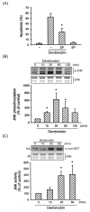
ASK1 activation is involved in denbinobin-induced apoptosis in A549 cells. (A) A549 cells were transiently transfected with pcDNA (mock) or ASK1DN for 6 h. Following transfection, cells were replaced with fresh culture medium for 24 h and treated with vehicle or 20 μM denbinobin for another 24 h. The percentage of apoptotic cells was then determined using flow cytometric analysis of PI-stained cells as described in "Materials and methods". Each column represents the mean ± S.E.M. of at least three independent experiments. *p < 0.05, compared to the mock transfection group in the presence of denbinobin. Cells were treated with 20 μM denbinobin (B) or 5 mM H2O2 (C) for the indicated time intervals. Cell lysates were then immunoprecipitated with the anti-ASK1 antibody, and the kinase activity was measured as described in "Materials and methods". Equal loading in each lane is reflected by similar intensities of α-tubulin. Compiled results are shown in the lower panel. Each column represents the mean ± S.E.M. of at least three independent experiments. * p < 0.05, compared to the control group without denbinobin or H2O2 treatment.
Figure 2
Figure 2
ROS generation mediates denbinobin-induced apoptosis in A549 cells. (A) Following pretreatment with NAC (1 mM) or GSH (100 μM) for 30 min, cells were incubated with the vehicle or 20 μM denbinobin for another 24 h. Cells then were harvested, and apoptosis was analyzed by flow cytometry as described in "Materials and methods". Each column represents the mean ± S.E.M. of at least three independent experiments performed in triplicate. * p < 0.05, compared to the group treated with denbinobin. (B) Cells were treated with 20 μM denbinobin, and DCF fluorescence was monitored by flow cytometry for up to 120 min as described in "Materials and methods". Results are plotted as the mean fluorescence intensity (MFI) ± S.E.M. of five independent experiments. * p < 0.05, compared to the control group. (C) Cells were pretreated with NAC (1 mM) or GSH (100 μM) for 30 min prior to denbinobin (20 μM) stimulation for 10 min. ROS generation was detected by H2DCFDA using flow cytometry. Data are represented as the MFI ± S.E.M. of three independent experiments. * p < 0.05, compared to the group treated with denbinobin. (D) Cells were pretreated with NAC (1 mM) or GSH (100 μM) for 30 min before treatment with 20 μM denbinobin for another 10 min. ASK1 kinase activity was then measured. Equal loading in each lane is reflected by similar intensities of α-tubulin. Compiled results are shown in the lower panel. Each column represents the mean ± S.E.M. of at least three independent experiments. * p < 0.05, compared to the control group in the presence of denbinobin. Cells were transiently transfected with pcDNA (mock), AktDN (E) or ASK1DN (F) for 6 h. Following transfection, cells were replaced with fresh culture medium for 24 h and treated with vehicle or 20 μM denbinobin for another 10 min (for ASK1 kinase activity) or 6 h (for Akt phosphorylation). Cell lysates were then prepared and subjected to ASK1 kinase activity or Akt phosphorylation as described in "Materials and methods". Equal loading in each lane is demonstrated by similar intensities of α-tubulin or Akt1/2, respectively. Compiled results are shown in the lower panel. Typical traces are representative of two experiments with similar results.
Figure 3
Figure 3
Involvement of JNK activation in denbinobin-induced apoptosis in A549 cells. (A) Cells were pretreated with SP600125 (10 μM) for 30 min before incubation with 20 μM denbinobin for 24 h, and then apoptosis was detected using flow cytometry of PI-stained cells as described in "Materials and methods". Each column represents the mean ± SEM of at least three independent experiments. *p < 0.05, compared to the denbinobin group. Cells were treated with 20 μM denbinobin for the indicated time intervals. Cell lysates were then prepared and subjected to JNK phosphorylation (B) or JNK kinase activity (C) as described in "Materials and methods". Equal loading in each lane is demonstrated by similar intensities of JNK. Compiled results are shown in the lower panel. Each column represents the mean ± S.E.M. of at least three independent experiments. * p < 0.05, compared to the control group.
Figure 4
Figure 4
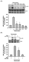
Involvement of ROS and ASK1 in denbinobin-induced JNK activation in A549 cells. Cells were pretreated with 1 mM NAC or 100 μM GSH (A) or transiently transfected with pcDNA or ASK1DN for 6 h (B) before incubation with 20 μM denbinobin for 30 min. Cells were then harvested and subjected to immunoblotting for JNK phosphorylation as described in "Materials and methods". Equal loading in each lane is shown by the similar intensities of JNK. Compiled results are shown in the lower panel. Each column represents the mean ± S.E.M. of at least three independent experiments. * p < 0.05, compared to the denbinobin group.
Figure 5
Figure 5
Involvement of AP-1 activation in denbinobin-induced apoptosis in A549 cells. (A) Cells were pretreated with curcumin (1 μM) for 30 min before incubation with 20 μM denbinobin for another 24 h, and then apoptosis was detected using flow cytometry of PI-stained cells as described in "Materials and methods". Each column represents the mean ± SEM of at least three independent experiments. *p < 0.05, compared to the denbinobin group. (B) Cells were treated with 20 μM denbinobin for 10, 30, 60, and 120 min. Cells were then harvested and subjected to immunoblotting with an anti-phospho-c-Jun antibody as described in "Materials and methods". Equal loading in each lane is shown by the similar intensities of c-Jun. Compiled results are shown in the lower panel. Each column represents the mean ± S.E.M. of at least three independent experiments. * p < 0.05, compared to the control group. (C) Cells were incubated with 20 μM denbinobin for various time intervals. Following incubation, the nuclear protein fraction was prepared, and AP-1-specific DNA protein complex formation was analyzed by an EMSA as described in "Materials and methods". The antibodies of c-Jun and c-Fos were incubated prior to detecting the specificity of AP-1 binding activity. Fifty-fold concentrations of unlabeled AP-1 or NF-κB oligonucleotides (50× cold) were used for the competition experiment. Data shown are representative of three independent experiments with similar results.
Figure 6
Figure 6
Involvement of ROS, ASK1, and JNK in denbinobin-induced c-Jun phosphorylation in A549 cells. Cells were pretreated with 1 mM NAC, 100 μM GSH (A), or 10 μM SP600125 (B), or transiently transfected with pcDNA, ASK1DN, JNK1DN, or JNK2DN for 6 h (C). After treatment, cells were treated with vehicle or 20 μM denbinobin for 30 min. Cells were then harvested for immunoblotting to assess the level of c-Jun phosphorylation as described in "Materials and methods". Equal loading in each lane is reflected by approximately similar intensities of the c-Jun bands. Compiled results are shown in the lower panel. Each column represents the mean ± S.E.M. of at least three independent experiments. * p < 0.05, compared to the denbinobin group. * p < 0.05, compared to the control group (A and B) or the pcDNA (mock) group (C) in the presence of denbinobin.
Figure 7
Figure 7
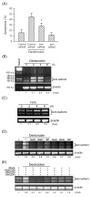
Bim expression is involved in denbinobin-induced apoptosis in A549 cells. (A) Following transfection with control siRNA or bim siRNA for 6 h, cells were treated with vehicle or 20 μM denbinobin for another 24 h. The DNA content was then analyzed by flow cytometry of PI-stained cells as described in "Materials and methods". Each column represents the mean ± S.E.M. of at least three independent experiments. * p < 0.05, compared to the control siRNA group in the presence of denbinobin. Cells were incubated with 20 μM denbinobin (B) or 5 mM H2O2 (C) for the indicated intervals, pretreated with 1 mM NAC, 100 μM GSH, or 10 μM SP600125 (D), or transiently transfected with pcDNA, ASK1DN, JNK1DN, or JNK2DN for 6 h (E). Cells were then treated with vehicle or 20 μM denbinobin for 2 h. Following treatment, RNA was collected to assess bim expression by semiquantitative RT-PCR as described in "Materials and methods". Samples were normalized for β-actin intensities. Typical traces are representative of three experiments with similar results.
Figure 8
Figure 8
Schematic summary of the apoptotic pathway involved in denbinobin-induced A549 cell apoptosis. Denbinobin caused Akt inactivation, leading to Bad dephosphorylation, mitochondrial dysfunction, and subsequent cell apoptosis. Denbinobin also activated ASK1 through ROS generation to cause JNK/AP-1 activation, which in turn induced Bim expression, ultimately resulting in A549 cell apoptosis.

Similar articles

Cited by

References

    1. Lee YH, Park JD, Baek NI, Kim SI, Ahn BZ. In vitro and in vivo antitumoral phenanthrenes from the aerial parts of Dendrobium nobile. Planta Med. 1995;61(2):178–180. doi: 10.1055/s-2006-958043. - DOI - PubMed
    1. Lin TH, Chang SJ, Chen CC, Wang JP, Tsao LT. Two phenanthraquinones from Dendrobium moniliforme. J Nat Prod. 2001;64(8):1084–1086. doi: 10.1021/np010016i. - DOI - PubMed
    1. Chen CC, Huang YL, Teng CM. Antiplatelet aggregation principles from Ephemerantha lonchophylla. Planta Med. 2000;66(4):372–374. doi: 10.1055/s-2000-8553. - DOI - PubMed
    1. Chen HY, Shiao MS, Huang YL, Shen CC, Lin YL, Kuo YH, Chen CC. Antioxidant principles from Ephemerantha lonchophylla. J Nat Prod. 1999;62(9):1225–1227. doi: 10.1021/np990025f. - DOI - PubMed
    1. Huang YC, Guh JH, Teng CM. Denbinobin-mediated anticancer effect in human K562 leukemia cells: role in tubulin polymerization and Bcr-Abl activity. J Biomed Sci. 2005;12(1):113–121. doi: 10.1007/s11373-004-8171-y. - DOI - PubMed

Publication types