

Arginine 383 is a crucial residue in ABCG2 biogenesis
- PMID: 19406100
- PMCID: PMC4163909
- DOI: 10.1016/j.bbamem.2009.04.016
Arginine 383 is a crucial residue in ABCG2 biogenesis
Abstract
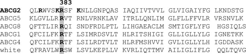
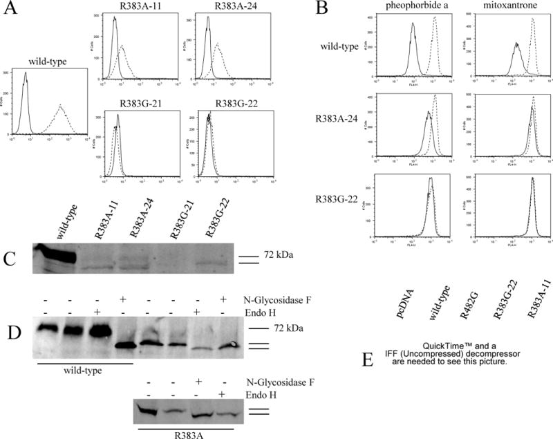
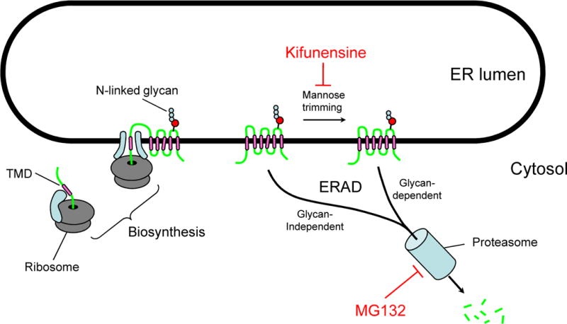
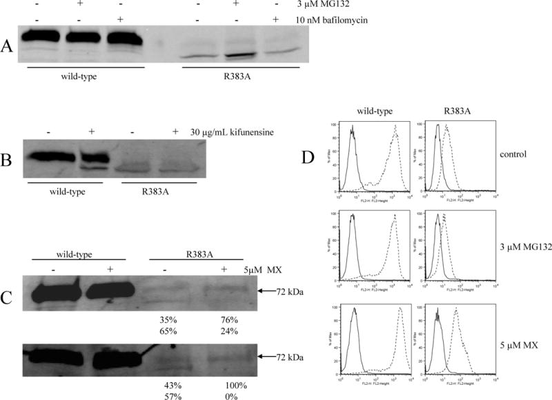
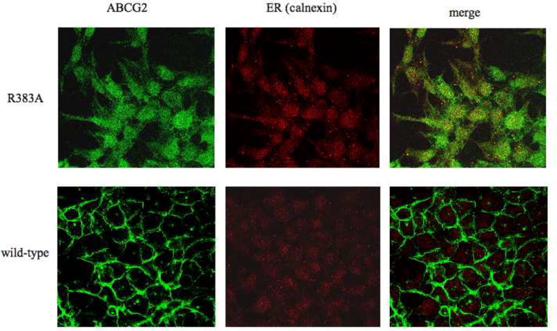
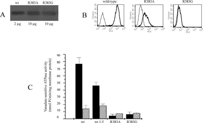
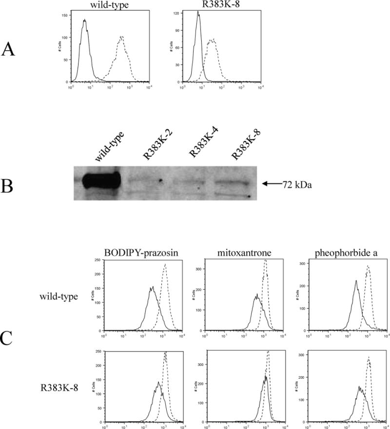
ABCG2 is an ATP-binding cassette half-transporter initially identified in multidrug-resistant cancer cell lines and recently suggested to play an important role in pharmacokinetics. Here we report studies of a conserved arginine predicted to localize near the cytoplasmic side of TM1. First, we determined the effect of losing charge and bulk at this position via substitutions with glycine and alanine. The R383G mutant when transfected into HEK cells was not detectable on immunoblot or by functional assay, while the R383A mutant exhibited detectable but significantly decreased levels compared to wild-type, partial retention in the ER and altered glycosylation. Efflux of the ABCG2-substrates mitoxantrone and pheophorbide a was observed. Our experiments suggested rapid degradation of the R383A mutant by the proteasome via a kifunensine-insensitive pathway. Interestingly, overnight treatment of the R383A mutant with mitoxantrone assisted in protein maturation as evidenced by a shift to the N-glycosylated form. The R383A mutant when expressed in insect cells, though detected on the surface, had no measurable ATPase activity. In addition, substitution with the positively charged lysine resulted in significantly decreased protein expression levels in HEK cells, while retaining function. In conclusion, arginine 383 is a crucial residue for ABCG2 biogenesis, where even the most conservative mutations have a large impact.
Figures
Similar articles
-
Mutational analysis of threonine 402 adjacent to the GXXXG dimerization motif in transmembrane segment 1 of ABCG2.Biochemistry. 2010 Mar 16;49(10):2235-45. doi: 10.1021/bi902085q. Biochemistry. 2010. PMID: 20088606 Free PMC article.
-
Mutational studies of G553 in TM5 of ABCG2: a residue potentially involved in dimerization.Biochemistry. 2006 Apr 25;45(16):5251-60. doi: 10.1021/bi0521590. Biochemistry. 2006. PMID: 16618113 Free PMC article.
-
Single amino acid (482) variants of the ABCG2 multidrug transporter: major differences in transport capacity and substrate recognition.Biochim Biophys Acta. 2005 Feb 1;1668(1):53-63. doi: 10.1016/j.bbamem.2004.11.005. Biochim Biophys Acta. 2005. PMID: 15670731
-
Multidrug resistance in cancer chemotherapy and xenobiotic protection mediated by the half ATP-binding cassette transporter ABCG2.Curr Med Chem Anticancer Agents. 2004 Jan;4(1):31-42. doi: 10.2174/1568011043482205. Curr Med Chem Anticancer Agents. 2004. PMID: 14754410 Review.
-
Human ABC transporter ABCG2 in xenobiotic protection and redox biology.Drug Metab Rev. 2006;38(3):371-91. doi: 10.1080/03602530600727947. Drug Metab Rev. 2006. PMID: 16877258 Review.
Cited by
-
Cellular Processing of the ABCG2 Transporter-Potential Effects on Gout and Drug Metabolism.Cells. 2019 Oct 8;8(10):1215. doi: 10.3390/cells8101215. Cells. 2019. PMID: 31597297 Free PMC article. Review.
-
Posttranslational negative regulation of glycosylated and non-glycosylated BCRP expression by Derlin-1.Biochem Biophys Res Commun. 2011 Jan 21;404(3):853-8. doi: 10.1016/j.bbrc.2010.12.074. Epub 2010 Dec 22. Biochem Biophys Res Commun. 2011. PMID: 21184741 Free PMC article.
-
Structure and function of the human breast cancer resistance protein (BCRP/ABCG2).Curr Drug Metab. 2010 Sep;11(7):603-17. doi: 10.2174/138920010792927325. Curr Drug Metab. 2010. PMID: 20812902 Free PMC article. Review.
-
Jump into a New Fold-A Homology Based Model for the ABCG2/BCRP Multidrug Transporter.PLoS One. 2016 Oct 14;11(10):e0164426. doi: 10.1371/journal.pone.0164426. eCollection 2016. PLoS One. 2016. PMID: 27741279 Free PMC article.
-
Medically Important Alterations in Transport Function and Trafficking of ABCG2.Int J Mol Sci. 2021 Mar 10;22(6):2786. doi: 10.3390/ijms22062786. Int J Mol Sci. 2021. PMID: 33801813 Free PMC article. Review.
References
-
- Dean M, Hamon Y, Chimini G. The human ATP-binding cassette (ABC) transporter superfamily. J Lipid Res. 2001;42:1007–1017. - PubMed
-
- Allikmets R, Schriml LM, Hutchinson A, Romano-Spica V, Dean M. A human placenta-specific ATP-binding cassette gene (ABCP) on chromosome 4q22 that is involved in multidrug resistance. Cancer Res. 1998;58:5337–5339. - PubMed
-
- Miyake K, Mickley L, Litman T, Zhan Z, Robey R, Cristensen B, Brangi M, Greenberger L, Dean M, Fojo T, Bates SE. Molecular cloning of cDNAs which are highly overexpressed in mitoxantrone-resistant cells: demonstration of homology to ABC transport genes. Cancer Res. 1999;59:8–13. - PubMed
-
- Robey RW, Medina-Perez WY, Nishiyama K, Lahusen T, Miyake K, Litman T, Senderowicz AM, Ross DD, Bates SE. Overexpression of the ATP-binding cassette half-transporter, ABCG2 (MXR/BCRP/ABCP1), in flavopiridol-resistant human breast cancer cells. Clin Cancer Res. 2001;7:145–152. - PubMed
Publication types
MeSH terms
Substances
Grants and funding
LinkOut - more resources
Full Text Sources
Miscellaneous
