. 2009 Sep;47(3):352-8.
doi: 10.1016/j.yjmcc.2009.04.014. Epub 2009 May 4.

Stress-induced dilated cardiomyopathy in a knock-in mouse model mimicking human titin-based disease

Affiliations

Stress-induced dilated cardiomyopathy in a knock-in mouse model mimicking human titin-based disease

Michael Gramlich et al. J Mol Cell Cardiol. 2009 Sep.

Abstract

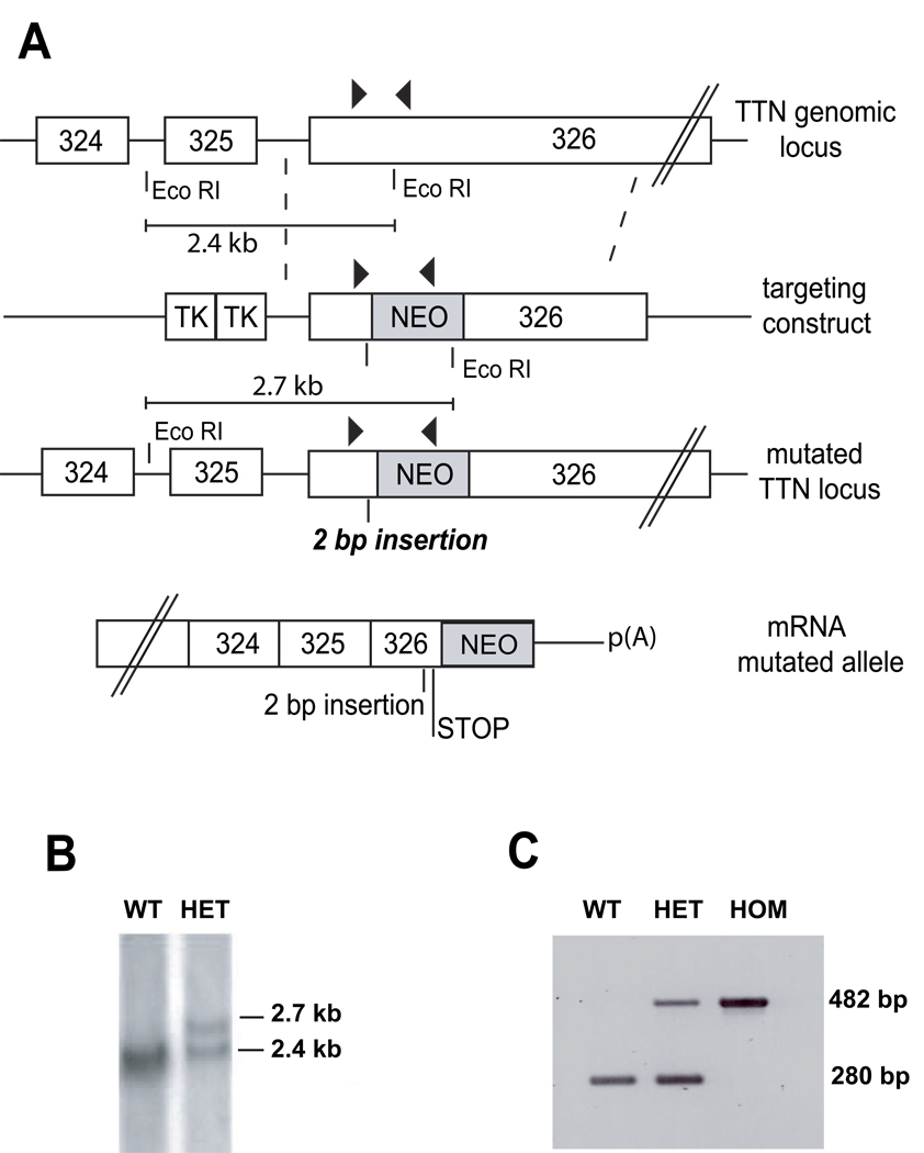
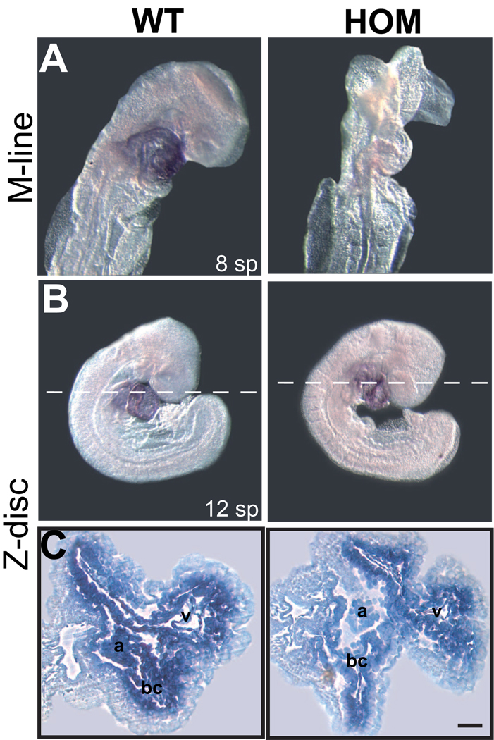
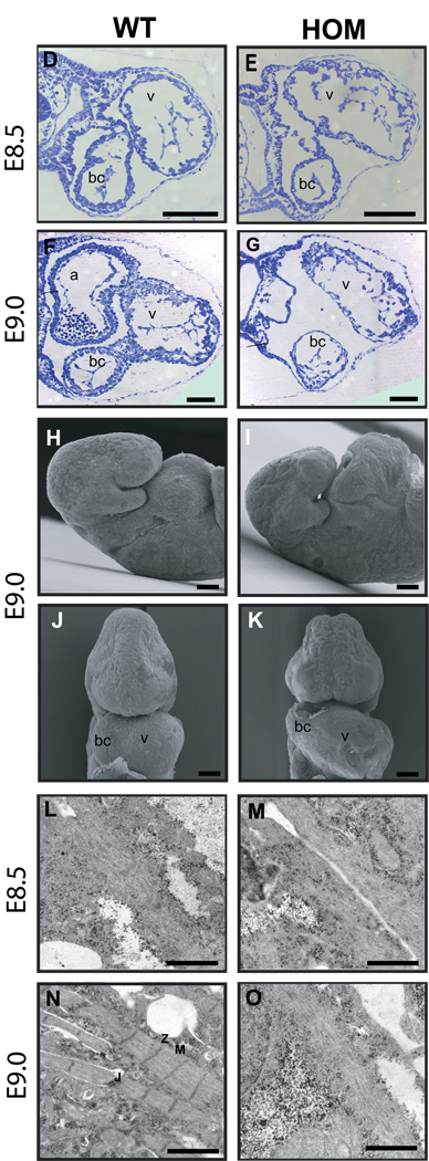
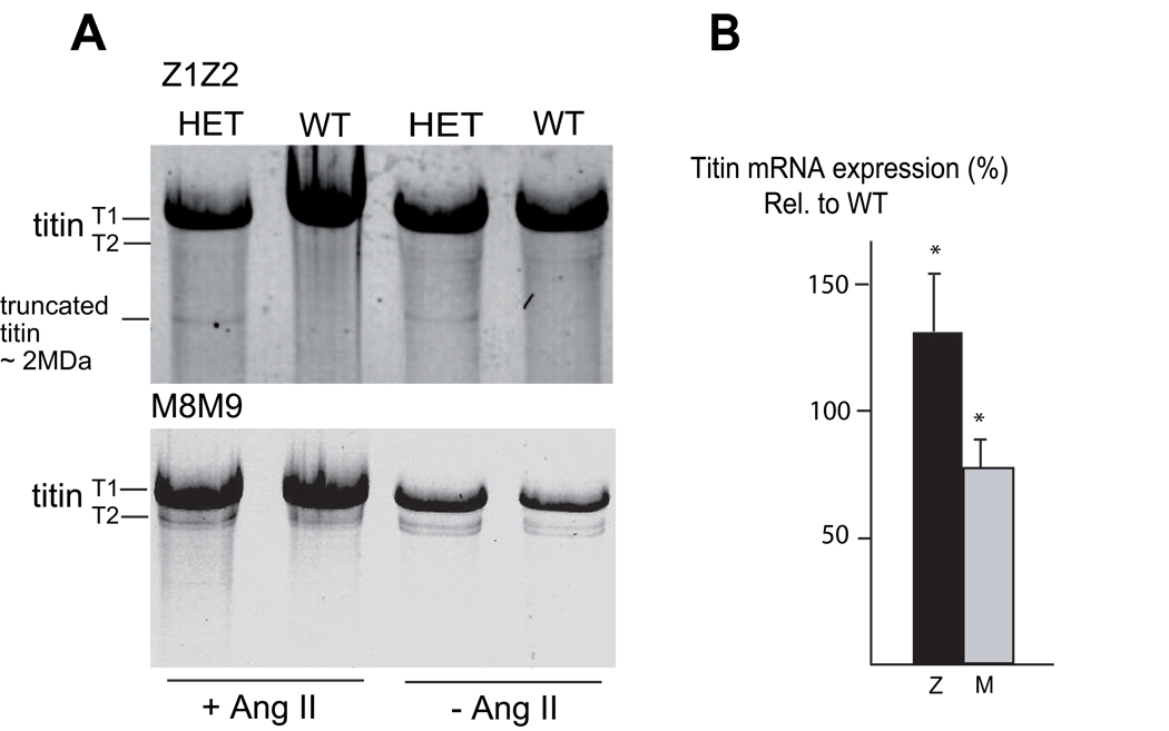
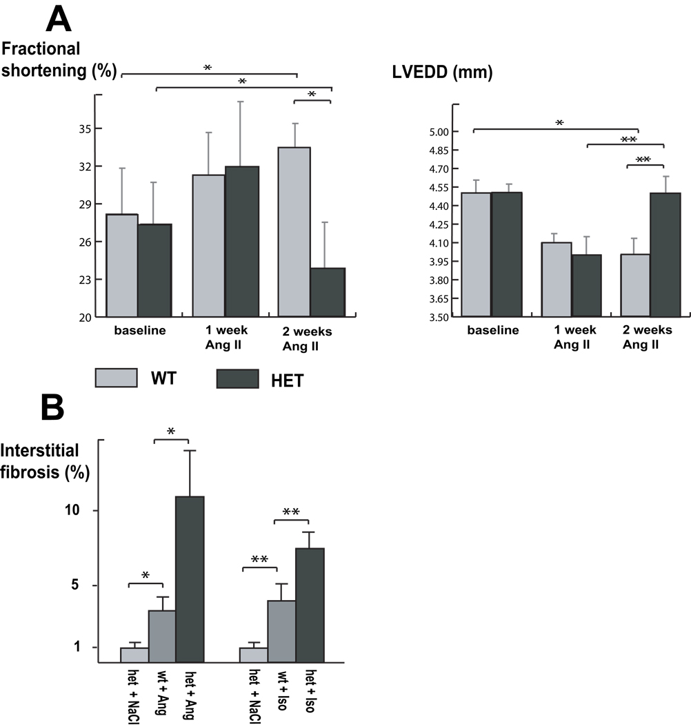
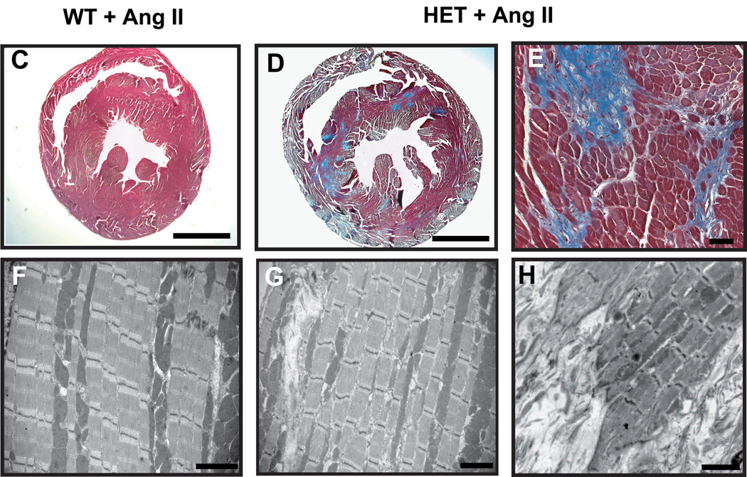
Mutations in a variety of myofibrillar genes cause dilated cardiomyopathy (DCM) in humans, usually with dominant inheritance and incomplete penetrance. Here, we sought to clarify the functional effects of the previously identified DCM-causing TTN 2-bp insertion mutation (c.43628insAT) and generated a titin knock-in mouse model mimicking the c.43628insAT allele. Mutant embryos homozygous for the Ttn knock-in mutation developed defects in sarcomere formation and consequently died before E9.5. Heterozygous mice were viable and demonstrated normal cardiac morphology, function and muscle mechanics. mRNA and protein expression studies on heterozygous hearts demonstrated elevated wild-type titin mRNA under resting conditions, suggesting that up-regulation of the wild-type titin allele compensates for the unstable mutated titin under these conditions. When chronically exposed to angiotensin II or isoproterenol, heterozygous mice developed marked left ventricular dilatation (p<0.05) with impaired fractional shortening (p<0.001) and diffuse myocardial fibrosis (11.95+/-2.8% vs. 3.7+/-1.1%). Thus, this model mimics typical features of human dilated cardiomyopathy and may further our understanding of how titin mutations perturb cardiac function and remodel the heart.


Conflict of interest statement

Conflict of interest: none declared.

Figures

Figure 1
Figure 1. Generation of Ttn knock-in mouse by gene targeting
(A) Depiction of the mouse Ttn genomic locus, the targeting vector, the allele resulting from homologous recombination, and the mutant mRNA allele. Exons are indicated as white boxes, and 324–326 (above) refers to the numbers of titin exons in the human TTN sequence. Arrowheads denote the positions of primers used for PCR genotyping. The position of the 2-bp (AT) insertion mutation is designated. (B) Southern blot analysis of genomic DNA digested with EcoRI produced bands of the expected sizes (2.7 kb and 2.4 kb) in heterozygous animals. (C) PCR-based genotyping. PCR products derived from the wild-type (WT) and homozygous (HOM) alleles were 280bp and 482bp, respectively. Heterozygous animals (HET) showed both the WT and HOM alleles. Embryos were genotyped from the yolk sack.
Figure 2
Figure 2. Defects in heart development at E9.0 caused by unformed sarcomeres in homozygous Ttn knock-in embryos
(A–C) Whole-mount in situ hybridization for Ttn Z-disc (A) and Ttn M-line (B) in wild-type (WT) and homozygous (HOM) embryos. Hearts are stained in all wild-type embryos, whereas the M-line probe in homozygous embryos does not show a signal. Note the intact cardiac looping in the homozygous embryo (A). Transverse sections (C) in two sections planes with dotted lines in B. (D–G) Histological analysis of transverse sections through the forming heart of WT and HOM embryos at E8.5 and E9.0 demonstrates an enlarged common ventricle (v) with reduced ventricular wall thickness, loosely packed endocardial cells, and pericardial edema (E,G) in HOM embryos. (H–K) Scanning electron microscopy of WT and HOM animals at E9.0 confirmed the observation of an enlarged heart region in HOM animals (I,K). (L–O) Ultrastructural analysis of cardiac sarcomere maturation in WT and HOM hearts showed appropriate myofibrils at E8.5. At E9.0, sarcomeres had been assembled in WT hearts (N), in contrast to titin-deficient hearts, which demonstrated no striation or sarcomere formation (O). sp, somite pairs; v, common ventricular chamber; bc, bulbus cordis; a, atrial chamber; Z, Z-disc; M, M-band; J, cellular junctions. Scale bars, (C) 100 µm (D)–(K) 200µm; (L)–(O) 1 µm.
Figure 3
Figure 3. Titin expression analysis in heterozygous mice
(A) SDS agarose gel analysis of heart muscle tissue from unstressed (− Ang II) and stressed (+ Ang II) heterozygous (HET) and wild-type (WT) mice revealed in HET’s of both groups an extra band consistent with a small amount of a truncated protein (~2.0 MDa) which is detectable with anti-Z1/Z2, but not with anti-M8M9. Note no obvious difference in wild-type titin protein amounts between HET’s and WT’s. T1 undegraded titin, T2 degraded titin, Ang II angiotensin II. (B) Real-time PCR analysis of titin mRNA expression in hearts of heterozygous mice compared to their wild-type littermates. Primer pairs specific for Z-disc titin (amplifying both alleles) and M-line titin (amplifying only the wild-type allele) were used for PCR. Titin mRNA levels were 76±11% measured with the M-line probe and 131±24% measured with the Z-disc probe (*p<0.05), indicating a partial compensation (full compensation would result in 100% mRNA expression with the M-line and 150% with the Z-disc probe relative to mRNA expression levels of wild-type littermates).
Figure 4
Figure 4. Stressed heterozygous Ttn knock-in mice develop features of DCM
(A) Echocardiographic assessment before, after one week, and after two weeks of angiotensin II (Ang II) application. After one week, both genotypes showed cardiac hypertrophy, as observed in end-diastolic dimensions (LVEDD) and enhanced systolic contractility (fractional shortening). After two weeks of Ang II application, the hypertrophy in wild-type (WT) animals is increased, whereas heterozygous mice (HET) develop left ventricular dilatation (**p<0.05) with impaired systolic function (*p<0.001). Compared to baseline conditions fractional shortening is decreased (*p<0.001) in heterozygous animals. (B) Quantification of interstitial myocardial fibrosis after application of two weeks of Ang II or one week of isoproterenol (ISO). The development of fibrosis after Ang II/ISO application administration is pronounced in heterozygous animals (*p<0.01; † p<0.05) compared with their wild-type littermates. (C–H) Myocardial histology and electron microscopy of mouse hearts after Ang II application. Representative sections from hearts of wild-type and heterozygous animals after two weeks of Ang II application stained with Masson’s trichrome (C–E). Note the increased level of fibrosis in heterozygous hearts. Electron microscopy of heterozygous ventricles demonstrates regions of preserved sarcomere assembly (G) and interstitial fibrosis (G, arrowhead and H). Note areas of increased fibrosis and infiltration into the sarcomere (H). Scale bars: (C) and (D) 2 mm; (E) 40µm; (F–H) 2 µm

Comment in

References

    1. Dec GW, Fuster V. Idiopathic dilated cardiomyopathy. N Engl J Med. 1994;331:1564–1575. - PubMed
    1. Morita H, Seidman J, Seidman CE. Genetic causes of human heart failure. J Clin Invest. 2005;115:518–526. - PMC - PubMed
    1. Gerull B, Gramlich M, Atherton J, McNabb M, Trombitás K, Sasse-Klaassen S, et al. Mutations of TTN, encoding the giant muscle filament titin, cause familial dilated cardiomyopathy. Nat Genet. 2002;30:201–204. - PubMed
    1. Gerull B, Atherton J, Geupel A, Sasse-Klaassen S, Heuser A, Frenneaux M, et al. Identification of a novel frameshift mutation in the giant muscle filament titin in a larger Australian family with dilated cardiomyopathy. J Mol M. 2006;84:478–483. - PubMed
    1. Satoh M, Takahashi M, Sakamoto T, Hiroe M, Marumo F, Kimura A. Structural analysis of the titin gene in hypertrophic cardiomyopathy: identification of a novel disease gene. Biochem Biophys Res Commun. 1999;262(2):411–417. - PubMed

Publication types