. 2009 Aug;1793(8):1379-86.
doi: 10.1016/j.bbamcr.2009.04.007. Epub 2009 May 4.

A dual mechanism of cytoprotection afforded by M-LDH in embryonic heart H9C2 cells

Affiliations

A dual mechanism of cytoprotection afforded by M-LDH in embryonic heart H9C2 cells

Sofija Jovanović et al. Biochim Biophys Acta. 2009 Aug.

Abstract

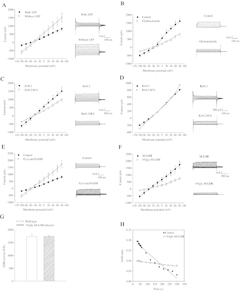
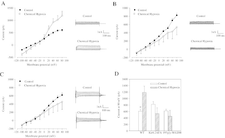
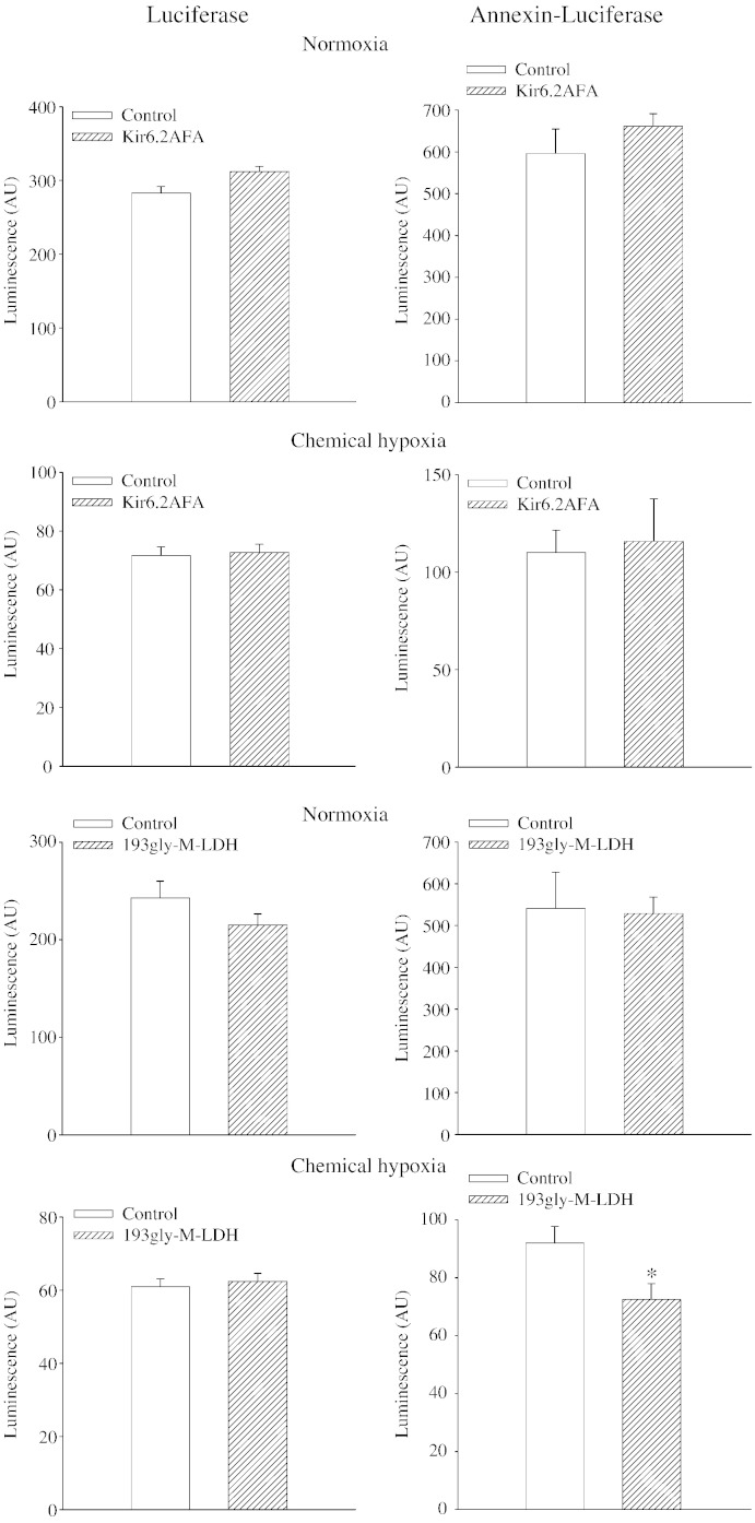
Muscle form of lactate dehydrogenase (M-LDH), a minor LDH form in cardiomyocytes, physically interacts with ATP-sensitive K+ (K ATP) channel-forming subunits. Here, we have shown that expression of 193gly-M-LDH, an inactive mutant of M-LDH, inhibit regulation of the K ATP channels activity by LDH substrates in embryonic rat heart H9C2 cells. In cells expressing 193gly-M-LDH chemical hypoxia has failed to activate K ATP channels. The similar results were obtained in H9C2 cells expressing Kir6.2AFA, a mutant form of Kir6.2 with largely decreased K+ conductance. Kir6.2AFA has slightly, but significantly, reduced cellular survival under chemical hypoxia while the deleterious effect of 193gly-M-LDH was significantly more pronounced. The levels of total and subsarcolemmal ATP in H9C2 cells were not affected by Kir6.2AFA, but the expression of 193gly-M-LDH led to lower levels of subsarcolemmal ATP during chemical hypoxia. We conclude that M-LDH regulates both the channel activity and the levels of subsarcolemmal ATP and that both mechanism contribute to the M-LDH-mediated cytoprotection.


Figures

Fig. 1
Fig. 1
Kir6.2AFA and 193gly-M-LDH are efficient dominant negatives of Kir6.2 and M-LDH in H9C2 cells. A. Current–voltage relationships with corresponding original membrane currents in cells filled with pipette solution with (3 mM) and without ATP. Each point represents mean ± SEM (n = 5–6). B. Current–voltage relationships with corresponding original membrane currents in cells filled with ATP-free pipette solution in the absence (control) and presence of glybenclamide (30 μM). Each point represents mean ± SEM (n = 5–6). C. Current–voltage relationships with corresponding original membrane currents in control cells (Kir6.2) and cells infected with Kir6.2AFA filled with ATP-free pipette solution. Each point represents mean ± SEM (n = 5–6). D. Current–voltage relationships with corresponding original membrane currents in control cells (Kir6.2) and cells infected with Kir6.2AFA filled with pipette solution containing ATP (3 mM). Each point represents mean (n = 5–6). E. Current–voltage relationships with corresponding original membrane currents in cells filled with pipette solution containing 3 mM ATP (control) and ATP (3 mM) plus pyruvate (20 mM) plus NADH (20 mM). Each point represents mean ± SEM (n = 5–6). F. Current–voltage relationships with corresponding original membrane currents in control cells (M-LDH) and cells infected with 193gly-M-LDH filled with pipette solution containing ATP (3 mM) plus pyruvate (20 mM) plus NADH (20 mM). Each point represents mean ± SEM (n = 5–6). G. LDH activity in control cells and cells infected with 193gly-M-LDH. Each bar represents mean ± SEM (n = 4). H. LDH assay with anti-Kir6.2 immunoprecipitate of membrane fraction of control cells (control) and cells infected with 193gly-M-LDH.
Fig. 2
Fig. 2
Intact Kir6.2 and M-LDH are required for the opening of KATP channels induced by chemical hypoxia. (A–C) Current–voltage relationships with corresponding original membrane currents in control cells (A), cells infected with Kir6.2AFA (B) and cells infected with 193gly-M-LDH (C) under control conditions and when exposed to DNP (10 mM). Each point represents mean ± SEM (n = 5). (D) Membrane current at 80 mV under conditions in A–C. Each bar represents mean ± SEM (n = 5).
Fig. 3
Fig. 3
Catalytic activity of M-LDH is crucial for cell survival in chemical hypoxia. A bar graph showing a percentage of control cells, and cells infected with Kir6.2 AFA and 193gly-M-LDH that survived treatment with DNP (10 mM). Each bar represents mean ± SEM (n = 6–11). ⁎P < 0.05 when compared to the control and +P < 0.05 when Kir6.2AFA were compared with 193gly-M-LDH.
Fig. 4
Fig. 4
M-LDH catalytic activity, but not Kir6.2 K+ conductance, is required for counteracting chemical hypoxia-induced decrease in subsarcolemmal ATP. Bar graphs showing luciferase (graphs on the left) and annexin-luciferase (graphs on the right) luminescence in cells infected with Kir6.2AFA under control conditions (normoxia) and after treatment with 10 mM DNP (chemical hypoxia), and in cells infected with 193gly-M-LDH under control conditions (normoxia) and after treatment with 10 mM DNP (chemical hypoxia). Each bar represents mean ± SEM (n = 5). ⁎P < 0.05.

References

    1. Crawford R.M., Budas G.R., Jovanović S., Ranki H.J., Wilson T.J., Davies A.M., Jovanović A. M-LDH serves as a sarcolemmal KATP channel subunit essential for cell protection against ischemia. EMBO J. 2002;21:3936–3948. - PMC - PubMed
    1. Zingman L.V., Alekseev A.E., Hodgson-Zingman D., Terzic A. , ATP-sensitive K+ channels: metabolic sensing and cardioprotection. J. Appl. Physiol. 2007;103:1888–1893. - PubMed
    1. Budas G.R., Jovanović S., Crawford R.M., Jovanović A. Hypoxia-induced preconditioning in adult stimulated cardiomyocytes is mediated by the opening and trafficking of sarcolemmal KATP channels. FASEB J. 2004;18:1046–1048. - PMC - PubMed
    1. Inagaki N., Gonoi T., Clement J.P., Wang C.Z., Aguilar-Bryan L., Bryan J., Seino S. A family of sulfonylurea receptors determines the pharmacological properties of ATP-sensitive K+ channels. Neuron. 1996;16:1011–1017. - PubMed
    1. Carrasco A.J., Dzeja P.P., Alekseev A.E., Pucar D., Zingman L.V., Abraham M.R., Hodgson D., Bienengraeber M., Puceat M., Janssen E., Wieringa B., Terzic A. Adenylate kinase phosphotransfer communicates cellular energetic signals to ATP-sensitive potassium channels. Proc. Natl. Acad. Sci. U. S. A. 2001;98:7623–7628. - PMC - PubMed

Publication types

MeSH terms

LinkOut - more resources