Review
. 2009 Jul;20(5):543-56.
doi: 10.1016/j.semcdb.2009.04.013. Epub 2009 May 3.

Bioelectric mechanisms in regeneration: Unique aspects and future perspectives

Affiliations
Review

Bioelectric mechanisms in regeneration: Unique aspects and future perspectives

Michael Levin. Semin Cell Dev Biol. 2009 Jul.

Abstract

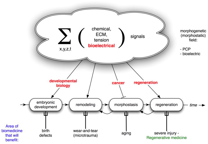
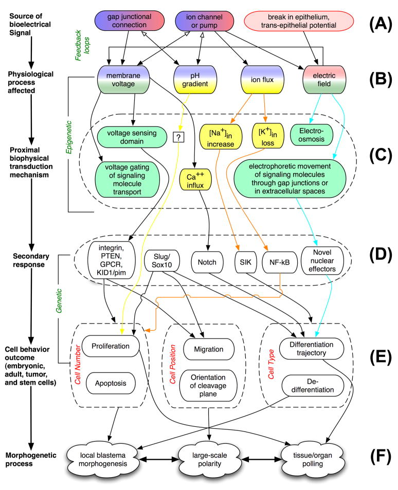
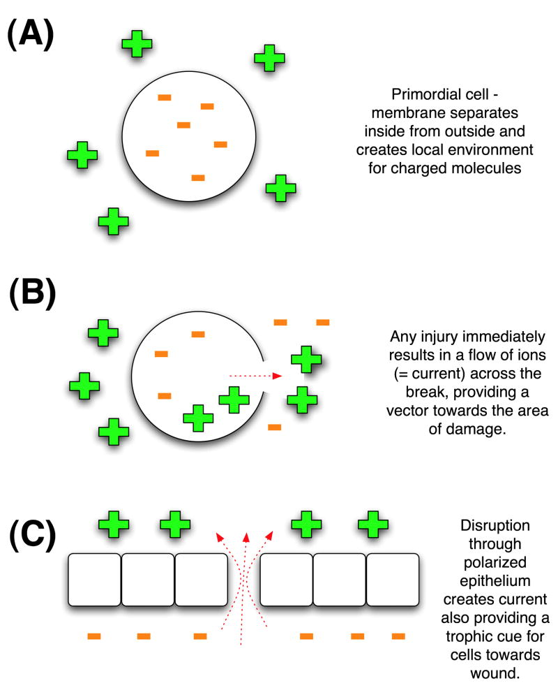
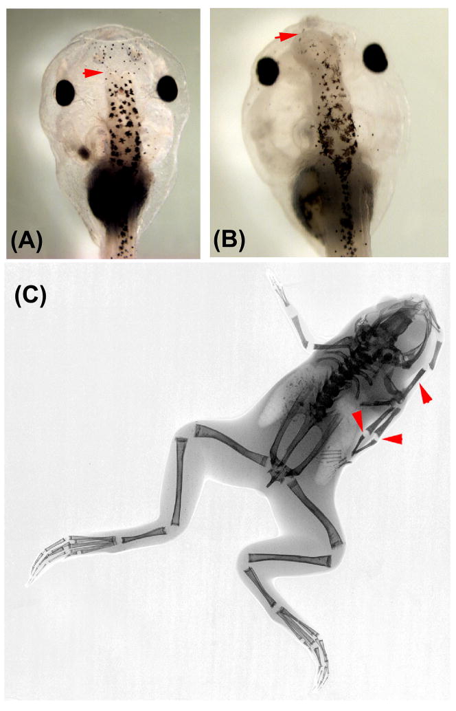
Regenerative biology has focused largely on chemical factors and transcriptional networks. However, endogenous ion flows serve as key epigenetic regulators of cell behavior. Bioelectric signaling involves feedback loops, long-range communication, polarity, and information transfer over multiple size scales. Understanding the roles of endogenous voltage gradients, ion flows, and electric fields will contribute to the basic understanding of numerous morphogenetic processes and the means by which they can robustly restore pattern after perturbation. By learning to modulate the bioelectrical signals that control cell proliferation, migration, and differentiation, we gain a powerful set of new techniques with which to manipulate growth and patterning in biomedical contexts. This chapter reviews the unique properties of bioelectric signaling, surveys molecular strategies and reagents for its investigation, and discusses the opportunities made available for regenerative medicine.


Figures

Figure 1
Figure 1. morphogenetic fields and biomedicine
The morphogenetic field can be defined as the sum, integrated over 3 spatial, and 1 temporal dimensions, of all non-local signals impinging on cells and cell groups in an organism. Functionally, these signals carry information about the current and desired pattern of the organism. This allows the initial development of complex form from a single fertilized egg cell, as well as the subsequent maintenance of form in adulthood against trauma and individual cell loss. Errors in various aspects of the establishment and interpretation of this field result in birth defects, cancer, aging, and failure to regenerate after injury. Thus, almost every area of biomedicine is impacted by our knowledge of how cells interact with this set of complex signals. Bioelectrical aspects of the morphogenetic field are crucial, although planar polarity systems and chemical gradients also form components of this information field. ECM = extracellular matrix.
Figure 2
Figure 2. integration of bioelectric signals with canonical pathways
(A) Expression of ion channels or pumps, gap junctional connections, or epithelial damage all give rise to bioelectric signals. (B) These signals manifest as changes in transmembrane potential, pH gradients, specific ion flows, or electric fields. In the first two rows, pink shading indicates non-cell-autonomous signals while purple indicates cell-autonomous cues. Some nodes are both. (C) These processes are transduced via a variety of proximal epigenetic mechanisms including voltage-sensing domains on proteins, electro-osmosis, gating of morphogen transporters, and movement of specific ions like calcium. Green indicates a true electrical effect, while yellow indicates a biochemical effect due to ion identity. (D) These processes feed into several known genetic signaling pathways, including NF-kB, Notch, PTEN, Slug/Sox10, and Integrins. (E) Downstream of these signaling molecules are changes in cell cycle, apoptosis, position, orientation, and differentiation. (F) The ultimate result of orchestrated changes in cell behavior are morphogenetic processes including patterning of blastemas and embryonic fields, polarity decisions on several scales, and polling of remote tissues that enable wounds to decide what already exists and what must be recreated. The arrows indicate sample cases where the whole pathway has been traced for bioelectrical control of patterning.
Figure 3
Figure 3. bioelectrical signals leverage the laws of physics into information for living systems
(A) In an early primordial cell, which has only a membrane separating inside from outside, a separation of charges will occur. When the membrane is damaged (B), the flow of ions that occurs (as the gradient tries to equalize across the break) provides a vector cue indicating the direction of damage to intracellular components. This occurs “for free” – it does not require the cell to have any specific machinery for this purpose. In more complex systems, an epithelium (C) maintains a transepithelial potential due to the segregation of charges by the component cells and their apical-basal polarization. When this is broken, nearby cells likewise experience electric fields which direct them towards the damage; this is especially useful for migratory cell types such as neoblasts and homing mesenchymal stem cells. Interestingly, this can be co-opted by normal developmental mechanisms; Borgens has proposed that a programmed tight-junctional breakdown in the flank results in “injury” currents that guide migratory cells to the right place during limb induction in embryonic development [166]. A final layer of complexity can be added to the passive fields that occur from breaks in epithelia by directing specific up-regulation of channels and pumps in wound cells (e.g., the V-ATPase in the regeneration bud of the amputated tadpole tail), thus shaping necessary fields further. These targeting cues, meant for the organism’s own cells, could potentially be capitalized upon by galvanotactic fungi/bacteria and metastatic cells to identify areas of weak epithelialization that can be more easily attacked.
Figure 4
Figure 4. sample phenotypes arising from molecular-genetic modulation of bioelectrical cues in Xenopus laevis
Unpublished data from our lab showing that misexpression of ion channel constructs during embryogenesis can make coherent changes in pattern. Experiments performed with potassium channels by Sherry Aw result in the normal forebrain (A) being drastically increased (B); red arrow indicates anterior border of forebrain. Similarly, entire limbs can be induced (C), with X-ray imaging revealing the normal skeletal pattern in the ectopic limbs in this adult frog.
Figure 5
Figure 5. bioelectric state space
Cells live in a state space with a number of orthogonal axes corresponding to physiological properties. Here are shown only 3 (membrane voltage, Vmem, internal pH, and K+ content). A more detailed dataset will contain additional semi-independent metrics such as the content of other ions, nuclear potential, surface charge, etc. One hypothesis is that cell types (e.g., stem cells, cancer cells, non-proliferative cells) will be seen to cluster in different regions of this space. If true, this will be not only a useful diagnostic framework but can also, when coupled with quantitative data and mathematical modeling, be used for rational modulation of cell behavior. Using well-characterized transporters in gene therapy, and pharmacological reagents targeting endogenous transporters in damaged tissue, bioelectrical properties can be specifically changed to move wound cells from a non-proliferative state towards a more plastic, regenerative condition.
Figure 6
Figure 6. missing the physiological forest for the mRNA/protein expression trees
Analysis of gene or protein expression can often be very misleading with respect to physiological state. (A) Two hypothetical cell types have very different expression patterns. A microarray or differential analysis characterizes them as different, since one cell has low expression of Na,K-ATPase and high expression of V-ATPase, while the other cell is the opposite. However (B), analysis of pH and membrane potential may reveal that the cells actually have a similar proliferative potential because both pumps are hyperpolarizing (although genetically distinct), and the cells may in fact be similar from the point of view of bioelectrical controls.
Figure 7
Figure 7. a schematic of the Regeneration Sleeve: application to limb regeneration
Once sufficient quantitative data are available about the specific bioelectric states that promote regeneration, it will be necessary to develop sophisticated bioreactors, such as that pictured on the limb amputation wound in the rat model. These bioreactors will use microfluidics and light delivery to control, using pharmacological, genetic, and optical means the physiological properties of the wound. This is one vision of how information on bioelectrical controls of cell behavior can be transitioned into applications in regenerative biomedicine.

Comment in

Similar articles

Cited by

References

    1. Borgens RB. The role of natural and applied electric fields in neuronal regeneration and development. Progress in Clinical & Biological Research. 1986;210:239–50. - PubMed
    1. Borgens RB, Blight AR, Murphy DJ. Axonal regeneration in spinal cord injury: a perspective and new technique. Journal of Comparative Neurology. 1986;250:157–67. - PubMed
    1. Borgens RB, Blight AR, McGinnis ME. Functional recovery after spinal cord hemisection in guinea pigs: the effects of applied electric fields. J Comp Neurol. 1990;296:634–53. - PubMed
    1. Bentrup F, Sandan T, Jaffe L. Induction of Polarity in Fucus Eggs by Potassium Ion Gradients. Protoplasma. 1967;64:254.
    1. Novák B, Bentrup FW. An electrophysiological study of regeneration in Acetabularia Mediterranea. Planta. 1972;108:227–44. - PubMed

Publication types

LinkOut - more resources