

Peripapillary retinal nerve fiber layer thinning in patients with autosomal recessive cone-rod dystrophy
- PMID: 19406377
- PMCID: PMC2976650
- DOI: 10.1016/j.ajo.2009.03.001
Peripapillary retinal nerve fiber layer thinning in patients with autosomal recessive cone-rod dystrophy
Abstract
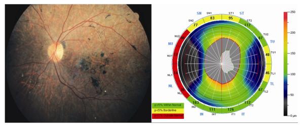
Purpose: To evaluate peripapillary retinal nerve fiber layer (RNFL) thickness using spectral-domain optical coherence tomography in patients with autosomal recessive cone-rod dystrophy (CRD).
Design: Cross-sectional study.
Methods: Eleven patients (22 eyes) with CRD were studied, including 4 patients with identified ABCA4 gene mutations. Peripapillary RNFL thickness was measured in 16 segments from 4 quadrants. The analyses were based on age and disc size-adjusted normative data. An abnormal thinning was considered when RNFL thickness measurements were less than the fifth percentile in at least 2 of 4 segments in a quadrant. Mean RNFL thickness was compared quantitatively with normative data obtained from 134 subjects.
Results: Eight patients (73%) had peripapillary RNFL thinning in at least 1 quadrant of at least 1 eye, including 3 of 4 patients with known ABCA4 gene mutations. Peripapillary RNFL thinning in the temporal quadrant was seen most commonly in 11 (79%) of 14 eyes with thinning in at least 1 quadrant. Significant thinning of the overall peripapillary RNFL was observed in CRD patients compared with controls (P = .0002). Subgroup analysis showed that 8 (89%) of 9 patients who were older than 40 years had thinning in at least 1 quadrant of at least 1 eye.
Conclusions: Peripapillary RNFL thinning was observed commonly in our patients with autosomal recessive CRD. The results confirm that the inner retinal structures can be affected in outer retinal disease. Careful evaluation of the inner retina may be important in determining the success rate of potential treatments for predominantly outer retinal diseases.
Figures
References
-
- Yagasaki K, Jacobson SG. Cone-rod dystrophy. Phenotypic diversity by retinal function testing. Arch Ophthalmol. 1989;107:701–708. - PubMed
-
- Szlyk JP, Fishman GA, Alexander KR, Peachey NS, Derlacki DJ. Clinical subtypes of cone-rod dystrophy. Arch Ophthalmol. 1993;111:781–788. - PubMed
-
- Krill AE, Deutman AF, Fishman M. The cone degenerations. Doc Ophthalmol. 1973;35:1–80. - PubMed
-
- Fishman GA, Stone EM, Eliason DA, Taylor CM, Lindeman M, Derlacki DJ. ABCA4 gene sequence variations in patients with autosomal recessive cone-rod dystrophy. Arch Ophthalmol. 2003;121:851–855. - PubMed
Publication types
MeSH terms
Substances
Grants and funding
LinkOut - more resources
Full Text Sources
