Review
. 2009 Jun;19(6):245-52.
doi: 10.1016/j.tcb.2009.03.003. Epub 2009 May 4.

Unconventional myosins acting unconventionally

Affiliations
Review

Unconventional myosins acting unconventionally

Sarah Woolner et al. Trends Cell Biol. 2009 Jun.

Abstract

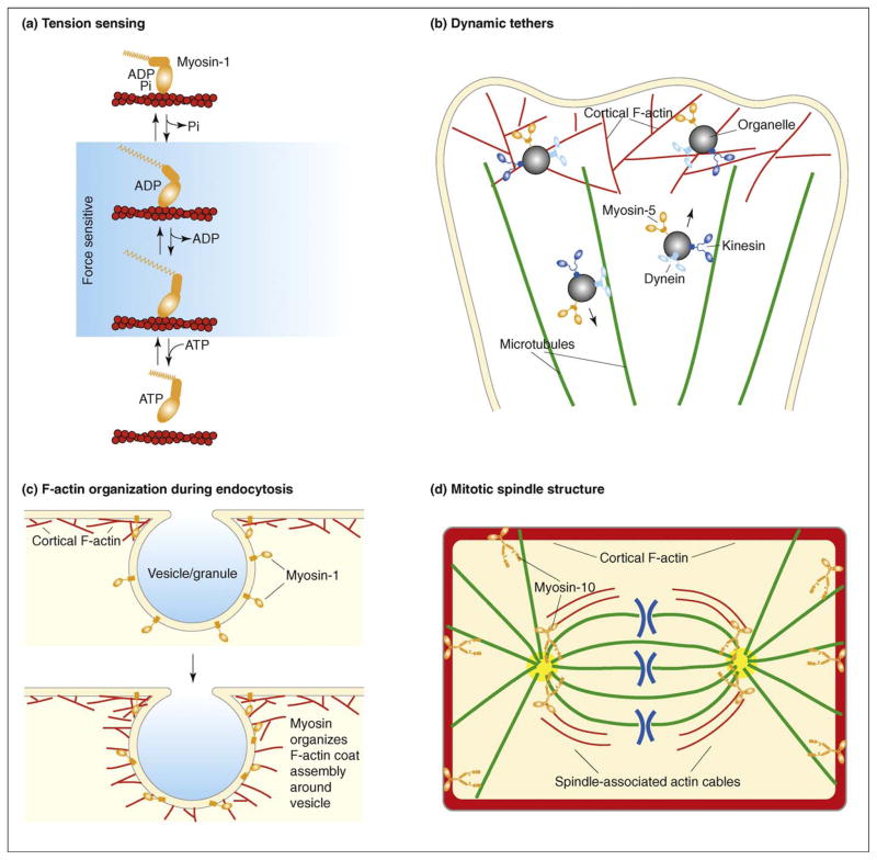
Unconventional myosins are proteins that bind actin filaments in an ATP-regulated manner. Because of their association with membranes, they have traditionally been viewed as motors that function primarily to transport membranous organelles along actin filaments. Recently, however, a wealth of roles for myosins that are not obviously related to organelle transport have been uncovered, including organization of F-actin, mitotic spindle regulation and gene transcription. Furthermore, it has also become apparent that the motor domains of different myosins vary strikingly in their biophysical attributes. We suggest that the assumption that most unconventional myosins function primarily as organelle transporters might be misguided.


Figures

Figure I
Figure I
Myosin-2 and the sliding filament hypothesis. Myosins-2 can form bipolar filaments through their extended coiled-coil domains. Here, just two dimers are shown in a filament, but muscle myosin-2 can form large filaments containing many myosin molecules. When the myosin-2 heads ratchet along the actin filaments towards the barbed ends (+), the actin filaments will slide in opposite directions.
Figure 1
Figure 1
Examples of unconventional roles for unconventional myosins. (a) Tension sensing: members of the myosin-1 family can act as tension sensors owing to their structural and kinetic properties. Myosins-1 have a two-step working stroke, the second step of which is sensitive to applied force [21]. When tension is applied to the myosin, it is less likely to release ADP and remains bound to the actin filament for longer periods of time. (b) Dynamic tethers: rather than acting to transport organelles along F-actin tracks, there is growing evidence that unconventional myosins (such as myosins-5a and -5b) instead function to tether organelles to cortical F-actin after their delivery to the cell periphery along microtubules by kinesin [23,25]. These myosin tethers are proposed to be very dynamic and their effect reversible, such that organelles or vesicles can be released and sent back down microtubules to the cell interior via dynein-mediated transport [27]. (c) F-actin organization during endocytosis: myosins-1c and -1e have been shown to organize the assembly of F-actin during endo- and exocytosis [27,35,36]; here, compensatory endocytosis is shown. Myosin-1 localizes to the vesicle or granule before the formation of the F-actin coat and is required to organize the F-actin to facilitate its endocytosis. (d) Mitotic spindle structure: myosins-10 and -15 have been found to be involved in maintaining mitotic spindle structure [–57]; here, for simplicity, just myosin-10 is shown. Myosin-10 localizes to the actin-rich cell cortex and to the mitotic spindle and functions in spindle length regulation, spindle pole structure and spindle anchoring [57]. These functions could be partly mediated through possible connections between cortical myosin-10 and astral microtubules or through separate functions by myosin-10 in the spindle interior.

References

    1. Sokac AM, Bement WM. Regulation and expression of metazoan unconventional myosins. Int Rev Cytol. 2000;200:197–304. - PubMed
    1. Foth BJ, et al. New insights into myosin evolution and classification. Proc Natl Acad Sci U S A. 2006;103:3681–3686. - PMC - PubMed
    1. Odronitz F, Kollmar M. Drawing the tree of eukaryotic life based on the analysis of 2,269 manually annotated myosins from 328 species. Genome Biol. 2007;8:R196. - PMC - PubMed
    1. Richards TA, Cavalier-Smith T. Myosin domain evolution and the primary divergence of eukaryotes. Nature. 2005;436:1113–1118. - PubMed
    1. Lodish H, et al. Molecular Cell Biology. 6. W.H. Freeman & Co; 2008.

Publication types