. 2009 Sep;23(9):2844-54.
doi: 10.1096/fj.08-128843. Epub 2009 Apr 30.

Statin-induced muscle damage and atrogin-1 induction is the result of a geranylgeranylation defect

Affiliations

Statin-induced muscle damage and atrogin-1 induction is the result of a geranylgeranylation defect

Peirang Cao et al. FASEB J. 2009 Sep.

Abstract

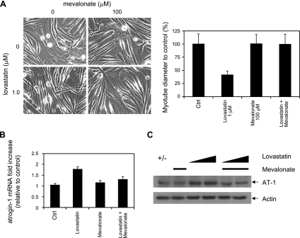
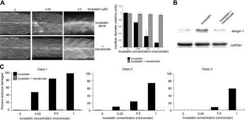
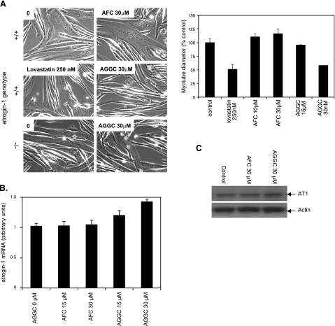
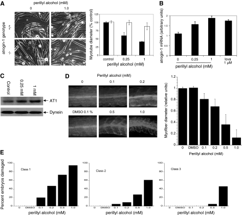
Statins are widely used to treat hypercholesterolemia but can lead to a number of side effects in muscle, including rhabdomyolysis. Our recent findings implicated the induction of atrogin-1, a gene required for the development of muscle atrophy, in statin-induced muscle damage. Since statins inhibit many biochemical reactions besides cholesterol synthesis, we sought to define the statin-inhibited pathways responsible for atrogin-1 expression and muscle damage. We report here that lovastatin-induced atrogin-1 expression and muscle damage in cultured mouse myotubes and zebrafish can be prevented in the presence of geranylgeranol but not farnesol. Further, inhibitors of the transfer of geranylgeranyl isoprene units to protein targets cause statin muscle damage and atrogin-1 induction in cultured cells and in fish. These findings support the concept that dysfunction of small GTP-binding proteins lead to statin-induced muscle damage since these molecules require modification by geranylgeranyl moieties for their cellular localization and activity. Collectively, our animal and in vitro findings shed light on the molecular mechanism of statin-induced myopathy and suggest that atrogin-1 may be regulated by novel signaling pathways.


Figures

Figure 1.
Figure 1.
Lovastatin damage in cultured myotubes. Myoblasts derived from atrogin-1-knockout mice (−/−) and corresponding wild-type littermates (+/+) were differentiated into myotubes. Cultures were treated with varying concentrations of lovastatin for 24 h. A) Morphology (left), myotube diameter (right). Solid bars, +/+ cells; open bars, −/− cells. B) Atrogin-1 mRNA levels in +/+ cells assessed by real-time PCR. C) Atrogin-1 protein levels in +/+ cells assessed by immunoblot.
Figure 2.
Figure 2.
Mevalonate rescues lovastatin-induced myotube damage. Primary myotubes from wild-type mice were treated with lovastatin or lovastatin and mevalonate for 24 h. A) Morphology (left), myotube diameter (right). B) Atrogin-1 mRNA. C) Atrogin-1 protein. Mevalonate concentration was 100 μM throughout. Lovastatin concentration was 1.0 μM (A, B); 0.5 and 1.0 μM (C).
Figure 3.
Figure 3.
Mevalonate rescues lovastatin-induced myofiber damage in zebrafish embryos. A) Zebrafish embryos (20 hpf) were treated with concentrations of lovastatin ranging from 0.05 to 0.5 μM and 100 μM mevalonate for 12 h. Embryos were fixed and stained with anti-myosin heavy-chain antibody (F59) as described in Materials and Methods. Representative somite phenotypes are shown. All panels are side views, anterior, left. Muscle fiber diameter was measured following myosin heavy-chain staining as described in Materials and Methods. At least 500 fibers were measured at each concentration. Results were graphed as the ratio of mean ± se experimental fiber size/control fiber size. Control fiber size: 7.60 ± 0.19 μM. B) Immunoblot of zebrafish atrogin-1. C) Quantitation of muscle damage. Morphological phenotypes shown in panel A were grouped into three classes. Class 1 changes include bowing, gap formation, and blocked/disrupted fibers. Class 2 changes include irregular fibers and diffuse appearance. Class 3 changes are typified by irregular somite boundaries. Values are percentages of embryos displaying specific class defects as a function of lovastatin concentration; 100 embryos/group.
Figure 4.
Figure 4.
Geranylgeranol but not farnesol rescues lovastatin-induced myotube damage in cultured myotubes. Primary myotubes from wild-type mice were treated with lovastatin (1.0 μM) or lovastatin (1.0 μM) and either farnesol (2.5 μM) or geranylgeranol (2.5 μM) for 24 h. A) Morphology (left), myotube diameter (right). B) Atrogin-1 mRNA. C) Atrogin-1 protein.
Figure 5.
Figure 5.
Geranylgeranol but not farnesol rescues lovastatin-induced muscle damage in zebrafish embryos. A) Zebrafish embryos (20 hpf) were treated with concentrations of lovastatin ranging from 0.05 to 0.5 μM and 10 μM farnesol or geranylgeranol for 12 h. Embryos were fixed and stained as in Fig. 3. Muscle fiber diameter was measured as described in Fig. 3. B) Immunoblot of zebrafish atrogin-1. Lovastatin, 0.5 mM; geranylgeranol and farnesol, 10 mM. C) Quantitation of muscle damage; 100 embryos/group.
Figure 6.
Figure 6.
AGGC but not AFC mimics lovastatin-induced myotube damage. Myotubes derived from atrogin-1-knockout mice (−/−) and corresponding wild-type littermates (+/+) were treated with lovastatin (250 nM) and AGGC (15–30 μM) or AFC (10–30 μM) for 24 h. A) Morphology (left); myotube diameter, +/+ cells only (right). B) Atrogin-1 mRNA levels in +/+ cells assessed by real-time PCR. C) Atrogin-1 protein levels in +/+ cells assessed by immunoblot.
Figure 7.
Figure 7.
Perillyl alcohol mimics lovastatin-induced myotube damage. Myotubes derived from atrogin-1-knockout mice (−/−) and corresponding wild-type littermates (+/+) were treated with perillyl alcohol (0.25–1.0 mM) for 24 h. A) Morphology (left), myotube diameter. Solid bars, +/+ cells; open bars, −/− cells. B) Atrogin-1 mRNA levels in +/+ cells assessed by real-time PCR. C) Atrogin-1 protein levels in +/+ cells assessed by immunoblot. D) Zebrafish embryos (20 hpf) were treated with concentrations of perillyl alcohol ranging from 0.1 to 1.0 mM for 12 h. Embryos were fixed and stained as in Fig. 3. Representative myofiber morphology (top) and myofiber diameter (bottom). E) Quantitation of muscle damage. Embryos/group: 73 for 0.1% DMSO and 71, 93, 87, 81, and 85 for 0, 0.1, 0.2. 0.5, and 1.0 mM perillyl alcohol, respectively.
Figure 8.
Figure 8.
Knockdown of geranylgeranyl transferase II in zebrafish mimics lovastatin-induced myofiber damage. A) Myosin heavy-chain staining of representative control morpholino-injected or z-GGTase I or II-injected zebrafish embryos (top), and myofiber diameter (bottom). B) Quantitation of muscle damage. Embryos/group: 100, 98, 102 for controls and 98, 96, 100 for z-GGT I knockdowns at 0, 8, 16 ng morpholino/embryo, respectively; 75, 91, 83 for controls and 75, 88, 95 for z-GGT II knockdowns at 0, 8, 16 ng morpholino/embryo, respectively.

References

    1. Staffa J A, Chang J, Green L. Cerivastatin and reports of fatal rhabdomyolysis. N Engl J Med. 2002;346:539–540. - PubMed
    1. Thompson P D, Clarkson P, Karas R H. Statin-associated myopathy. JAMA. 2003;289:1681–1690. - PubMed
    1. Ballantyne C M, Corsini A, Davidson M H, Holdaas H, Jacobson T A, Leitersdorf E, Marz W, Reckless J P, Stein E A. Risk for myopathy with statin therapy in high-risk patients. Arch Intern Med. 2003;163:553–564. - PubMed
    1. Graham D J, Staffa J A, Shatin D, Andrade S E, Schech S D, La Grenade L, Gurwitz J H, Chan K A, Goodman M J, Platt R. Incidence of hospitalized rhabdomyolysis in patients treated with lipid-lowering drugs. JAMA. 2004;292:2585–2590. - PubMed
    1. Phillips P S, Haas R H, Bannykh S, Hathaway S, Gray N L, Kimura B J, Vladutiu G D, England J D. Statin-associated myopathy with normal creatine kinase levels. Ann Intern Med. 2002;137:581–585. - PubMed

Publication types

MeSH terms