A glycolytic burst drives glucose induction of global histone acetylation by picNuA4 and SAGA
- PMID: 19406923
- PMCID: PMC2709565
- DOI: 10.1093/nar/gkp270
A glycolytic burst drives glucose induction of global histone acetylation by picNuA4 and SAGA
Abstract
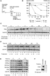
Little is known about what enzyme complexes or mechanisms control global lysine acetylation in the amino-terminal tails of the histones. Here, we show that glucose induces overall acetylation of H3 K9, 18, 27 and H4 K5, 8, 12 in quiescent yeast cells mainly by stimulating two KATs, Gcn5 and Esa1. Genetic and pharmacological perturbation of carbon metabolism, combined with (1)H-NMR metabolic profiling, revealed that glucose induction of KAT activity directly depends on increased glucose catabolism. Glucose-inducible Esa1 and Gcn5 activities predominantly reside in the picNuA4 and SAGA complexes, respectively, and act on chromatin by an untargeted mechanism. We conclude that direct metabolic regulation of globally acting KATs can be a potent driving force for reconfiguration of overall histone acetylation in response to a physiological cue.
Figures


Similar articles
-
NuA4 links methylation of histone H3 lysines 4 and 36 to acetylation of histones H4 and H3.J Biol Chem. 2014 Nov 21;289(47):32656-70. doi: 10.1074/jbc.M114.585588. Epub 2014 Oct 9. J Biol Chem. 2014. PMID: 25301943 Free PMC article.
-
Sgf29 binds histone H3K4me2/3 and is required for SAGA complex recruitment and histone H3 acetylation.EMBO J. 2011 Jun 17;30(14):2829-42. doi: 10.1038/emboj.2011.193. EMBO J. 2011. PMID: 21685874 Free PMC article.
-
The Gcn5 bromodomain of the SAGA complex facilitates cooperative and cross-tail acetylation of nucleosomes.J Biol Chem. 2009 Apr 3;284(14):9411-7. doi: 10.1074/jbc.M809617200. Epub 2009 Feb 13. J Biol Chem. 2009. PMID: 19218239 Free PMC article.
-
The SAGA continues: The rise of cis- and trans-histone crosstalk pathways.Biochim Biophys Acta Gene Regul Mech. 2021 Feb;1864(2):194600. doi: 10.1016/j.bbagrm.2020.194600. Epub 2020 Jul 6. Biochim Biophys Acta Gene Regul Mech. 2021. PMID: 32645359 Free PMC article. Review.
-
Untargeted tail acetylation of histones in chromatin: lessons from yeast.Biochem Cell Biol. 2009 Feb;87(1):107-16. doi: 10.1139/O08-097. Biochem Cell Biol. 2009. PMID: 19234527 Review.
Cited by
-
Acetyl-CoA carboxylase regulates global histone acetylation.J Biol Chem. 2012 Jul 6;287(28):23865-76. doi: 10.1074/jbc.M112.380519. Epub 2012 May 11. J Biol Chem. 2012. PMID: 22580297 Free PMC article.
-
NSUN2 is a glucose sensor suppressing cGAS/STING to maintain tumorigenesis and immunotherapy resistance.Cell Metab. 2023 Oct 3;35(10):1782-1798.e8. doi: 10.1016/j.cmet.2023.07.009. Epub 2023 Aug 15. Cell Metab. 2023. PMID: 37586363 Free PMC article.
-
Metabolic Intermediates in Tumorigenesis and Progression.Int J Biol Sci. 2019 May 7;15(6):1187-1199. doi: 10.7150/ijbs.33496. eCollection 2019. Int J Biol Sci. 2019. PMID: 31223279 Free PMC article. Review.
-
Cancer stem cell theory and the warburg effect, two sides of the same coin?Int J Mol Sci. 2014 May 19;15(5):8893-930. doi: 10.3390/ijms15058893. Int J Mol Sci. 2014. PMID: 24857919 Free PMC article.
-
Metabolic Signaling to Epigenetic Alterations in Cancer.Biomol Ther (Seoul). 2018 Jan 1;26(1):69-80. doi: 10.4062/biomolther.2017.185. Biomol Ther (Seoul). 2018. PMID: 29212308 Free PMC article. Review.
References
-
- Millar CB, Grunstein M. Genome-wide patterns of histone modifications in yeast. Nat. Rev. Mol. Cell Biol. 2006;7:657–666. - PubMed
-
- Friis RM, Schultz MC. Untargeted tail acetylation of histones in chromatin: lessons from yeast. Biochem. Cell Biol. 2009;87:107–116. - PubMed
-
- Zaman S, Lippman SI, Zhao X, Broach JR. How Saccharomyces responds to nutrients. Annu. Rev. Genet. 2008;42:27–81. - PubMed
Publication types
MeSH terms
Substances
LinkOut - more resources
Full Text Sources
Other Literature Sources
Molecular Biology Databases
Research Materials

