. 2009 Jul;202(1):87-97.
doi: 10.1677/JOE-08-0509. Epub 2009 Apr 30.

Acute or chronic stress induce cell compartment-specific phosphorylation of glucocorticoid receptor and alter its transcriptional activity in Wistar rat brain

Affiliations

Acute or chronic stress induce cell compartment-specific phosphorylation of glucocorticoid receptor and alter its transcriptional activity in Wistar rat brain

Miroslav Adzic et al. J Endocrinol. 2009 Jul.

Abstract

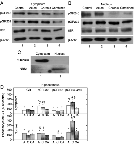
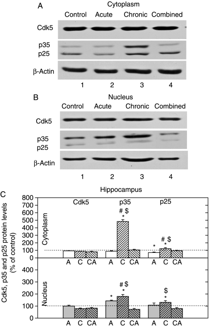
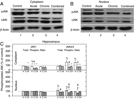
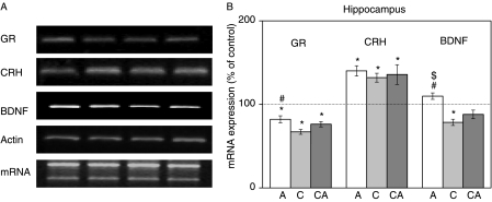
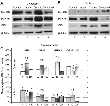
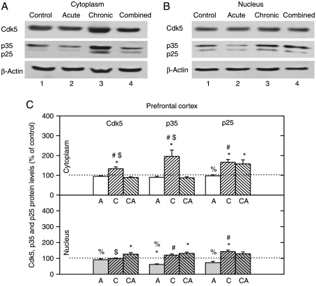
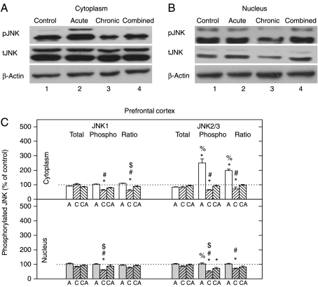
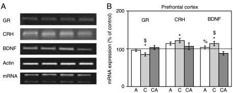
Chronic stress and impaired glucocorticoid receptor (GR) feedback are important factors for the compromised hypothalamic-pituitary-adrenal (HPA) axis activity. We investigated the effects of chronic 21 day isolation of Wistar rats on the extrinsic negative feedback part of HPA axis: hippocampus (HIPPO) and prefrontal cortex (PFC). In addition to serum corticosterone (CORT), we followed GR subcellular localization, GR phosphorylation at serine 232 and serine 246, expression of GR regulated genes: GR, CRF and brain-derived neurotropic factor (BDNF), and activity of c-Jun N-terminal kinase (JNK) and Cdk5 kinases that phosphorylate GR. These parameters were also determined in animals subjected to acute 30 min immobilization, which was taken as 'normal' adaptive response to stress. In isolated animals, we found decreased CORT, whereas in animals exposed to acute immobilization, CORT was markedly increased. Even though the GR was predominantly localized in the nucleus of HIPPO and PFC in acute, but not in chronic stress, the expression of GR, CRF, and BDNF genes was similarly regulated under both acute and chronic stresses. Thus, the transcriptional activity of GR under chronic isolation did not seem to be exclusively dependent on high serum CORT levels nor on the subcellular location of the GR protein. Rather, it resulted from the increased Cdk5 activation and phosphorylation of the nuclear GR at serine 232 and the decreased JNK activity reflected in decreased phosphorylation of the nuclear GR at serine 246. Our study suggests that this nuclear isoform of hippocampal and cortical GR may be related to hypocorticism i.e. HPA axis hypoactivity under chronic isolation stress.


Figures

Figure 1
Figure 1
Western-blot (WB) experiment demonstrating the effects of acute immobilization (A), chronic isolation (C), or combined stress (CA) on the levels of glucocorticoid receptor (GR) and its phosphoisoforms in the cytoplasm and nucleus of hippocampus: (A and B) GR phosphorylation at S246 (pGR246) or S232 (pGR232). Cell lysates were resolved by SDS-PAGE and probed with antibodies against total GR (tGR), pGR246, pGR232, or actin as a loading control. (C) The purity of subcellular fractions was assayed by anti-α-tubulin or anti-NBS1 antibodies for the cytoplasmic or nuclear compartments respectively. (D) Immunoreactivities of the cytoplasmic and nuclear GR, pGR232, and pGR246 (normalized to actin) and ratio of pGR232/pGR246 (normalized to GR in each compartment) are expressed as mean±s.e.m. (as described under Materials and Methods); n=15 animals per experimental group. Asterisks indicate significant differences between treated groups. Statistical analysis was performed using one-way ANOVA followed by the Tukey's post hoc test (*P<0·05, stress versus control; #P<0·05, acute versus chronic; %P<0·05, acute versus combined; $P<0·05 chronic versus combined).
Figure 2
Figure 2
Western-blot (WB) experiment demonstrating the effects of acute immobilization (A), chronic isolation (C), or combined stress (CA) on the levels of Cdk5, its activators p35 and p25 in the cytoplasm and nucleus of hippocampus: (A and B) Cdk5, p35, and p25 proteins in the cytoplasm (A) and nucleus (B) of hippocampus. Hippocampal lysates were resolved by SDS-PAGE and membranes were probed with respective antibodies that recognized indicated proteins and with anti-actin antibody as a loading control. (C) Immunoreactivities of Cdk5, p35, and p25 proteins in the cytoplasm and nucleus of hippocampus are given as mean±s.e.m. (as described under Materials and Methods); n=15 animals per experimental group. Statistical analysis was performed using one-way ANOVA followed by the Tukey's post hoc test and labelled as in the Fig. 1.
Figure 3
Figure 3
Western-blot (WB) experiment demonstrating the effects of acute immobilization (A), chronic isolation (C), or combined stress (CA) on the levels of JNKs and their phosphoisoforms in the cytoplasm and nucleus of hippocampus: (A and B) JNK proteins (tJNK1 at 46 kDa and tJNK2 and 3 at 54 kDa) and their phosphoisoforms (pJNK1, pJNK2 and 3) in the cytoplasm (A) and nucleus (B) of hippocampus. Hippocampal lysates were resolved by SDS-PAGE and membranes were probed with antibody that recognized all three tJNKs, or the specific JNK phosphoisoforms (pJNK1, 2, and 3) and with anti-actin antibody as a loading control. (C) Immunoreactivities of tJNK1, tJNK2/3, pJNK1, pJNK2/3, and their ratios (pJNK1/tJNK1 and pJNK2/3/tJNK2/3) in the cytoplasm and nucleus of hippocampus are given as mean±s.e.m. (as described under Materials and Methods); n=15 animals per experimental group. Statistical analysis was performed using one-way ANOVA followed by the Tukey's post hoc test and labelled as in Fig. 1.
Figure 4
Figure 4
Effects of acute, chronic, and combined stresses on GR, CRH, and BDNF gene expressions in the rat hippocampus measured by RT-PCR (A). A semi-quantitative evaluation of gene expressions from six independent experiments is shown in (B). Data are presented as mean±s.e.m., n=15 animals per experimental group. Statistical analysis was performed using one-way ANOVA followed by the Tukey's post hoc test and labelled as in Fig. 1.
Figure 5
Figure 5
Western-blot (WB) experiment demonstrating the effects of acute immobilization (A), chronic isolation (C), or combined stress (CA) on the levels of glucocorticoid receptor (GR) and its phosphoisoforms in the cytoplasm and nucleus of prefrontal cortex: (A and B) GR phosphorylated at S246 (pGR246) or S232 (pGR232). Cell lysates were resolved by SDS-PAGE and probed with antibodies against total GR (tGR), pGR246, pGR232, or actin as a loading control. (C) Immunoreactivities of the cytoplasmic and nuclear GR, pGR232, and pGR246 (normalized to actin) and ratio of pGR232/pGR246 (normalized to GR in each compartment) are expressed as mean±s.e.m. (as described under Materials and Methods); n=15 animals per experimental group. Asterisks indicate significant differences between treated groups. Statistical analysis was performed using one-way ANOVA followed by the Tukey's post hoc test and labelled as in Fig. 1.
Figure 6
Figure 6
Western-blot (WB) experiment demonstrating the effects of acute immobilization (A), chronic isolation (C), or combined stress (CA) on the levels of Cdk5, its activators p35 and p25 in the cytoplasm and nucleus of prefrontal cortex: (A and B) Cdk5, p35, and p25 proteins in the cytoplasm (A) and nucleus (B) of prefrontal cortex. Prefrontal cortex lysates were resolved by SDS-PAGE and membranes were probed with respective antibodies that recognized these proteins and with anti-actin antibody as a loading control. (C) Immunoreactivities of Cdk5, p35, and p25 proteins in the cytoplasm and nucleus of prefrontal cortex are given as mean±s.e.m. (as described under Materials and Methods); n=15 animals per experimental group. Statistical analysis was performed using one-way ANOVA followed by the Tukey's post hoc test and labelled as in the Fig. 1.
Figure 7
Figure 7
Western-blot (WB) experiment demonstrating the effects of acute immobilization, chronic isolation, or combined stress on the levels of JNKs and their phosphoisoforms in the cytoplasm and nucleus of prefrontal cortex (PFC). (A and B) JNK proteins (tJNK1 at 46 kDa and tJNK2 and 3 at 54 kDa) and their phosphoisoforms (pJNK1, pJNK2, and 3) in cytoplasm (A) and nucleus (B) of prefrontal cortex. Cell lysates were resolved by SDS-PAGE and membranes were probed with antibody that recognized all three tJNKs, or the specific JNK phosphoisoforms (pJNK1, 2 and 3) and with anti-actin antibody as a loading control. (C) Immunoreactivities of tJNK1, tJNK2/3, pJNK1, pJNK2/3, and their ratios (pJNK1/tJNK1 and pJNK2/3/tJNK2/3) in the cytoplasm and nucleus of prefrontal cortex are given as mean±s.e.m. (as described under Materials and Methods); n=15 animals per experimental group. Statistical analysis was performed using one-way ANOVA followed by the Tukey's post hoc test and labelled as in the Fig. 1.
Figure 8
Figure 8
Effects of acute, chronic, and combined stresses on GR, CRH, and BDNF gene expressions in the rat prefrontal cortex measured by semi-quantitative PCR (A). A semi-quantitative evaluation of gene expressions from six independent experiments is shown in (B). Data are presented as mean±s.e.m.; n=15 animals per experimental group. Statistical analysis was performed using one-way ANOVA followed by the Tukey's post hoc test and labelled as in the Fig. 1.

References

    1. Baigent SM, Lowry PJ. mRNA expression profiles for corticotrophin-releasing factor (CRF), urocortin, CRF receptors and CRF-binding protein in peripheral rat tissues. Journal of Molecular Endocrinology. 2000;25:43–52. - PubMed
    1. Blind RD, Garabedian MJ. Differential recruitment of glucocorticoid receptor phospho-isoforms to glucocorticoid-indued genes. Journal of Steroid Biochemistry Molecular Biology. 2008;109:150–157. - PMC - PubMed
    1. Cerqueira JJ, Pêgo JM, Taipa R, Bessa JM, Almeida OF, Sousa N. Morphological correlates of corticosteroid-induced changes in prefrontal cortex-dependent behaviors. Journal of Neuroscience. 2005;25:7792–7800. - PMC - PubMed
    1. Chen W, Dang T, Blind RD, Wang Z, Cavasotto CN, Hittelman AB, Rogatsky I, Logan SK, Garabedian MJ. Glucocorticoid receptor phosphorylation differentially affects target gene expression. Molecular Endocrinology. 2008;22:1754–1766. - PMC - PubMed
    1. Chrousos GP, Kino T. Glucocorticoid action networks and complex psychiatric and/or somatic disorders. Stress. 2007;10:213–219. - PubMed

Publication types

MeSH terms