. 2009 Jun;20(6):1223-35.
doi: 10.1681/ASN.2008050492. Epub 2009 Apr 30.

Loss of TIMP3 enhances interstitial nephritis and fibrosis

Affiliations

Loss of TIMP3 enhances interstitial nephritis and fibrosis

Zamaneh Kassiri et al. J Am Soc Nephrol. 2009 Jun.

Abstract

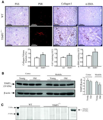
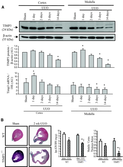
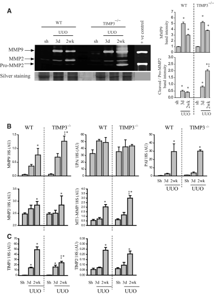
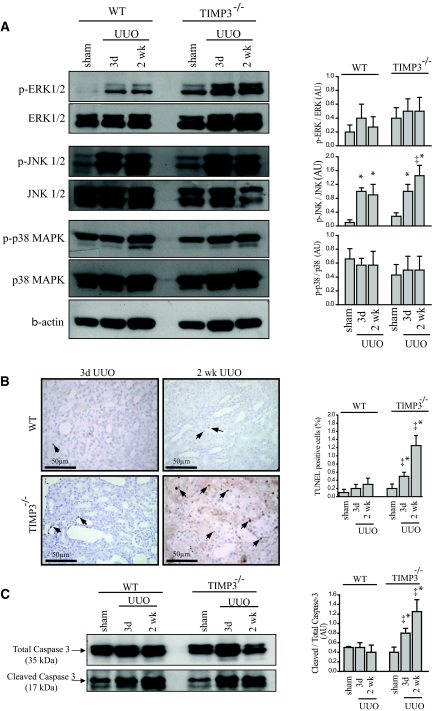
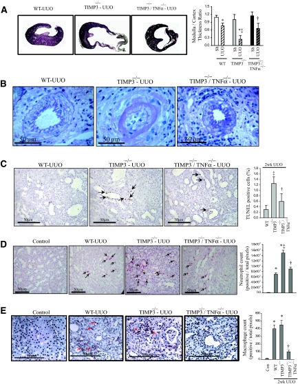
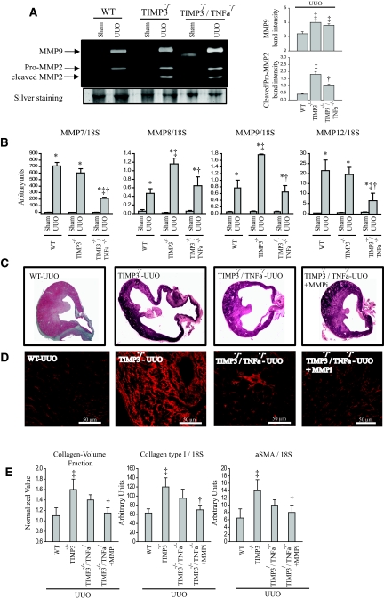
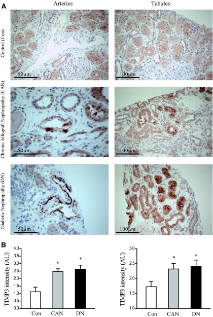
The balance of matrix metalloproteinases (MMPs) and tissue inhibitors of matrix metalloproteinases (TIMPs) determines the integrity of the extracellular matrix. TIMP3 is the most highly expressed tissue inhibitor of metalloproteinase (TIMP) in the kidney, but its function in renal disease is incompletely understood. In this study, TIMP3-/- mice demonstrated an age-dependent chronic tubulointerstitial fibrosis. After unilateral ureteral obstruction (UUO), young TIMP3-/- mice exhibited increased renal injury (tubular atrophy, cortical and medullary thinning, and vascular damage) compared with wild-type mice. In addition, TIMP3-/- mice had greater interstitial fibrosis; increased synthesis and deposition of type I collagen; increased activation of fibroblasts; enhanced apoptosis; and greater activation of MMP2, but not MMP9, after UUO. TIMP3 deficiency also led to accelerated processing of TNFalpha, demonstrated by significantly higher TACE activity and greater soluble TNFalpha levels by 3 d after UUO. The additional deletion of TNFalpha markedly reduced inflammation, apoptosis, and induction of a number of MMPs. Moreover, inhibition of MMPs in TIMP3-/-/TNFalpha-/- mice further abrogated postobstructive injury and prevented tubulointerestitial fibrosis. In humans, TIMP3 expression increased in the renal arteries and proximal tubules of subjects with diabetic nephropathy or chronic allograft nephropathy. Taken together, these results provide evidence that TIMP3 is an important mediator of kidney injury, and regulating its activity may have therapeutic benefit for patients with kidney disease.


Figures

Figure 1.
Figure 1.
Loss of tissue inhibitor of matrix metalloproteinases 3 (TIMP3) enhances age-dependent tubulointerstitial fibrosis. (A) Histologic analyses of 2-yr-old TIMP3−/− show enhanced tubulointerstitial injury, shrunken glomerular tufts, and increased fibrosis compared with wild-type (WT) mice. Collagen/volume fraction (Picrosirius red [PSR] staining), collagen type-I, and α-smooth muscle actin (αSMA) quantification are shown for WT (white) and TIMP3−/− kidneys (gray) (n = 5 per genotype; *P < 0.05 compared with WT group). (B) TIMP3 protein levels in young and old mice show a significant reduction in the medullar of the old kidneys (n = 4 per group; *P < 0.05 compared with young group). (C) No protein was detected in the urine of old TIMP3−/− compared with old WT mice. Chemical-grade bovine serum albumin was used as positive control. AU, arbitrary units; PAS, periodic acid-Schiff.
Figure 2.
Figure 2.
Tissue inhibitor of matrix metalloproteinases 3 (TIMP3) deficiency exacerbates tubulointerstitial injury after unilateral ureteral obstruction (UUO) in young mice. (A) In wild-type (WT) mice, TIMP3 protein and mRNA levels are increased early after UUO, followed by a reduction at 3d and 14 d post-UUO. Protein values are normalized to sham levels. (B) Young TIMP3−/− mice show exacerbated renal injury after UUO, as shown by severe tubular atrophy, and cortical and medullary thinning compared with WT kidneys at 2 wk post-UUO. Ipsilateral kidney-to-body weight ratio (Ipsi KW/BW) and medulla-to-cortex thickness ratio are significantly reduced in TIMP3−/−-UUO compared with WT-UUO kidneys (n = 6 per group). *P < 0.05 compared with corresponding sham-operated group; ‡P < 0.05 compared with corresponding WT group. AU, arbitrary units.
Figure 3.
Figure 3.
Increased interstitial fibrosis, enhanced collagen synthesis, and α-smooth muscle actin (αSMA) expression in response to tubulointerstitial injury in tissue inhibitor of matrix metalloproteinases 3 (TIMP3)−/− mice. (A) Picrosirius red staining and confocal microscopy shows marked thickening and disorganization of the peritubular fibrosis in TIMP3-deficient kidneys compared with wild-type (WT) kidneys at 2 wk after unilateral ureteral obstruction (UUO). (B) Enhanced collagen synthesis and deposition as shown by immunostaining for collagen type I (arrows indicate areas of collagen deposition), as well as TaqMan RNA analysis for collagen type I normalized to 18S. (C) αSMA shows a significantly higher degree of staining and mRNA expression in TIMP3−/− kidneys at 3 d and 2 wk post-UUO. Arrows indicate regions of interstitial staining for αSMA. *P < 0.05 compared with sham-operated group; ‡P < 0.05 compared with corresponding WT group.
Figure 4.
Figure 4.
Altered activation and expression of matrix metalloproteinases (MMP) in tissue inhibitor of MMPs 3 (TIMP3)-deficient mice in response to tubulointerstitial injury. (A) Gelatin zymography shows that pro-MMP9 (92 kD) levels are significantly increased after unilateral ureteral obstruction (UUO) in both genotypes, while cleavage of pro-MMP2 (68 kD) to its cleaved form (62 kD) is markedly higher in TIMP3−/−-UUO kidneys. Averaged band densities for MMP9 and cleaved/pro-MMP2 from 5 zymograms are shown on the right. (B) TaqMan real-time PCR shows a significantly greater increase in MMP9 mRNA at 2 wk post-UUO in TIMP3−/− kidneys, whereas expression of urokinase plasminogen activator was not altered, and plasminogen activator inhibitor was significantly increased in both genotypes (n = 10 per group). MMP2 expression increased similarly in both genotypes, but MT1-MMP expression increased more significantly in TIMP3−/− kidneys at 2 wk post-UUO. (C) TIMP1 levels increased significantly at 3 d and 2 wk post-UUO in both genotypes, with a markedly greater increase in the wild-type (WT) group at 2 wk post-UUO. TIMP2 levels increased similarly in WT and TIMP3−/− mice, reaching statistical significance at 2 wk post-UUO (n = 10 per group). AU, arbitrary unit. *P < 0.05 compared with sham (Sh)-operated group; ‡P < 0.05 compared with corresponding WT group.
Figure 5.
Figure 5.
Increased activation of the maladaptive mitogen-activated protein kinase (MAPK) and enhanced apoptosis in tissue inhibitor of matrix metalloproteinases 3 (TIMP3)−/− mice after tubulointerstitial injury. (A) Western blots for phosphorylated and total extracellular-activated kinase (ERK) 1 and 2, c-Jun activated kinase (JNK) 1 and 2, p38, and phospho-to-total ratio for each MAPK are shown on the right. Phospho-to-total ratio for JNK1/2 shows a significant increase in TIMP3−/− compared with that of the wild-type (WT) group, but similar changes for ERK1/2 and p38 between the two genotypes after unilateral ureteral obstruction (UUO) (n = 5 per group). Phospho-to-total ratios for ERK1/2 and p38 show similar changes between the two genotypes after UUO. The right panels show the quantitative analysis for the western blots. (B) TUNEL staining shows a larger population of apoptotic cells in the TIMP3−/− kidneys within 3 d of UUO that amplifies by 2 wk. Arrows indicate TUNEL positive cells. (C) Western blots show cleaved and total caspase 3, a molecular marker for apoptosis. Cleaved-to-total ratio for caspase 3 is enhanced in TIMP3−/− kidneys, as evident from the higher levels of cleaved-to-total caspase 3 ratio at 3 d and 2 wks post-UUO (n = 5). AU, arbitrary unit; p, phosphorylated. *P < 0.05 compared with sham-operated group; ‡P < 0.05 compared with corresponding WT group.
Figure 6.
Figure 6.
TNF-α is a key molecule mediating the severe renal injury in tissue inhibitor of matrix metalloproteinases 3 (TIMP3)−/− mice in response to unilateral ureteral obstruction (UUO). (A) Activity of TNFα-converting enzyme (TACE) is significantly increased in (TIMP3)−/− kidneys at 3 d post-UUO but comparable to WT-UUO at 2 wk post-UUO (n = 5). (B) Western blot for TNFα shows the membrane-bound (27 kD) and soluble TNFα (17 kD). Strikingly higher levels of soluble TNFα are detected in TIMP3−/− kidneys at 3 d post-UUO, suggesting enhanced processing of TNFα protein by TACE early after disease in these mice compared with WT (n = 5). TNFα mRNA production is increased similarly in TIMP3−/− and WT kidneys (n = 10 per group). (C) Western blots for NFκB (nuclear factor Kappa-β) on the nuclear and cytosolic fractions show greater nuclear translocation of NFκB in TIMP3−/−-UUO kidneys, as indicated by a higher nuclear-to-cytosolic ratio shown on the right (n = 5). Histone was used as a loading control for the nuclear fraction and tubulin for the cytosolic fraction. Averaged nuclear-to-cytosolic ratio from four blots is shown on the right. *P < 0.05 compared with corresponding sham group. RFU, relative fluorescence units.
Figure 7.
Figure 7.
Loss of TNFα in tissue inhibitor of matrix metalloproteinases 3 (TIMP3)-deficient mice markedly improves renal injury, apoptosis, and inflammation after unilateral ureteral obstruction (UUO). (A) Marked improvement in cortical and medullary thinning in TIMP3−/−/TNFα−/− compared with TIMP3−/− kidneys after UUO (n = 6 per genotype). (B) Interlobular arteries show post-UUO disruption of normal vasculature in TIMP3−/− mice, which was largely prevented by the loss of TNFα. (C) TUNEL staining shows a drastic reduction in the degree of apoptosis in TIMP3−/−/TNFα−/− kidneys. (D) Immunostaining for neutrophils shows that the significantly greater infiltration in TIMP3−/−-UUO kidneys is markedly reduced in the TIMP3−/−/TNFα−/− kidneys, to levels comparable to WT-UUO. Neutrophil counts per heart are shown on the right. (E) Immunostaining for macrophages (F4/80) shows a similar post-UUO increase in macrophage population in wild-type (WT) and TIMP3−/− groups, which is clearly reduced in the TIMP3−/−/TNFα−/− kidneys. Averaged macrophage numbers per heart are shown on the right. Arrows indicate positive stainings. *P < 0.05 compared with sham-operated group; ‡P < 0.05 compared with corresponding WT-UUO group; †P < 0.05 compared with TIMP3−/− group.
Figure 8.
Figure 8.
Inhibition of residual matrix metalloproteinases (MMP) activities in tissue inhibitor of MMP 3 (TIMP3)−/−/TNFα−/− mice further improves after unilateral ureteral obstruction (UUO) injury and fibrosis. (A) Gelatin zymography shows comparable levels of MMP9 and pro-MMP2, but reduced cleaved MMP2 in TIMP3−/−/TNFα−/−-UUO kidneys compared with TIMP3−/−-UUO kidneys, although still higher than WT-UUO group. Averaged band densities for five zymograms are shown on the right. (B) TaqMan real-time PCR analysis of candidate MMPs shows significant reductions in expression of MMP7, MMP8, MMP9, and MMP12 in the TIMP3−/−/TNFα−/− compared with TIMP3−/−-UUO. (C) Representative cross-sectional images of wild-type (WT), TIMP3−/−, TIMP3−/−/TNFα−/−, and TIMP3−/−/TNFα−/− + MMPi (PD166793, a broad-spectrum MMP inhibitor) show a clear improvement in the medullary thickness in the last group. (D) Picrosirius red (PSR) staining and confocal imaging show that tubulointerstitial fibrosis is significantly improved in the double-knock out kidneys and completely blocked with additional MMP inhibition. Collagen-volume fraction (calculated on the basis of the PSR-stained section), production of collagen type I and α-SMA, and markers of fibrosis at 2wk post-UUO are significantly reduced in TIMP3−/−/TNFα−/− + MMPi compared with TIMP3−/− kidneys. *P < 0.05 compared with sham-operated group; ‡P < 0.05 compared with corresponding WT group. †P < 0.05 compared with TIMP3−/− group.
Figure 9.
Figure 9.
Increased tissue inhibitor of matrix metalloproteinases 3 (TIMP3) expression in human kidney disease based on immunohistochemical staining in human kidney biopsies. (A) Increased TIMP3 staining in kidney biopsy samples from patients with diabetic nephropathy (DN) (n = 10) and chronic allograft nephropathy (CAN) (n = 10) compared with controls (Con) (n = 9) particularly confined to the proximal tubular and vascular compartments. (B) Averaged semiquantitative analysis shows significantly increased TIMP3 staining in kidney biopsies from patients with type 2 diabetes mellitus and chronic allograft nephropathy. *P < 0.05 compared with the control group.

Similar articles

Cited by

References

    1. Remuzzi G, Bertani T: Pathophysiology of progressive nephropathies. N Engl J Med 339: 1448–1456, 1998 - PubMed
    1. Eddy AA: Molecular basis of renal fibrosis. Pediatr Nephrol 15: 290–301, 2000 - PubMed
    1. Schrier RW: Cancer therapy and renal injury. J Clin Invest 110: 743–745, 2002 - PMC - PubMed
    1. Moe OW: Kidney stones: Pathophysiology and medical management. Lancet 367: 333–344, 2006 - PubMed
    1. Nuttall RK, Sampieri CL, Pennington CJ, Gill SE, Schultz GA, Edwards DR: Expression analysis of the entire MMP and TIMP gene families during mouse tissue development. FEBS Lett 563: 129–134, 2004 - PubMed

Publication types

MeSH terms

Substances