. 2009 Jul 15;180(2):159-66.
doi: 10.1164/rccm.200808-1250OC. Epub 2009 Apr 30.

Human neutrophil peptides and phagocytic deficiency in bronchiectatic lungs

Affiliations

Human neutrophil peptides and phagocytic deficiency in bronchiectatic lungs

Stefanos Voglis et al. Am J Respir Crit Care Med. .

Abstract

Rationale: A well-known clinical paradox is that severe bacterial infections persist in the lungs of patients with cystic fibrosis (CF) despite the abundance of polymorphonuclear neutrophils (PMN) and the presence of a high concentration of human neutrophil peptides (HNP), both of which are expected to kill the bacteria but fail to do so. The mechanisms remain unknown.

Objectives: This study examined several possible mechanisms to understand this paradox.

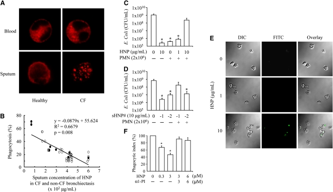
Methods: PMN were isolated from sputum and blood of subjects with and without CF or non-CF bronchiectasis for phagocytic assays. HNP isolated from patients with CF were used to stimulate healthy PMN followed by phagocytic tests.

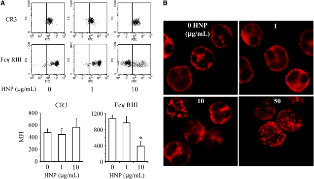
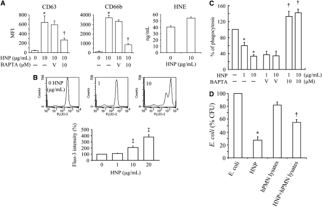
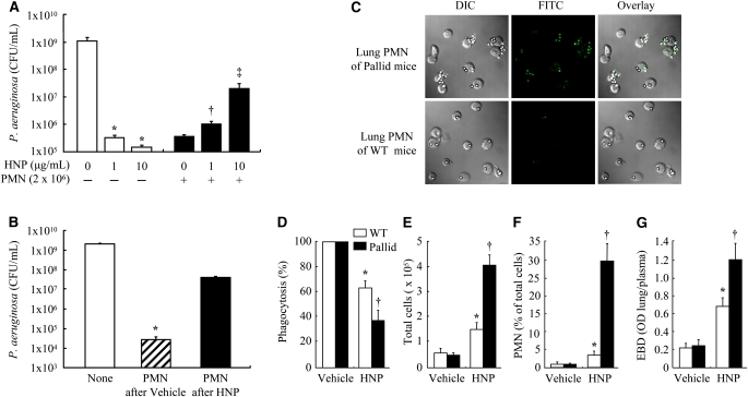
Measurements and main results: PMN isolated from the sputum of the bronchiectatic patients display defective phagocytosis that correlated with high concentrations of HNP in the lung. When healthy PMN were incubated with HNP, decreased phagocytic capacity was observed in association with depressed surface Fc gamma RIII, actin-filament remodeling, enhanced intracellular Ca(2+), and degranulation. Treatment of PMN with an intracellular Ca(2+) blocker or alpha1-proteinase inhibitor to attenuate the activity of HNP largely prevented the HNP-induced phagocytic deficiency. Intratracheal instillation of HNP in Pallid mice (genetically deficient in alpha1-proteinase inhibitor) resulted in a greater PMN lung infiltration and phagocytic deficiency compared with wild-type mice.

Conclusions: HNP or PMN alone exert antimicrobial ability, which was lost as a result of their interaction. These effects of HNP may help explain the clinical paradox seen in patients with inflammatory lung diseases, suggesting HNP as a novel target for clinical therapy.


Figures

<b>Figure 1.</b>
Figure 1.
Human neutrophil peptides (HNP) inhibited phagocytosis of polymorphonuclear neutrophils (PMN). (A) Altered distribution of F-actin in PMN isolated from cystic fibrosis (CF) sputum. The PMN from blood and sputum of healthy donors and patients with CF were seeded on fibronectin-coated coverslips, fixed and permeabilized before staining with rhodamine-phalloidin (red). n = 9; a representative image is shown in each group. (B) Inverse correlation between PMN phagocytosis and HNP concentration in sputum of CF (open circle) and non-CF bronchiectasis (filled circle), where the x represents HNP concentration, and y the percentage of phagocytosis in the regression equation. (C) PMN isolated from blood of healthy donors were incubated with HNP for 30 minutes before Escherichia coli exposure for 2 hours. The supernatants were plated for E. coli colony counting. *P < 0.05 versus the far left and the right bar, respectively. (D) PMN isolated from blood of healthy donors were incubated with synthetic HNP-1 or HNP-2 for 30 minutes before E. coli exposure for 2 hours. The supernatants were plated for E. coli colony counting. sHNP = synthetic HNP. *P < 0.05 versus the far left bar, respectively. (E) Healthy human PMN were incubated with HNP for 30 minutes, followed by exposure to IgG-opsonized latex beads for 60 minutes. The extracellular beads were stained with fluorescein isothiocyanate anti-IgG antibody (green). DIC = differential interference contrast. (F) Phagocytic index defined as the number of intracellular beads per cell. A complex of HNP (10 μg/ml HNP is equal to 2.8 μM) and an equimolar Prolastin was used to treat PMN 30 minutes before exposure to the beads. n = 10 per group, *P < 0.05 versus 0 μM HNP and the two groups at the far right, respectively.
<b>Figure 2.</b>
Figure 2.
Effects of human neutrophil peptides (HNP) on surface expression of CD markers and F-actin distribution of polymorphonuclear neutrophils (PMN). (A) HNP decreased surface Fcγ RIII expression. Healthy PMN were treated with the indicated concentrations of HNP for 1 hour and stained with anti-human CR3–fluorescein isothiocyanate or Fcγ RIII–fluorescein isothiocyanate antibodies for 30 minutes, followed by analysis with flow cytometry. Analysis markers in lines indicate gating for unstained cells. MFI = mean fluorescence intensity. n = 10, *P < 0.05 versus other groups, respectively, at identical conditions. (B) HNP altered F-actin distribution. PMN from healthy donors were incubated with indicated concentrations of HNP for 1 hour. Cells were washed, fixed, and permeabilized with 0.2% Triton-X for 15 minutes before staining of F-actin with rhodamine-phalloidin (red). A representative image of F-actin is illustrated from seven experiments per group.
<b>Figure 3.</b>
Figure 3.
Mechanisms of human neutrophil peptides (HNP)–induced deficiency of polymorphonuclear neutrophils (PMN) phagocytosis. (A) HNP increased PMN degranulation. PMN from healthy donors were incubated with HNP for 30 minutes and stained with anti-human antibody against CD63 or CD66b, in the presence and absence of an intracellular Ca2+ chelator BAPTA. MFI = mean fluorescence intensity. n = 10. Healthy PMN (1 × 10⁶/ml) in 24-well plates were treated with HNP at 10 μg/ml or vehicle control solution for 1 hour. The concentrations of human neutrophil elastase (HNE) were measured in cell culture medium. (B) HNP increased intracellular Ca2+. Healthy PMN were loaded with fluo-3 AM for 1 hour in phosphate-buffered saline free of Ca2+ and Mg2+, exposed to HNP followed by analysis over 60 seconds (n = 5). (C) Effects of BAPTA on the phagocytic index. Healthy PMN were incubated with BAPTA and HNP for 30 minutes, followed by exposure to IgG opsonized latex beads for 1 hour. *P < 0.05 versus 0 HNP, P < 0.05 versus vehicle (V, 0.01% dimethyl sulfoxide) in the presence of HNP at identical conditions, respectively. P < 0.05 versus 0 and 1 μg/ml of HNP at identical conditions, respectively. (D) PMN disables the antibacterial activity of HNP. Escherichia coli was incubated for 3 hours with and without HNP (10 μg/ml), with and without PMN lysates (1 × 10⁶ cells isolated from blood of healthy volunteers [hPMN lysates] or bone marrow of C57BL/6J mice [mPMN lysates]), or with and without the combination of HNP and the human or murine PMN lysates, respectively. n = 3 experiments. *P < 0.05 versus E. coli, P < 0.05 versus E. coli and HNP, respectively.
<b>Figure 4.</b>
Figure 4.
Effects of human neutrophil peptides (HNP) on polymorphonuclear neutrophils (PMN) phagocytosis in mice. (A) PMN isolated from bone marrow of C57BL/6J mice (n = 5) were incubated with the indicated concentrations of HNP for 30 minutes before Pseudomonas aeruginosa exposure for 3 hours for bacterial killing assay. *P < 0.05 versus 0 HNP in the absence of PMN; P < 0.05 versus 0 HNP in the presence of PMN; P < 0.05 versus other groups in the presence of PMN, respectively. (B) Mice received intratracheal instillation of HNP (200 pg) or vehicle control in 30 μl phosphate-buffered saline and observed for 5 hours (n = 7 each). PMN were then isolated from lung lavage fluids and exposed to P. aeruginosa for 3 hours for bacterial killing assay. *P < 0.05 versus other groups. (C and D) In a separate assay to evaluate phagocytic function, PMN recovered from lavage fluids of the wild-type (WT) and the Pallid mice (n = 7 each group) 5 hours after HNP instillation were exposed to IgG opsonized beads for 30 minutes. After fixation, the extracellular beads were labeled with fluorescein isothiocyanate (green). Thus for a given number of beads and PMN in the assays, the greater the number of extracellular green beads, the less efficient was phagocytosis (as quantified by the phagocytic index) as seen in the Pallid mice. Phagocytosis was defined by the number of beads internalized per cell was calculated as a percentage of controls. (E and F) Total cell count and differentiation in lung lavage fluids was performed in the wild-type (WT) and Pallid mice. (G) In separate experiments, mice (n = 5 each) received Evans blue dye (EBD) via tail vein 30 minutes before the end of a 5-hour experiment after HNP instillation. EBD concentration in lung homogenate supernatant was quantified by a dual wavelength spectrophotometer for correction of contaminating heme pigments. *P < 0.05 versus WT in Vehicle control; P < 0.05 versus Pallid in Vehicle and WT in HNP group, respectively.

References

    1. Elston C, Geddes D. Inflammation in cystic fibrosis–when and why? Friend or foe? Semin Respir Crit Care Med 2007;28:286–294. - PubMed
    1. Sagel SD, Chmiel JF, Konstan MW. Sputum biomarkers of inflammation in cystic fibrosis lung disease. Proc Am Thorac Soc 2007;4:406–417. - PMC - PubMed
    1. Terheggen-Lagro SW, Rijkers GT, van der Ent CK. The role of airway epithelium and blood neutrophils in the inflammatory response in cystic fibrosis. J Cyst Fibros 2005;4:15–23. - PubMed
    1. Tirouvanziam R, Gernez Y, Conrad CK, Moss RB, Schrijver I, Dunn CE, Davies ZA, Herzenberg LA. Profound functional and signaling changes in viable inflammatory neutrophils homing to cystic fibrosis airways. Proc Natl Acad Sci USA 2008;105:4335–4339. - PMC - PubMed
    1. Ganz T. Defensins: antimicrobial peptides of innate immunity. Nat Rev Immunol 2003;3:710–720. - PubMed

Publication types

MeSH terms