. 2009 Jun 25;113(26):6593-602.
doi: 10.1182/blood-2009-01-201467. Epub 2009 Apr 30.

NK-cell-mediated killing of target cells triggers robust antigen-specific T-cell-mediated and humoral responses

Affiliations

NK-cell-mediated killing of target cells triggers robust antigen-specific T-cell-mediated and humoral responses

Philippe Krebs et al. Blood. .

Abstract

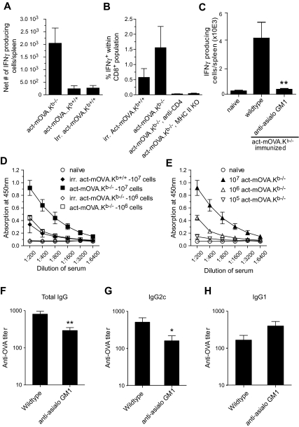
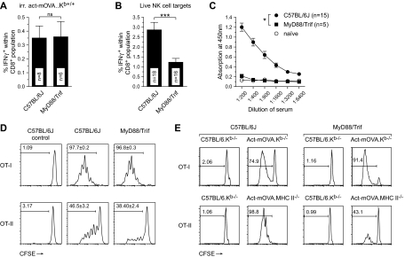
Previous work showed that administration of antigen-expressing apoptotic cells in vivo results in antigen-specific CD8+ T-cell responses independent of Toll-like receptor signaling. We report here that natural killer (NK) cells can serve a function directly upstream of this pathway and initiate robust adaptive immune responses via killing of antigen-expressing target cells. This pathway is highly sensitive, in that administration of as few as 10(4) target cells induced detectable antigen-specific CD8+ T-cell responses. Importantly, NK cell-mediated cytotoxicity of target cells could also induce robust antigen-specific CD4+ T-cell responses, which were critical for subsequent CD8+ T-cell priming and IgG responses. Unlike adaptive immune responses induced by gamma-irradiated cells, the NK-cell pathway required myeloid differentiating factor 88 (MyD88) and Toll/interleukin-1 receptor domain-containing adapter-inducing interferon-beta (Trif) signaling. NK cells have previously been shown to detect and kill pathogen-infected host cells, as well as neoplastic cells and tissue allografts. The present data provide further evidence that they also discharge a strong tie with their relatives in the adaptive immune system. We think that the recognition and killing of target cells by NK cells represents an important pathway for the generation of robust CD8+ T and humoral responses that may be exploited for vaccine development.


Figures

Figure 1
Figure 1
Administration of antigen-expressing NK-cell targets results in early clearance and robust CD8+ T-cell responses. (A) In vivo clearance of CFSE-labeled Kb-deficient splenocytes in wild-type but not Kb-deficient hosts. Twenty-four hours after transfer, blood samples were collected and analyzed for the presence of Kb-sufficient (high CFSE) and Kb-deficient splenocytes (low CFSE). Numbers in graph represent percentage killing ± SD (n = 3). (B,C) CD8+ T-cell responses measured 8 days after immunization with either 107 live Kb-sufficient or -deficient act-mOVA cells or with 107 irradiated act-mOVA.Kb+/+ cells. Responses were quantified by IFN-γ production after stimulation with OVA peptide ex vivo (B) or the ability to kill OVA-expressing target cells in vitro (C). (D) Survival of Kb-deficient splenocytes in wild-type and anti-asialo GM1-treated recipient mice (percentage is calculated from the ratio between Kb-deficient and Kb-sufficient cells in Kb-deficient recipients for each day). (E) Frequency of antigen-specific CD8+ T cells in wild-type or mice receiving anti-asialo GM1 treatment before and after immunization. (F) Association curve between NK-cell presence and antigen-specific CD8+ T-cell responses measured after mice received different doses of anti-asialo GM1 antibody 1 day before and 1 day after vaccination with 106 Kb−/− act-mOVA splenocytes.
Figure 2
Figure 2
CD8+ T-cell priming upon administration of Kb-deficient or allogeneic NK-cell targets is highly sensitive. CD8+ T-cell responses upon administration of various doses of Kb-deficient (A,B) or allogeneic (C-D) NK-cell targets. CD8+ T-cell responses were quantified 8 days after immunization by measuring IFN-γ production upon restimulation with OVA257-264 ex vivo (A) or the ability of CD8+ T cells to kill OVA-expressing target cells in vitro (B). (C,D) C57BL/6 mice were either immunized with act-mOVA targets on a mixed H-2b × H-2k (closed bars) or H-2b × H-2d (open bars) background (± SEM; n = 3). (E) Survival of allogeneic C3H/HeN-act-mOVA and β2m-deficient splenocytes in C57BL/6J recipient mice (percentage is calculated from the ratio between β2m-deficient and C57BL/6J cells in β2m-deficient recipients for each day). (F) CD8+ T-cell responses upon administration of 106 allogeneic C3H/HeN.act-mOVA target cells in C57BL/6J control, C57BL/6J-Prf1tm1Sdz, and C57BL/6Jgld/gld recipient mice (n = 6 for each group).
Figure 3
Figure 3
Administration of NK-cell targets induces strong CD4+ T- and B-cell responses. (A) OVA323-339-specific IFN-γ production as measured by ELISPOT by isolated splenocytes from mice immunized with either 106 live Kb-sufficient or -deficient act-mOVA cells or with 106 irradiated act-mOVA.Kb+/+ cells. Data are expressed as the average number of IFN-γ-producing cells per spleen (n = 4 per group). (B) Absence of CD4 help either through CD4 depletion via Ab treatment or using class II MHC-deficient recipient mice leads to complete abrogation of CD8+ T-cell responses as measured by antigen-specific intracellular IFN-γ production. (C) Anti-asialo GM1 treatment abrogates CD4+ T-cell response in mice immunized with 107 act-mOVA.Kb−/−-deficient target cells. (D,E) OVA-specific total IgG titers after immunization of mice with various doses of live Kb-sufficient or -deficient act-mOVA cells or irradiated act-mOVA.Kb+/+ cells. (F-H) Polarization of antibody responses as measured by OVA-specific IgG1 and IgG2c levels induced by 106 act-mOVA.Kb−/− in the presence or absence of NK cells. Sera are collected at day 14 after immunization. Data represent mean values ± SEM (n = 8). * P < .05; ** P < .01.
Figure 4
Figure 4
IFNs are key components of the NK cell–mediated CD8+ T-cell responses. (A-C) CD8+ T-cell responses as measured by IFN-γ production upon restimulation with OVA257-264 peptide ex vivo (day 8) and OVA-specific total IgG (measured day 14) in wild-type, IFN-αR, IFN-γR, and IFN-α/IFN-γR double-deficient mice. Values represent the mean ± SEM of pooled data from 3 to 4 independent experiments (*P ≤ .05).
Figure 5
Figure 5
Involvement of MyD88/Trif signaling in the amplification of CD8+ T-cell responses induced by NK-cell targets. (A,B) Frequency of CD8+IFN-γ+ T cells as measured by restimulation ex vivo 8 days after immunization of wild-type and MyD88/Trif double-deficient mice with either 106 irradiated cells or 106 live act-mOVA.Kb−/− splenocytes (n ≥ 8 for each group). (C) B-cell responses in wild-type and MyD88/Trif double-deficient mice immunized with 106 live act-mOVA.Kb−/− cells as measured by OVA-specific total IgG levels in sera collected 14 days after immunization. (D,E) Activation of OT-I and OT-II cells in wild-type and MyD88/Trif double-deficient mice either in vivo (D) or in vitro (E). (D) Mice were injected with 106 live act-mOVA.Kb−/− cells followed by administration of CD8+ OT-I or CD4+ OT-II cells intravenously after 3 days. (E) Activation of OT-I and OT-II cells by Flt3L-treated bone marrow–derived DCs from either wild-type or MyD88/Trif double-deficient mice. Proliferation of OT-I and OT-II cells is measured by flow cytometry measuring the dilution of CFSE upon each mother cell division (n = 4). *P ≤ .05; ***P ≤ .001.
Figure 6
Figure 6
Vaccination with NK-cell targets results in effective protection against OVA-expressing L monocytogenes. C57BL/6 mice were immunized with either 107 irradiated cells or 107 or 106 live act-mOVA.Kb−/− splenocytes. As a positive control, mice were immunized with a low dose (103) of L monocytogenes. On day 35 after immunization, mice were challenged with 105 CFU of LM-OVA and splenic titers were determined after an additional 3 days (n = 8 for each group). *P ≤ .05; **P ≤ .01 as determined by ANOVA.

Similar articles

Cited by

References

    1. Blankenstein T. The role of inflammation in tumour growth and tumour suppression. Novartis Found Symp. 2004;256:205–210. - PubMed
    1. Gallucci S, Lolkema M, Matzinger P. Natural adjuvants: endogenous activators of dendritic cells. Nat Med. 1999;5:1249–1255. - PubMed
    1. Degli-Esposti MA, Smyth MJ. Close encounters of different kinds: dendritic cells and NK cells take centre stage. Nat Rev Immunol. 2005;5:112–124. - PubMed
    1. Kelly JM, Darcy PK, Markby JL, et al. Induction of tumor-specific T cell memory by NK cell-mediated tumor rejection. Nat Immunol. 2002;3:83–90. - PubMed
    1. Levitsky HI, Lazenby A, Hayashi RJ, Pardoll DM. In vivo priming of two distinct antitumor effector populations: the role of MHC class I expression. J Exp Med. 1994;179:1215–1224. - PMC - PubMed

Publication types

MeSH terms