Role of NO synthase in the development of Trypanosoma cruzi-induced cardiomyopathy in mice
- PMID: 19407124
- PMCID: PMC2699411
Role of NO synthase in the development of Trypanosoma cruzi-induced cardiomyopathy in mice
Abstract
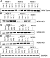
Trypanosoma cruzi infection results in an increase in myocardial NO and intense inflammation. NO modulates the T. cruzi-induced myocardial inflammatory reaction. NO synthase (NOS)1-, NOS2-, and NOS3-null mice were infected with T. cruzi (Brazil strain). Infected NOS1-null mice had increased parasitemia, mortality, and left ventricular inner diameter (LVID). Chronically infected NOS1- and NOS2-null and wild-type mice (WT) exhibited increased right ventricular internal diameter (RVID), although the fold increase in the NOS2-null mice was smaller. Infected NOS3-null mice exhibited a significant reduction both in LVID and RVID. Reverse transcriptase-polymerase chain reaction showed expression of NOS2 and NOS3 in hearts of infected NOS1-null and WT mice, whereas infected NOS2-null hearts showed little change in expression of other NOS isoforms. Infected NOS3-null hearts showed an increase only in NOS1 expression. These results may indicate different roles for NOS isoforms in T. cruzi-induced cardiomyopathy.
Figures


Similar articles
-
Nitric oxide synthase enzymes in the airways of mice exposed to ovalbumin: NOS2 expression is NOS3 dependent.Mediators Inflamm. 2010;2010:321061. doi: 10.1155/2010/321061. Epub 2010 Oct 5. Mediators Inflamm. 2010. PMID: 20953358 Free PMC article.
-
Inducible nitric oxide synthase in heart tissue and nitric oxide in serum of Trypanosoma cruzi-infected rhesus monkeys: association with heart injury.PLoS Negl Trop Dis. 2012;6(5):e1644. doi: 10.1371/journal.pntd.0001644. Epub 2012 May 8. PLoS Negl Trop Dis. 2012. PMID: 22590660 Free PMC article.
-
Differential regulation of nitric oxide synthase isoforms in experimental acute chagasic cardiomyopathy.Clin Exp Immunol. 2000 Jul;121(1):112-9. doi: 10.1046/j.1365-2249.2000.01258.x. Clin Exp Immunol. 2000. PMID: 10886247 Free PMC article.
-
Association between polymorphisms of NOS1, NOS2 and NOS3 genes and suicide behavior: a systematic review and meta-analysis.Metab Brain Dis. 2019 Aug;34(4):967-977. doi: 10.1007/s11011-019-00406-3. Epub 2019 Mar 21. Metab Brain Dis. 2019. PMID: 30900130
-
The Role of Single-Nucleotide Variants of NOS1, NOS2, and NOS3 Genes in the Comorbidity of Arterial Hypertension and Tension-Type Headache.Molecules. 2021 Mar 12;26(6):1556. doi: 10.3390/molecules26061556. Molecules. 2021. PMID: 33809023 Free PMC article. Review.
Cited by
-
Disease Tolerance and Pathogen Resistance Genes May Underlie Trypanosoma cruzi Persistence and Differential Progression to Chagas Disease Cardiomyopathy.Front Immunol. 2018 Dec 3;9:2791. doi: 10.3389/fimmu.2018.02791. eCollection 2018. Front Immunol. 2018. PMID: 30559742 Free PMC article.
-
Differences in cNOS/iNOS Activity during Resistance to Trypanosoma cruzi Infection in 5-Lipoxygenase Knockout Mice.Mediators Inflamm. 2019 Oct 24;2019:5091630. doi: 10.1155/2019/5091630. eCollection 2019. Mediators Inflamm. 2019. PMID: 31772504 Free PMC article.
-
Curcumin treatment provides protection against Trypanosoma cruzi infection.Parasitol Res. 2012 Jun;110(6):2491-9. doi: 10.1007/s00436-011-2790-9. Epub 2012 Jan 4. Parasitol Res. 2012. PMID: 22215192 Free PMC article.
-
NADPH phagocyte oxidase knockout mice control Trypanosoma cruzi proliferation, but develop circulatory collapse and succumb to infection.PLoS Negl Trop Dis. 2012;6(2):e1492. doi: 10.1371/journal.pntd.0001492. Epub 2012 Feb 14. PLoS Negl Trop Dis. 2012. PMID: 22348160 Free PMC article.
-
Trypanosoma cruzi antioxidant enzymes as virulence factors in Chagas disease.Antioxid Redox Signal. 2013 Sep 1;19(7):723-34. doi: 10.1089/ars.2012.4618. Epub 2012 May 21. Antioxid Redox Signal. 2013. PMID: 22458250 Free PMC article. Review.
References
-
- World Health Organization. Report on Chagas’ Disease. Geneva: World Health Organization; 2002.
-
- Carod-Artal FJ. Chagas cardiomyopathy and ischemic stroke. Expert Rev Cardiovasc Ther. 2006;4:119–130. - PubMed
-
- Jelicks LA, Shirani J, Wittner M, Chandra M, Weiss LM, Factor SM, Bekirov I, Braunstein VL, Chan J, Huang H, Tanowitz HB. Application of cardiac gated magnetic resonance imaging in murine Chagas’ disease. Am J Trop Med Hyg. 1999;61:207–214. - PubMed
-
- Huang H, Chan J, Wittner M, Jelicks LA, Morris SA, Factor SM, Weiss LM, Braunstein VL, Bacchi CJ, Yarlett N, Chandra M, Shirani J, Tanowitz HB. Expression of cardiac cytokines and inducible form of nitric oxide synthase (NOS2) in Trypanosoma cruzi-infected mice. J Mol Cell Cardiol. 1999;31:75–88. - PubMed
-
- Jelicks LA, Chandra M, Shirani J, Shtutin V, Tang B, Christ GJ, Factor SM, Wittner M, Huang H, Weiss LM, Mukherjee S, Bouzahzah B, Petkova SB, Teixeira MM, Douglas SA, Loredo ML, D’Orleans-Juste P, Tanowitz HB. Cardioprotective effects of phosphoramidon on myocardial structure and function in murine Chagas’ disease. Int J Parasitol. 2002;32:1497–1506. - PubMed
Publication types
MeSH terms
Substances
Grants and funding
LinkOut - more resources
Full Text Sources
