. 2009 Aug;23(8):1281-90.
doi: 10.1210/me.2008-0482. Epub 2009 Apr 30.

p38 MAPK is a major regulator of MafA protein stability under oxidative stress

Affiliations

p38 MAPK is a major regulator of MafA protein stability under oxidative stress

Takuma Kondo et al. Mol Endocrinol. 2009 Aug.

Abstract

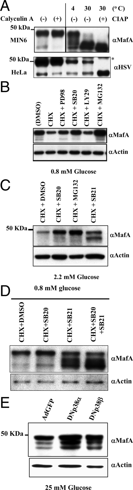
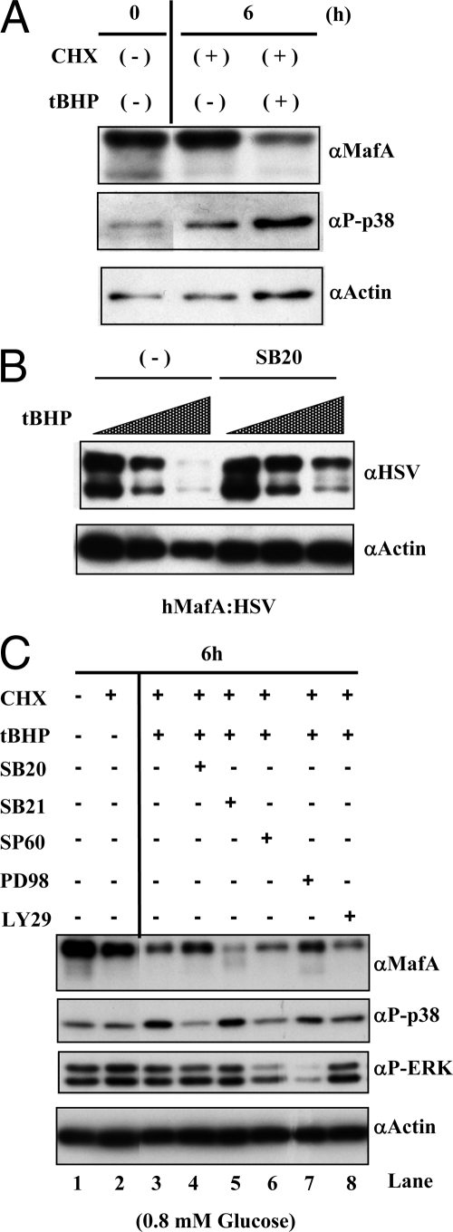
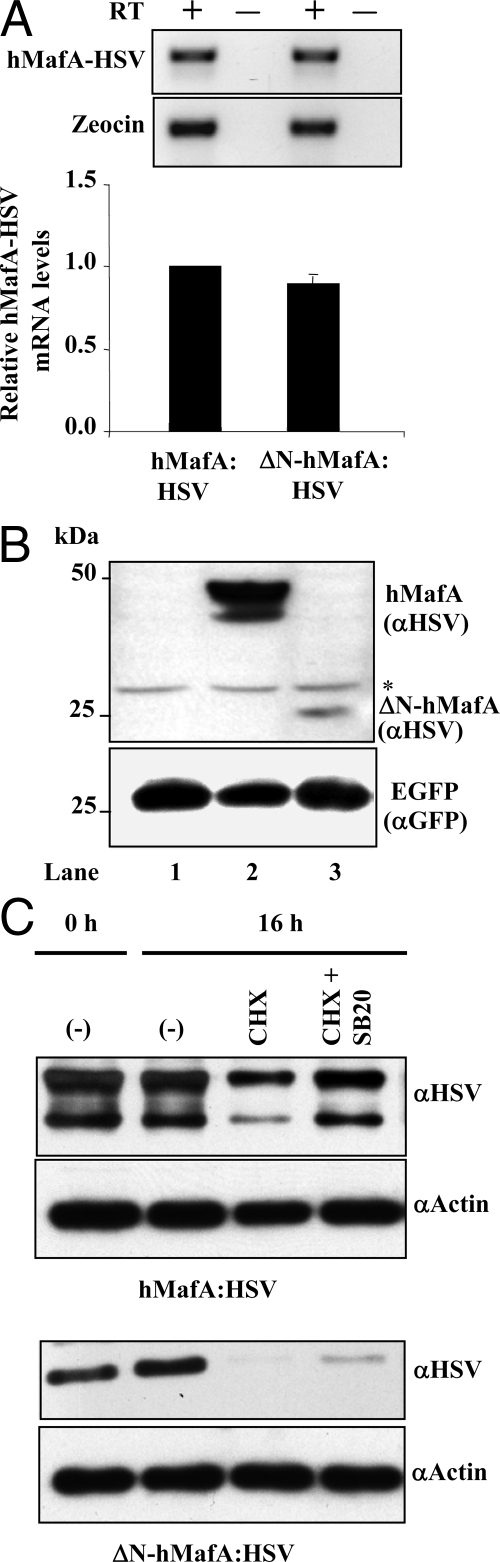
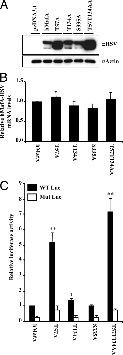
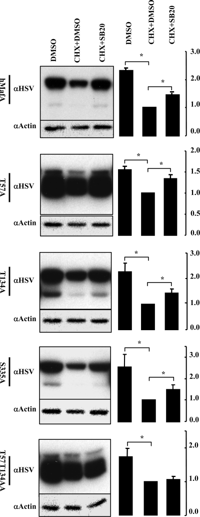
Mammalian MafA/RIPE3b1 is an important glucose-responsive transcription factor that regulates function, maturation, and survival of beta-cells. Increased expression of MafA results in improved glucose-stimulated insulin secretion and beta-cell function. Because MafA is a highly phosphorylated protein, we examined whether regulating activity of protein kinases can increase MafA expression by enhancing its stability. We demonstrate that MafA protein stability in MIN6 cells and isolated mouse islets is regulated by both p38 MAPK and glycogen synthase kinase 3. Inhibiting p38 MAPK enhanced MafA stability in cells grown under both low and high concentrations of glucose. We also show that the N-terminal domain of MafA plays a major role in p38 MAPK-mediated degradation; simultaneous mutation of both threonines 57 and 134 into alanines in MafA was sufficient to prevent this degradation. Under oxidative stress, a condition detrimental to beta-cell function, a decrease in MafA stability was associated with a concomitant increase in active p38 MAPK. Interestingly, inhibiting p38 MAPK but not glycogen synthase kinase 3 prevented oxidative stress-dependent degradation of MafA. These results suggest that the p38 MAPK pathway may represent a common mechanism for regulating MafA levels under oxidative stress and basal and stimulatory glucose concentrations. Therefore, preventing p38 MAPK-mediated degradation of MafA represents a novel approach to improve beta-cell function.


Figures

Figure 1
Figure 1
Role of protein kinases in regulating MafA stability. A, MafA is a phosphoprotein. Anti-MafA-specific antibody recognizes protein bands of apparent molecular mass of 40 and 48 kDa. MIN6 cells were treated with calyculin A (100 nm) or DMSO for 10 min before harvesting. Nuclear extracts (20 μg) were prepared, resolved on 7.5% SDS-PAGE, and immunoblotted with α-MafA antibody. To confirm the phosphorylation of MafA, nuclear extracts (20 μg) from MIN6 cells were incubated for 30 min at 4 or 30 C in the absence or presence of CIAP. Extracts were subsequently resolved on 7.5% SDS-PAGE and immunoblotted with α-MafA antibody. Increased levels of faster migrating MafA bands at 30 C in the absence of CIAP reflects endogenous phosphatase activity. Similar studies were performed using nuclear extracts (20 μg) from HeLa cells transfected with hMafA-HSV tag plasmid that were treated with either calyculin A (100 nm) or DMSO for 10 min before harvesting. Additionally, nuclear extracts (20 μg) from HeLa cells transfected with hMafA-HSV tag plasmid were incubated for 30 min at 4 or 30 C in the absence or presence of CIAP. The expression of hMafA was detected using anti-HSV antibody. *, Nonspecific band. B–E, p38 MAPK and GSK3 regulate MafA stability. B, MIN6 cells were cultured for 12 h with the indicated inhibitors [100 μm PD98059 (PD98), 20 μm SB203580 (SB20), 50 μm LY294002 (LY29), and 10 μm MG-132] in the absence or presence of 50 μm CHX in medium containing 0.8 mm glucose. Cell lysates were resolved on 10% SDS-PAGE and immunoblotted with α-MafA or α-actin antibody. C, Adult mouse islets were cultured for 12 h with 20 μm SB203580, 20 μm SB21673, or 10 μm MG-132 in the presence of 50 μm CHX in medium containing 2.2 mm glucose. Cell lysates were resolved on 10% SDS-PAGE and immunoblotted with α-MafA or α-actin antibody. D, MIN6 cells cultured as in B in the presence of DMSO or indicated kinase inhibitors [20 μm SB203580 (SB20) and 20 μm SB21673 (SB21)]. Cell lysates were resolved on 10% SDS-PAGE and immunoblotted with α-MafA or α-actin antibody. E, MIN6 cells cultured in 25 mm glucose and infected with GFP, DN p38α MAPK, or DN p38β MAPK-expressing adenoviruses. At 48 h after infection, whole-cell lysates were prepared and expression of endogenous MafA and actin were determined by immunoblotting using α-MafA or α-actin antibodies. Representative immunoblots are shown in each case from at least three independent experiments.
Figure 2
Figure 2
Effect of oxidative stress on p38 MAPK- and GSK3-mediated regulation of MafA stability. A, Oxidative stress triggers p38 MAPK activation. MIN6 cells were cultured for 6 h in the absence (−) or presence (+) of 50 μm CHX and 100 μm oxidant tBHP in medium containing 25 mm glucose. Cells lysates were resolved on 10% SDS-PAGE. A representative image of immunoblotted gel with α-MafA, α-phospho-p38 or α-actin antibody is shown. B, Inhibiting p38 MAPK pathway can enhance the stability of MafA in the presence of oxidative stress. MIN6 cells transfected with hMafA-HSV were cultured for 2 h in 0, 50, or 200 μm tBHP in the absence or presence of 20 μm SB203580 (SB20). Lysates were prepared and analyzed as above, and a representative image of immunoblotted gel with α-HSV or α-actin antibody is shown. C, p38 MAPK, but not GSK3, regulates MafA stability under oxidative stress. MIN6 cells were cultured for 6 h in 0.8 mm glucose in the absence (−) or presence (+) of 100 μm tBHP, 50 μm CHX, and either 20 μm SB203580 (SB20), 20 μm SB216763 (SB21), 20 μm SP600125 (SP60), 100 μm PD98059 (PD98), or 50 μm LY294002 (LY29). Lysates were analyzed by Western blotting as above. A representative image shows the expression of endogenous MafA, phospho-p38 MAPK, phospho-ERK, and actin.
Figure 3
Figure 3
Effect of chronic hyperglycemia on MafA expression. A, A representative image is shown of pancreatic sections from sham, Px, and Px plus phlorizin rats 4 wk after the surgery (n = 3) stained for MafA (green) and insulin (red). B, Relative expression of MafA mRNA. Pancreatic islets were isolated from rats 4 wk after Px or sham surgery with or without phlorizin treatment as described earlier (40). Total RNA was extracted from the islets, and real-time RT-PCR was performed using MafA and 18S rRNA-specific primers and probes. Results are presented as normalized MafA expression under different conditions relative to the expression in sham samples ± sem from at least three independent experiments. C, Representative images are shown of pancreatic sections from 12-wk-old db/+ and db/db (two images showing different islets) mice stained for MafA (brown). Magnification bar, 50 μm. D, Real-time RT-PCR was performed as described (42) to determine MafA expression in 12-wk-old db/+ and db/db islets. MafA expression was normalized to the expression of cyclophilin, and results are presented as MafA expression in db/db islets relative to its expression in db/+ samples ± sem; n ≥ 3.
Figure 4
Figure 4
N-terminal domain of MafA is required for efficient regulation of MafA stability by p38 MAPK. A, MIN6 cells were cotransfected with either hMafA-HSV- or ΔN-hMafA-HSV-expressing plasmid. At 48 h after transfection, cells were harvested and cDNA was prepared from the RNA and used to detect expression of hMafA and Zeocin (control). Real-time RT-PCR was performed using a conserved C-terminal hMafA-HSV primer set that recognizes both wild-type and ΔN-hMafA-HSV. Real-time PCR products after 40 amplification cycles were resolved on 2% agarose gel, and bands corresponding to HSV and Zeocin from a representative ethidium bromide-stained gel is shown (n = 3) (upper panel). The ΔΔCT method was use to quantify real-time RT-PCR results, and CT values for Zeocin were used to normalize hMafA-HSV expression. ΔN-hMafA-HSV mRNA expression is presented as relative to the expression of wild-type hMafA-HSV, mean ± sem from at least three independent experiments (lower panel). B, Whole-cell lysates from MIN6 cells cotransfected with pCMV-EGFP plasmid and pcDNA3.1, hMafA-HSV or ΔN-hMafA-HSV expression plasmids (lanes 1–3, respectively) were resolved on 10% SDS-PAGE and immunoblotted with α-HSV and α-GFP antibodies (n = 3). C, MIN6 cells transfected with hMafA-HSV or ΔN-hMafA-HSV were cultured for 16 h in the absence (−) or presence (+) of CHX or 20 μm SB203580 (SB20) in medium containing 25 mm glucose. Cell lysates were resolved on 10% SDS-PAGE and immunoblotted with α-HSV or α-actin antibodies, and a representative gel is shown (n = 3).
Figure 5
Figure 5
Role of p38 MAPK phospho-acceptor sites on MafA protein expression and insulin gene transcriptional activation. Designated mutants (T57A, T134A, S335A, or T57T134AA), wild-type hMafA, or pcDNA3.1 expression plasmids were individually cotransfected in HeLa cells along with luciferase reporter plasmids, −238 WT Luc or −109.110m Luc (44). A, Protein expression of either wild-type or different MafA derivatives was examined in HeLa cells. Cell lysates were resolved on 10% SDS-PAGE. A representative gel immunoblotted with α-HSV and α-actin antibodies is shown. B, Relative expression of hMafA-HSV mRNA in HeLa cells transfected with wild-type and different MafA derivatives. Real-time RT-PCR was performed using hMafA-HSV and Zeocin-specific primers as in Fig. 4. Results are presented as expression of different MafA derivatives relative to the expression of wild-type MafA, mean ± sem from at least three independent experiments. C, Relative luciferase activity of hMafA or different mutants (T57A, T134A, S335A, or T57T134AA) cotransfected with either WT Luc (black bars) or Mut Luc (white bars). Results are presented as relative to the activity of wild-type Luc in the presence of hMafA plasmid ± sem for n ≥ 3. *, P < 0.05.
Figure 6
Figure 6
Identification of phospho-acceptor sites essential for p38 MAPK-mediated degradation of MafA. MIN6 cells transfected with wild-type hMafA, T57A, T134A, S335A, or T57T134AA mutants were cultured in 0.8 mm glucose for 12 h in the presence of indicated combination of CHX, DMSO, and 20 μm SB203580 (SB20). Representative gels immunoblotted with α-HSV and α-actin antibodies is shown for each hMafA-HSV expression plasmid. Graphs on the right present results from quantification of band intensities from at least three independent experiments. Results are presented relative to the expression of MafA isoforms in the presence of CHX and DMSO as 1 ± sem. *, P < 0.05.

References

    1. Ohlsson H, Thor S, Edlund T 1991 Novel insulin promoter- and enhancer-binding proteins that discriminate between pancreatic α- and β-cells. Mol Endocrinol 5:897–904 - PubMed
    1. Karlsson O, Edlund T, Moss JB, Rutter WJ, Walker MD 1987 A mutational analysis of the insulin gene transcription control region: expression in β-cells is dependent on two related sequences within the enhancer. Proc Natl Acad Sci USA 84:8819–8823 - PMC - PubMed
    1. Shieh SY, Tsai MJ 1991 Cell-specific and ubiquitous factors are responsible for the enhancer activity of the rat insulin II gene. J Biol Chem 266:16708–16714 - PubMed
    1. Sharma A, Stein R 1994 Glucose-induced transcription of the insulin gene is mediated by factors required for B-cell-type-specific expression. Mol Cell Biol 14:871–879 - PMC - PubMed
    1. Ohlsson H, Karlsson K, Edlund T 1993 IPF1, a homeodomain-containing transactivator of the insulin gene. EMBO J 12:4251–4259 - PMC - PubMed

Publication types