

The picobirnavirus crystal structure provides functional insights into virion assembly and cell entry
- PMID: 19407816
- PMCID: PMC2693148
- DOI: 10.1038/emboj.2009.109
The picobirnavirus crystal structure provides functional insights into virion assembly and cell entry
Abstract
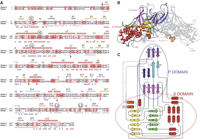
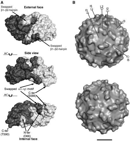
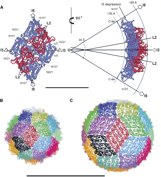
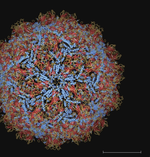
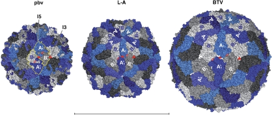
Double-stranded (ds) RNA virus particles are organized around a central icosahedral core capsid made of 120 identical subunits. This core capsid is unable to invade cells from outside, and animal dsRNA viruses have acquired surrounding capsid layers that are used to deliver a transcriptionally active core particle across the membrane during cell entry. In contrast, dsRNA viruses infecting primitive eukaryotes have only a simple core capsid, and as a consequence are transmitted only vertically. Here, we report the 3.4 A X-ray structure of a picobirnavirus--an animal dsRNA virus associated with diarrhoea and gastroenteritis in humans. The structure shows a simple core capsid with a distinctive icosahedral arrangement, displaying 60 two-fold symmetric dimers of a coat protein (CP) with a new 3D-fold. We show that, as many non-enveloped animal viruses, CP undergoes an autoproteolytic cleavage, releasing a post-translationally modified peptide that remains associated with nucleic acid within the capsid. Our data also show that picobirnavirus particles are capable of disrupting biological membranes in vitro, indicating that its simple 120-subunits capsid has evolved animal cell invasion properties.
Figures
Similar articles
-
Cryo-electron Microscopy Structure, Assembly, and Mechanics Show Morphogenesis and Evolution of Human Picobirnavirus.J Virol. 2020 Nov 23;94(24):e01542-20. doi: 10.1128/JVI.01542-20. Print 2020 Nov 23. J Virol. 2020. PMID: 32938763 Free PMC article.
-
The T=1 capsid protein of Penicillium chrysogenum virus is formed by a repeated helix-rich core indicative of gene duplication.J Virol. 2010 Jul;84(14):7256-66. doi: 10.1128/JVI.00432-10. Epub 2010 May 12. J Virol. 2010. PMID: 20463071 Free PMC article.
-
Heterodimers as the Structural Unit of the T=1 Capsid of the Fungal Double-Stranded RNA Rosellinia necatrix Quadrivirus 1.J Virol. 2016 Nov 28;90(24):11220-11230. doi: 10.1128/JVI.01013-16. Print 2016 Dec 15. J Virol. 2016. PMID: 27707923 Free PMC article.
-
Assembly of large icosahedral double-stranded RNA viruses.Adv Exp Med Biol. 2012;726:379-402. doi: 10.1007/978-1-4614-0980-9_17. Adv Exp Med Biol. 2012. PMID: 22297523 Review.
-
Architecture and Assembly of Structurally Complex Viruses.Subcell Biochem. 2024;105:431-467. doi: 10.1007/978-3-031-65187-8_12. Subcell Biochem. 2024. PMID: 39738954 Review.
Cited by
-
Plate tectonics of virus shell assembly and reorganization in phage φ8, a distant relative of mammalian reoviruses.Structure. 2013 Aug 6;21(8):1384-95. doi: 10.1016/j.str.2013.06.017. Epub 2013 Jul 25. Structure. 2013. PMID: 23891291 Free PMC article.
-
Structure of Fusarium poae virus 1 shows conserved and variable elements of partitivirus capsids and evolutionary relationships to picobirnavirus.J Struct Biol. 2010 Dec;172(3):363-71. doi: 10.1016/j.jsb.2010.06.022. Epub 2010 Jul 3. J Struct Biol. 2010. PMID: 20599510 Free PMC article.
-
Asparagine peptide lyases: a seventh catalytic type of proteolytic enzymes.J Biol Chem. 2011 Nov 4;286(44):38321-38328. doi: 10.1074/jbc.M111.260026. Epub 2011 Aug 8. J Biol Chem. 2011. PMID: 21832066 Free PMC article.
-
Epidemiology, phylogeny, and evolution of emerging enteric Picobirnaviruses of animal origin and their relationship to human strains.Biomed Res Int. 2014;2014:780752. doi: 10.1155/2014/780752. Epub 2014 Jul 17. Biomed Res Int. 2014. PMID: 25136620 Free PMC article. Review.
-
Structure of a protozoan virus from the human genitourinary parasite Trichomonas vaginalis.mBio. 2013 Apr 2;4(2):e00056-13. doi: 10.1128/mBio.00056-13. mBio. 2013. PMID: 23549915 Free PMC article.
References
-
- Agrawal DK, Johnson JE (1992) Sequence and analysis of the capsid protein of Nudaurelia capensis omega virus, an insect virus with T=4 icosahedral symmetry. Virology 190: 806–814 - PubMed
-
- An W (2007) Histone acetylation and methylation: combinatorial players for transcriptional regulation. Subcell Biochem 41: 351–369 - PubMed
-
- Bamford DH, Grimes JM, Stuart DI (2005) What does structure tell us about virus evolution? Curr Opin Struct Biol 15: 655–663 - PubMed
-
- Bhattacharya R, Sahoo GC, Nayak MK, Rajendran K, Dutta P, Mitra U, Bhattacharya MK, Naik TN, Bhattacharya SK, Krishnana T (2007) Detection of Genogroup I and II human picobirnaviruses showing small genomic RNA profile causing acute watery diarrhoea among children in Kolkata, India. Infect Genet Evol 7: 229–238 - PubMed
-
- Brunger AT, Adams PD, Clore GM, DeLano WL, Gros P, Grosse-Kunstleve RW, Jiang JS, Kuszewski J, Nilges M, Pannu NS, Read RJ, Rice LM, Simonson T, Warren GL (1998) Crystallography & NMR system: a new software suite for macromolecular structure determination. Acta Crystallogr 54: 905–921 - PubMed
Publication types
MeSH terms
Substances
Associated data
- Actions
LinkOut - more resources
Full Text Sources
Miscellaneous
