

Presence of cancer-associated fibroblasts inversely correlates with Schwannian stroma in neuroblastoma tumors
- PMID: 19407854
- PMCID: PMC3347894
- DOI: 10.1038/modpathol.2009.52
Presence of cancer-associated fibroblasts inversely correlates with Schwannian stroma in neuroblastoma tumors
Abstract
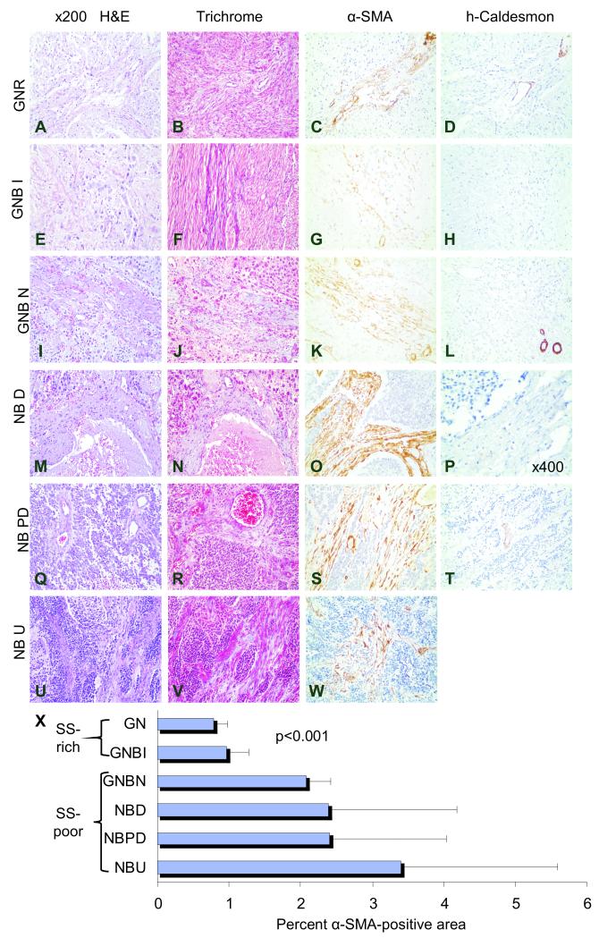
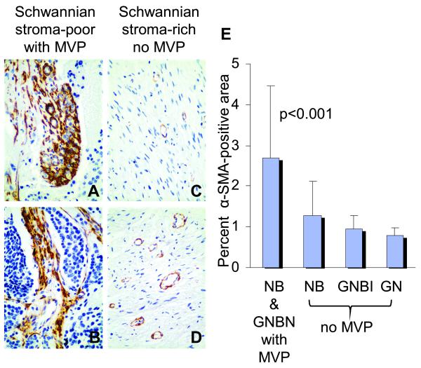
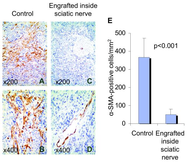
Stromal cells have a central function in the regulation of tumor angiogenesis. Recent studies have shown that stromal myofibroblasts (cancer-associated fibroblasts) actively promote tumor growth and enhance tumor angiogenesis in many types of adult carcinomas. To evaluate the function cancer-associated fibroblasts have in neuroblastoma angiogenesis and investigate their relationship to stromal Schwann cells, we quantified cancer-associated fibroblasts in 60 primary neuroblastoma tumors and in a novel neuroblastoma xenograft model in which murine Schwann cells were induced to infiltrate into the tumor stroma. Tumor sections were examined for presence of microvascular proliferation, a hallmark of tumor angiogenesis. Cancer-associated fibroblasts were characterized by positive immunostaining for alpha-smooth muscle actin (alpha-SMA) and were distinguished from pericytes by staining negatively for high-molecular-weight caldesmon. alpha-SMA-positive cells were quantified and their number was defined as high when >1.0% of the area was positive. Associations between high cancer-associated fibroblast number, microvascular proliferation and established prognosticators were analyzed. High numbers of cancer-associated fibroblasts were associated with Schwannian stroma-poor histopathology and microvascular proliferation. Thirty-seven (80%) of the 46 Schwannian stroma-poor tumors had high numbers of cancer-associated fibroblasts in the tumor stroma compared to only 2 (14%) of the 14 Schwannian stroma-rich/dominant tumors (P<0.001). Thirty-three (89%) of 37 tumors with microvascular proliferation had high numbers of cancer-associated fibroblasts compared to 9 (40%) of 22 tumors without microvascular proliferation (P<0.001). In the xenografts with infiltrating Schwann cells (n=10), the number of cancer-associated fibroblasts per mm(2) was approximately sevenfold less than in the control xenografts without stromal Schwann cells (n=9) (mean of 51+/-30 vs 368+/-105, respectively; P<0.001). Thus, cancer-associated fibroblasts were inversely associated with presence of Schwann cells, suggesting that Schwann cells may prevent the activation of fibroblasts. A deeper understanding of the function cancer-associated fibroblasts have in neuroblastoma angiogenesis may guide future development of stroma-directed therapeutic strategies.
Figures
Similar articles
-
'Cross-talk' between Schwannian stroma and neuroblasts promotes neuroblastoma tumor differentiation and inhibits angiogenesis.Cancer Lett. 2005 Oct 18;228(1-2):125-31. doi: 10.1016/j.canlet.2005.01.056. Cancer Lett. 2005. PMID: 15935552 Review.
-
Reconstitution of Schwannian stroma in neuroblastomas using human bone marrow stromal cells.Am J Pathol. 2008 Oct;173(4):1153-64. doi: 10.2353/ajpath.2008.070309. Epub 2008 Sep 4. Am J Pathol. 2008. PMID: 18772334 Free PMC article.
-
Schwann cell-conditioned medium inhibits angiogenesis.Cancer Res. 2000 Nov 1;60(21):5966-71. Cancer Res. 2000. PMID: 11085514
-
Cross-talk between Schwann cells and neuroblasts influences the biology of neuroblastoma xenografts.Am J Pathol. 2005 Mar;166(3):891-900. doi: 10.1016/S0002-9440(10)62309-7. Am J Pathol. 2005. PMID: 15743800 Free PMC article.
-
Fibrocytes: A Novel Stromal Cells to Regulate Resistance to Anti-Angiogenic Therapy and Cancer Progression.Int J Mol Sci. 2017 Dec 29;19(1):98. doi: 10.3390/ijms19010098. Int J Mol Sci. 2017. PMID: 29286323 Free PMC article. Review.
Cited by
-
A narrative review of the role of fibroblasts in the growth and development of neurogenic tumors.Ann Transl Med. 2020 Nov;8(21):1462. doi: 10.21037/atm-20-3218. Ann Transl Med. 2020. PMID: 33313207 Free PMC article. Review.
-
The Immunocytokine FAP-IL-2v Enhances Anti-Neuroblastoma Efficacy of the Anti-GD2 Antibody Dinutuximab Beta.Cancers (Basel). 2022 Oct 4;14(19):4842. doi: 10.3390/cancers14194842. Cancers (Basel). 2022. PMID: 36230765 Free PMC article.
-
COX/mPGES-1/PGE2 pathway depicts an inflammatory-dependent high-risk neuroblastoma subset.Proc Natl Acad Sci U S A. 2015 Jun 30;112(26):8070-5. doi: 10.1073/pnas.1424355112. Epub 2015 Jun 15. Proc Natl Acad Sci U S A. 2015. PMID: 26080408 Free PMC article.
-
Enhancing Neuroblastoma Immunotherapies by Engaging iNKT and NK Cells.Front Immunol. 2020 May 8;11:873. doi: 10.3389/fimmu.2020.00873. eCollection 2020. Front Immunol. 2020. PMID: 32457760 Free PMC article. Review.
-
Artificial Tumor Microenvironments in Neuroblastoma.Cancers (Basel). 2021 Apr 1;13(7):1629. doi: 10.3390/cancers13071629. Cancers (Basel). 2021. PMID: 33915765 Free PMC article. Review.
References
-
- Schurch W, Seemayer T, Hinz B, Gabbiani G. Myofibroblast. In: Mills S, editor. Histology for Pathologists. 3rd ed Lippincott Williams & Wilkins; Philadelphia: 2007. pp. 123–64.
-
- Kalluri R, Zeisberg M. Fibroblasts in cancer. Nat Rev Cancer. 6:392–401. - PubMed
-
- Sugimoto H, Mundel TM, Kieran MW, Kalluri R. Identification of fibroblast heterogeneity in the tumor microenvironment. Cancer Biol Ther. 5:1640–6. - PubMed
-
- Desmouliere A, Guyot C, Gabbiani G. The stroma reaction myofibroblast: a key player in the control of tumor cell behavior. Int J Dev Biol. 48:509–17. - PubMed
-
- Park JE, Lenter MC, Zimmermann RN, Garin-Chesa P, Old LJ, Rettig WJ. Fibroblast activation protein, a dual specificity serine protease expressed in reactive human tumor stromal fibroblasts. J Biol Chem. 274:36505–12. - PubMed
Publication types
MeSH terms
Substances
Grants and funding
LinkOut - more resources
Full Text Sources
Medical
