. 2009 Aug 15;125(4):868-78.
doi: 10.1002/ijc.24452.

CD4+ lymphocytes modulate prostate cancer progression in mice

Affiliations

CD4+ lymphocytes modulate prostate cancer progression in mice

Theofilos Poutahidis et al. Int J Cancer. .

Abstract

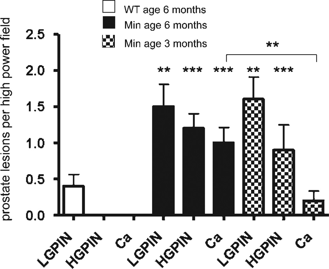
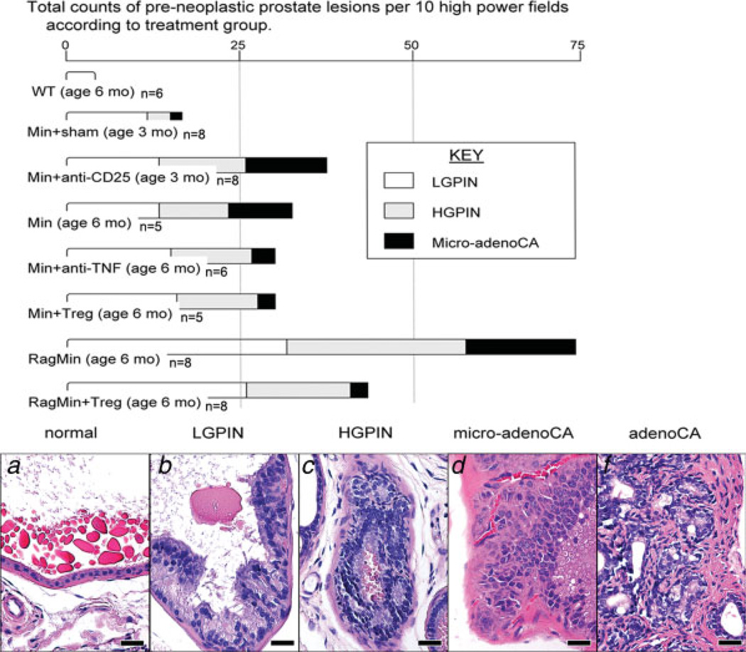
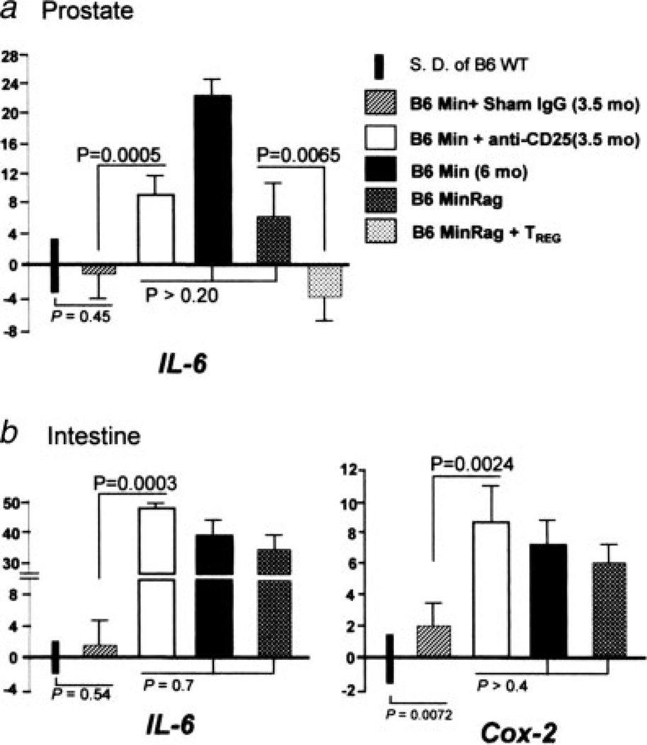
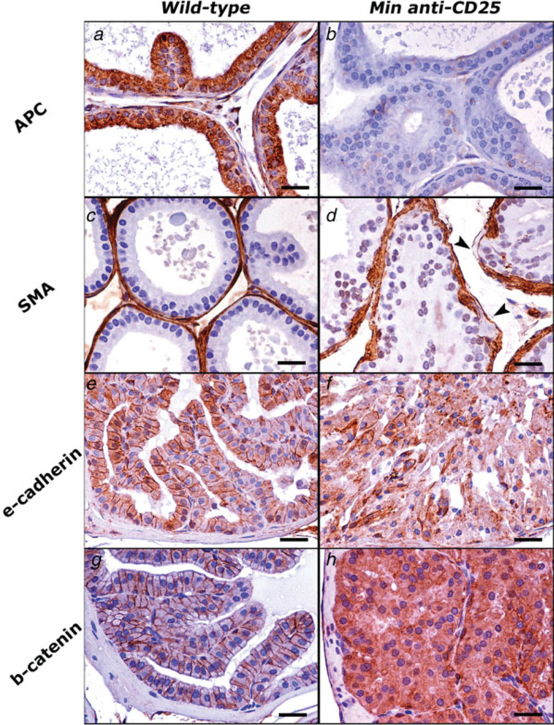
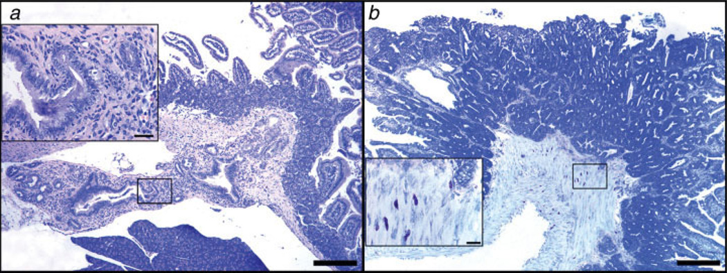
Chronic inflammation contributes to the development of prostate cancer in humans. Here, we show that male Apc(Min/+) mice also develop prostate carcinoma with increasing age, mimicking that seen in humans in their 5th or 6th decade of life. Proinflammatory cytokines were significantly linked with cancer and increasing age in our mouse model; however, prostate and bowel tissues lacked evidence of inflammatory cell infiltrates other than mast cells. Lymphocytes protected against cancer, and protection from prostate cancer resided in antiinflammatory CD4(+)CD25(+) regulatory (T(REG)) cells that downregulated inflammatory cytokines. Supplementation with syngeneic T(REG) cells collected from wild-type mice reduced the levels of interleukin (IL)-6 (p < 0.05) and IL-9 (p < 0.001) and lowered prostate cancer risk (p < 0.05). Depletion of CD25(+) cells in 2-month-old animals increased the expression of IL-6 (p < 0.005) within prostate and increased the frequency of high-grade prostatic intraepithelial neoplasia (p < 0.05) and microinvasive prostatic carcinoma (p < 0.05) in dorsolateral prostate. Depletion of CD25(+) cells in young animals also increased the frequency of intestinal cancer in Min mice. Taken together, chronically elevated proinflammatory cytokines promoted carcinoma in Apc(Min/+) mice. T(REG) lymphocytes downregulated inflammation-associated carcinogenic processes and contributed to immune and epithelial homeostasis.


Figures

Figure 1
Figure 1
Depletion of CD25+ cells increases the risk of prostate cancer in young mice, but complete lack of lymphocytes and increasing age are the major factors contributing to the progression of prostate neoplasia in ApcMin/+ mice. (a, b) HGPIN lesions in the anterior prostate of CD25-depleted Min mice at 3 months of age. Glands in a are distorted, surrounded by irregular fibromuscular sheaths and filled with atypical epithelial cells. The stroma adjacent to gland profile with PIN lesions in b contains neutrophils (open arrow-head) and mast cells (arrow-head). (c, d) Early invasive neoplastic lesions in the dorsolateral prostate of a RagMin mouse at 6 months of age. Highly irregular glands profiles with tufting epithelia and edematous stromal reaction (c). Higher magnification of the boxed area is shown in D. Early invasion of neoplastic epithelium is found in association with HGPIN. Note the hyperchromasia and the nuclear pleomorphism of the epithelial cells in PIN lesions (open arrow) and the large, euchromatinic nuclei bearing prominent nucleoli of invasive epithelial structures (arrow). (e, f) Prostate adenocarcinoma in the dorsolateral prostate of Min mice at 6 months of age. Moderately differentiated adenocarcinoma with desmoplastic reaction. Abnormal mitotic figure is indicated by arrow-head (e). Nuclear stabilization of β-catenin characterized advanced prostate adenocarcinoma lesions (f). (g, h) Prostate adenocarcinoma in the dorsolateral prostate of Min mice at 6 months of age. Well-differentiated adenocarcinoma with prominent desmoplasia (g). Increased numbers of mast cells (arrow-head) in stroma associate with abnormal glands (h). (a, b, c, d, e, g, h) Hematoxylin and Eosin. (f) β-catenin-specific immunohistochemistry; Hematoxylin counterstain, DAB chromogen. Bars a, c and g: 100 µm; b, d, e, f and h: 25 µm.
Figure 2
Figure 2
Frequency of prostate carcinoma lesions rises with increasing age. Prostate carcinoma arose spontaneously in male Min mice but not wt mice. Aged (6 months old) Min mice (N = 5 mice per trial) had greater frequency of early neoplastic prostate lesions, when compared to younger (3 months old) Min mice (N = 5 per trial). Pre-neoplastic and early neoplastic prostate lesions, which were detected based on recent consensus report criteria, were quantitatively assessed for each group of mice based upon review of 30 high-power fields ×20 (data presented as mean value) containing the glandular profiles low-grade PIN, high-grade PIN and microinvasive carcinoma as shown earlier. The most advanced preneoplastic and early neoplastic lesions were captured using a Nikon eclipse 50i microscope and a Nikon DS-5 M-L1 digital camera. Ten images were randomly selected per treatment group. Lesions were then quantified and recorded as the number of low-grade PIN, high-grade PIN and microinvasive carcinoma foci counted per image.
Figure 3
Figure 3
(a) Frequency of prostate cancer lesions in Min and Rag-deficient Min mice. Depletion of CD25+ cells (Min+anti-CD25) (N = 8 mice per trial) accelerated prostate carcinogenesis at age of 3 months, and the frequency of HGPIN and microinvasive adenocarcinoma was significantly higher than those of age-matched (N = 5 mice per trial) or sham-treated (N = 8) mice (p < 0.01 and <0.05, respectively). Compared to the age-matched Min mice (N = 5), RagMin mice (N = 8) had significantly higher frequency of prostate neoplasia at 6 months old (p < 0.05). Assays used tissues from 5 to 8 mice per treatment group (as shown) with review of microscopic fields as described in Material and methods. (b) Histology of dorsolateral prostate illustrating selected key intermediate steps in progression of carcinogenesis in ApcMin/+ mice. (a) Normal-appearing prostate gland. (b) Low-grade prostatic intraepithelial neoplasia (PIN) with epithelial tufting and nuclear stratification. Cellular atypia is evidenced by the presence of nuclear enlargement, increased but not severe nuclear pleomorphism, hyperchromasia and occasional prominent nucleoli. (c) High-grade PIN. Note the irregular contour of the prostate gland. Highly atypical cells with severe nuclear pleomorphism and hyperchromasia fill the lumen. (d) Microinvasive carcinoma. Epithelial cells with notably large, euchromatinic nuclei bearing prominently enlarged nucleoli with penetration through the basement membrane into surrounding stroma. (e) Invasive adenocarcinoma. Moderately differentiated small, irregular malignant glands are bounded by desmoplastic stroma. Hematoxylin and Eosin. Bars: 25 µm. [Color figure can be viewed in the online issue, which is available at www.interscience.wiley.com.]
Figure 4
Figure 4
mRNA expression levels of IL-6 and Cox-2 in the murine prostate and bowel. There were significant increases in expression of IL-6 in (a) prostate and (b) ileum of Min mice treated with anti-CD25 antibody, and also in Rag-deficient Min mice, when compared to sham-treated Min counterparts. Supplementation with TREG cells from wt donor mice decreased expression of IL-6 in prostate tissue, whereas depletion of CD25+ cells increased IL-6 gene expression in prostate tissue. Assay used tissues from 5 to 8 mice per treatment group. For comparison of mRNA levels, the target mRNA was normalized to that of the housekeeping gene GAPDH. Numbers on the y-axis represent mean fold change of target mRNA levels in reference to the control levels (B6 wt, defined as 0, standard deviation represented by solid bars). mos, age in months upon necropsy.
Figure 5
Figure 5
Immunohistochemical staining illustrates critical steps of carcinogenesis in the Min mouse model of prostate carcinoma. The aberrant immunostaining pattern observed in PIN lesions of Min mice that were depleted of CD25+ cells (right row) can be appreciated by comparison with normal prostate epithelia of wildtype mice (left row). (a–d) Dorsolateral prostate. (e–h) Anterior prostate. (a, b) The normal cytoplasmic expression of Apc (A) is absent in Min mouse prostate shown in b. (c, d) SMA immunohistochemistry highlights the fibromuscular sheath of prostate glands. Compare the smooth and continuous contour of normal prostate gland in c with the irregular and discontinuous (arrow-heads) sheath of prostate gland in d. (e, f) Normal lateral epithelial cell membrane staining pattern of E-cadherin in normal prostate (e) is by large lost in HGPIN shown in f. (g, h) Normal immunolabeling of β-catenin is largely restricted to adherence junctions in the prostate of wildtype mice (g). HGPIN lesions (h) instead of normal staining pattern show intense cytoplasmic staining, which indicates accumulation of β-catenin in the cytoplasm of abnormal prostate epithelial cells. Hematoxylin counterstain, DAB chromogen. Bars: 25 µm.
Figure 6
Figure 6
Depletion of lymphocytes correlates with an invasive neoplastic phenotype in the small intestine of Apcmin/+mice. (a) Ampullary cancer arising from pancreatic duct epithelium in 25% (2/8) of 3-month-old ApcMin/+ mice after undergoing depletion of CD25+ cells (N = 8 mice per trial). High magnification (inset in a) reveals features of the abnormal pancreatic duct epithelium including pseudostratification and nuclear pleomorphism with associated inflammation. (b) Ileum of Rag2−/− ApcMin/+ mouse at 6 months of age revealing submucosal and incipient tunica muscularis invasion of neoplastic glands in an adenomatous polyp. Large numbers of mast cells infiltrate the submucosa and muscle layers at the base of the polyp. The higher magnification (inset) reveals the close topographic association of mast cells with the invasive front of the tumor. (a) Hematoxylin and Eosin. (b) Toluidine Blue. Bars a and b: 250 µm; insets: 25 µm. [Color figure can be viewed in the online issue, which is available at www.interscience.wiley.com.]

Similar articles

Cited by

References

    1. Jemal A, Siegel R, Ward E, Murray T, Xu J, Smigal C, Thun MJ. Cancer statistics, 2006. CA Cancer J Clin. 2006;56:106–130. - PubMed
    1. Sakr WA, Grignon DJ, Crissman JD, Heilbrun LK, Cassin BJ, Pontes JJ, Haas GP. High grade prostatic intraepithelial neoplasia (HGPIN) and prostatic adenocarcinoma between the ages of 20–69: an autopsy study of 249 cases. In Vivo. 1994;8:439–443. - PubMed
    1. Nelson WG, De Marzo AM, Isaacs WB. Prostate cancer. N Engl J Med. 2003;349:366–381. - PubMed
    1. Balkwill F, Mantovani A. Inflammation and cancer: back to Virchow? Lancet. 2001;357:539–545. - PubMed
    1. Coussens LM, Werb Z. Inflammation and cancer. Nature. 2002;420:860–867. - PMC - PubMed

Publication types

MeSH terms