

doi: 10.1021/nl900437n.
Functionalized carbon nanotubes specifically bind to alpha-chymotrypsin's catalytic site and regulate its enzymatic function
Affiliations
- PMID: 19408924
- PMCID: PMC2814308
- DOI: 10.1021/nl900437n
Item in Clipboard
Functionalized carbon nanotubes specifically bind to alpha-chymotrypsin's catalytic site and regulate its enzymatic function
Nano Lett.
2009 Jun.
Abstract
Although carbon nanotubes (CNTs) have been shown to nonspecifically bind proteins through charge complementary, pi-pi stacking or hydrophobic interactions, they have not been shown to bind to a specific site on proteins. By generating surface molecular diversity, we created functionalized CNTs that recognize and bind to the catalytic site of alpha-chymotrypsin and inhibit its enzymatic activity competitively.
Figures
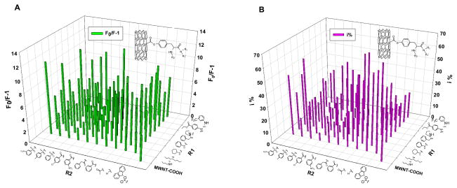
The quenching of ChT’s fluorescence and inhibition of its enzymatic activity by f-MWNTs. The fluorescence quenching expressed as F0/(F-1) and enzyme inhibition (i %) are presented in a library format arranged as two dimensions of diversities of the surface molecules. Steady fluorescence spectra experimental condition: ChT concentration was 2 μM in 25 mM Tris-HCl buffer solution (pH~7.6), and final concentration of f-MWNTs in ChT solutions was 10 μg/mL. Enzyme inhibition experimental condition: final f-MWNTs’ concentration was 40 μg/mL and the ChT concentration was 3.2 μM (i% = (1 − νi/ν0) × 100%, where νi, ν0 represent the enzyme reaction velocity initial and after adding f-MWNT respectively).
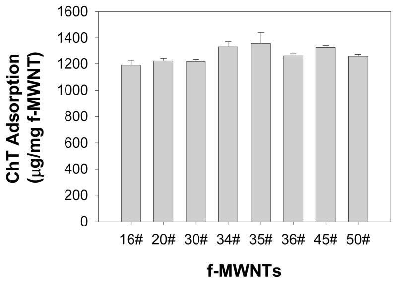
Quantification of adsorbed ChT onto MWNTs by HPLC assay. The free ChT was determined by HPLC based on a standard calibration curve. The adsorbed proteins were derived from the difference between the total proteins used in the assay and the free proteins. Error bars are the standard deviations from three measurements. The final f-MWNTs’ concentration was 150 μg/mL and the ChT concentration was 250 μg/mL. Experimental details are in section 1.5 of Supporting Information.
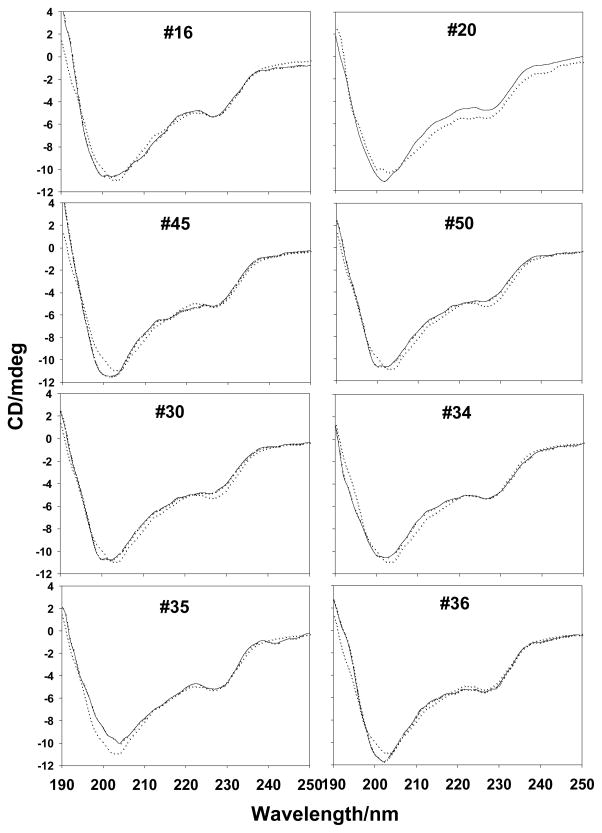
CD spectra of ChT in the absence or presence of f-MWNTs. The solid line was for ChT and doted line for ChT in the presence of f-MWNTs. The ChT concentration was 2μM in 25 mM Tris-HCl buffer solution (pH~7.6). The final concentration of f-MWNTs was 10 μg/mL.
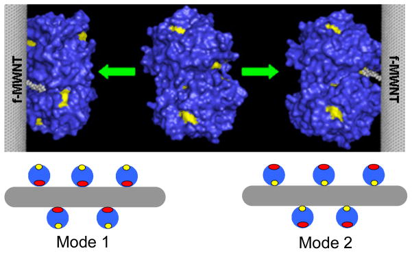
Models showing different ways f-MWNTs bind to ChT. ChT structure was taken from Protein Data Bank. Mode 1: competitive inhibition; mode 2: non-competitive inhibition. Red: catalytic site; yellow: tryptophan fluorophores.
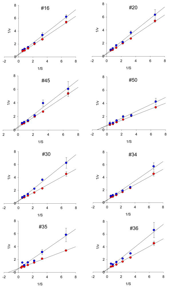
Lineweaver–Burk plots of ChT in the presence (red circle) and absence (blue circle) of f-MWNTs. Final concentration of f-MWNTs was 10 μg/mL, and concentrations of SPNA were 0.15, 0.45, 0.75, 1.2, 1.8, 2.4mM, respectively.
References
Publication types
MeSH terms
Substances
Grants and funding
LinkOut - more resources
Full Text Sources
Research Materials
Miscellaneous
