. 2009 May 1:6:20.
doi: 10.1186/1743-7075-6-20.

White Tea extract induces lipolytic activity and inhibits adipogenesis in human subcutaneous (pre)-adipocytes

Affiliations

White Tea extract induces lipolytic activity and inhibits adipogenesis in human subcutaneous (pre)-adipocytes

Jörn Söhle et al. Nutr Metab (Lond). .

Abstract

Background: The dramatic increase in obesity-related diseases emphasizes the need to elucidate the cellular and molecular mechanisms underlying fat metabolism. To investigate how natural substances influence lipolysis and adipogenesis, we determined the effects of White Tea extract on cultured human subcutaneous preadipocytes and adipocytes.

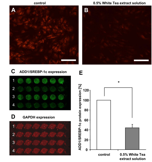
Methods: For our in vitro studies we used a White Tea extract solution that contained polyphenols and methylxanthines. Utilizing cultured human preadipocytes we investigated White Tea extract solution-induced inhibition of triglyceride incorporation during adipogenesis and possible effects on cell viability. In vitro studies on human adipocytes were performed aiming to elucidate the efficacy of White Tea extract solution to stimulate lipolytic activity. To characterize White Tea extract solution-mediated effects on a molecular level, we analyzed gene expression of essential adipogenesis-related transcription factors by qRT-PCR and determined the expression of the transcription factor ADD1/SREBP-1c on the protein level utilizing immunofluorescence analysis.

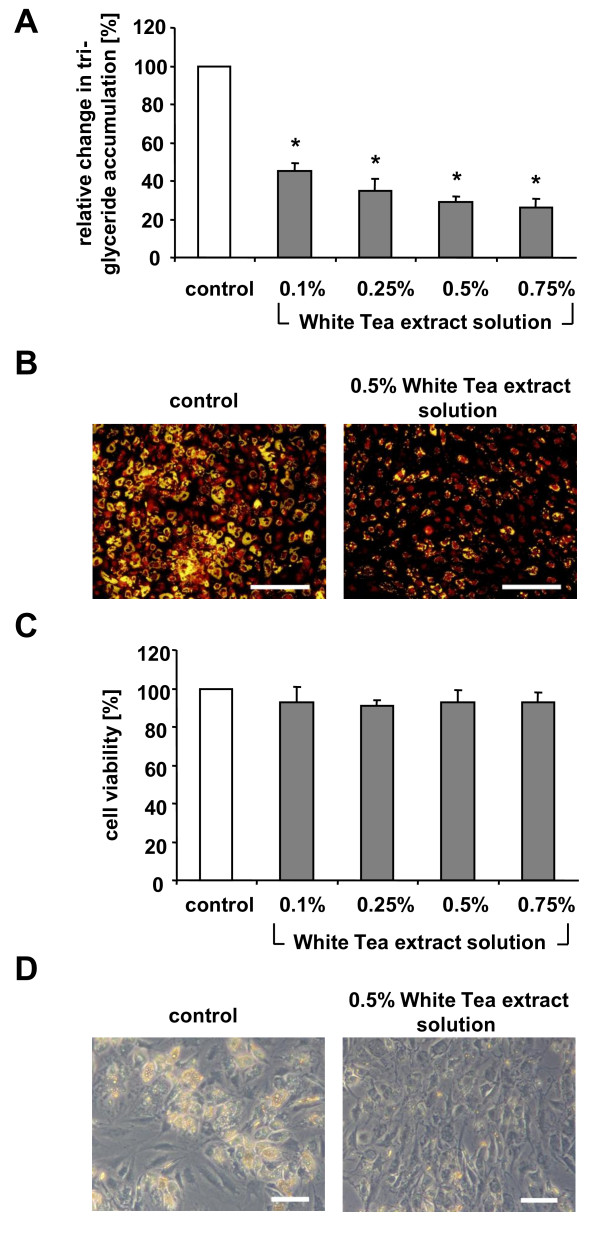
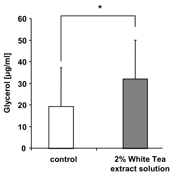
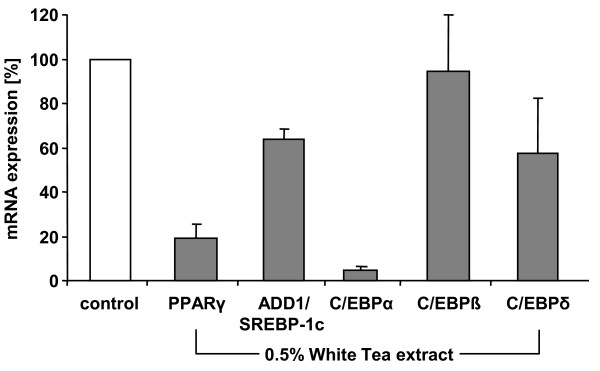
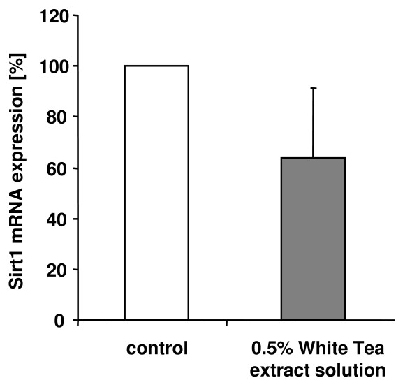
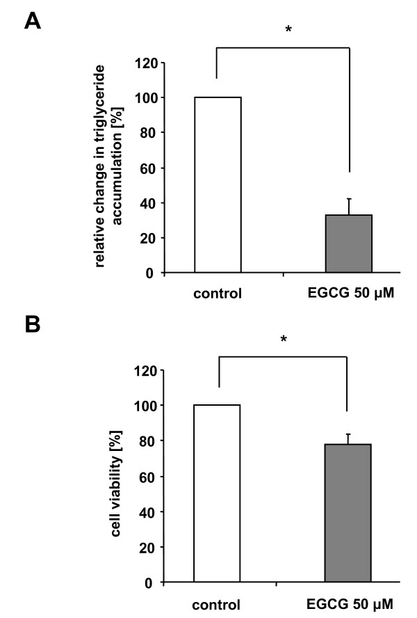
Results: Our data show that incubation of preadipocytes with White Tea extract solution significantly decreased triglyceride incorporation during adipogenesis in a dose-dependent manner (n = 10) without affecting cell viability (n = 10). These effects were, at least in part, mediated by EGCG (n = 10, 50 μM). In addition, White Tea extract solution also stimulated lipolytic activity in adipocytes (n = 7). Differentiating preadipocytes cultivated in the presence of 0.5% White Tea extract solution showed a decrease in PPARγ, ADD1/SREBP-1c, C/EBPα and C/EBPδ mRNA levels. Moreover, the expression of the transcription factor ADD1/SREBP-1c was not only decreased on the mRNA but also on the protein level.

Conclusion: White Tea extract is a natural source that effectively inhibits adipogenesis and stimulates lipolysis-activity. Therefore, it can be utilized to modulate different levels of the adipocyte life cycle.


Figures

Figure 1
Figure 1
Effects of White Tea extract solution on triglyceride accumulation during preadipocyte/adipocyte differentiation. (A) Triglyceride accumulation and (C) cell viability of maturing preadipocytes incubated with different amounts of White Tea extract solutions (0.1%, 0.25%, 0.5% and 0.75%) is shown relative to untreated control cells set as 100%. For experiments, ten independent cell cultures were prepared both for control and White Tea extract solution incubation (n = 10). Results are depicted as mean ± SD. Significant differences are marked with an asterisk (* for p < 0.0001). (B) Images after triglyceride staining (yellow) from cell populations incubated with and without 0.5% White Tea extract solution. Scale bar: 200 μm. (D) Displayed are phase contrast images from cell populations cultivated in the absence or presence of 0.5% White Tea extract solution. Scale bar: 50 μm. For our studies we used cells up to the third passage.
Figure 2
Figure 2
Determination of glycerol release in differentiated adipocytes after incubation with White Tea extract solution. Glycerol content is given in μg/ml. For experiments, seven independent cell cultures were prepared both for control and White Tea extract solution incubation (n = 7). Results are depicted as mean ± SD. Significant differences are marked with an asterisk (* for p ≤ 0.05). For our studies we used cells up to the third passage.
Figure 3
Figure 3
Effect of White Tea extract solution on ADD1/SREBP-1c expression during adipogenesis. Preadipocyte populations (0 d) were incubated with differentiation medium without (A) and with (B) 0.5% White Tea extract solution for 10 days. A and B: ADD1/SREBP-1c was detected by immunofluorescence microscopic analysis. Scale bar: 200 μm. C-E: Quantitative analyses were performed using the Odyssey Infrared Imager. 1 and 3: Cells incubated in control solution. 2 and 4: Cells incubated in the presence of White Tea extract solution. For experiments, 17 independent cell cultures were prepared both for control and White Tea extract solution incubation (n = 17). Results are depicted as mean ± SD. Significant differences are marked with an asterisk (* for p < 0.0001).
Figure 4
Figure 4
Effects of White Tea extract solution on gene expression of adipogenesis-associated transcription factors. Gene expression of PPARγ, ADD1/SREBP-1c, C/EBPα, C/EBPβ and C/EBPδ in differentiating preadipocytes after incubation with White Tea extract solution compared to control cells set as 100%. Expression of each gene is normalized to GAPDH. Three independent experiments were prepared both for control and White Tea extract solution incubation (n = 3). Results are depicted as mean ± SD. For our studies we used cells up to the third passage.
Figure 5
Figure 5
White Tea extract solution decreases Sirt1 gene expression. Sirt1 gene expression in differentiating preadipocytes after incubation with 0.5% White Tea extract solution is shown relative to untreated control cells set as 100%. Gene expression is normalized to GAPDH. Three independent experiments were prepared both for control and White Tea extract solution incubation (n = 3). Results are depicted as mean ± SD. For our studies we used cells up to the third passage.
Figure 6
Figure 6
Decrease in triglyceride concentration and cell viability in differentiating preadipocytes after stimulation with EGCG. (A) Triglyceride content in differentiating preadipocytes after stimulation with 50 μM EGCG is shown relative to untreated control cells set as 100%. For experiments, ten independent cell cultures were prepared both for control and EGCG incubation (n = 10). Results are depicted as mean ± SD. Significant differences are marked with an asterisk (* for p < 0.0001). (B) Decreased viability in differentiating preadipocytes after stimulation with 50 μM EGCG compared to control cells. Cell viability is shown relative to untreated control cells set as 100%. For experiments, ten independent cell cultures were prepared both for control and EGCG incubation (n = 10). Results are depicted as mean ± SD. Significant differences are marked with an asterisk (* for p < 0.0001). For our studies we used cells up to the third passage.

Similar articles

Cited by

References

    1. Gregoire FM. Adipocyte differentiation: From fibroblast to endocrine cell. Exp Biol Med (Maywood) . 2001;226:997–1002. - PubMed
    1. Rosen ED, Walkey CJ, Puigserver P, Spiegelman BM. Transcriptional regulation of adipogenesis. Genes De. 2000;14:1293–1307. - PubMed
    1. Avram MM, Sharpe Avram A, James WJ. Subcutaneous fat in normal and diseased states 3. Adipogenesis: From stem cell to fat cell. J Am Acad Dermatol. 2007;56:472–492. doi: 10.1016/j.jaad.2006.06.022. - DOI - PubMed
    1. Hirsch J, Batchelor B. Adipose tissue cellularity in human obesity. Clin Endocrinol Metab. 1976;5:299–311. doi: 10.1016/S0300-595X(76)80023-0. - DOI - PubMed
    1. Rodeheffer MS, Birsoy K, Friedman JM. Identification of white adipocyte progenitor cells in vivo. Cell. 2008;135:240–249. doi: 10.1016/j.cell.2008.09.036. - DOI - PubMed

LinkOut - more resources