. 2009 May 1;16(1):44.
doi: 10.1186/1423-0127-16-44.

Sympathetic-correlated c-Fos expression in the neonatal rat spinal cord in vitro

Affiliations

Sympathetic-correlated c-Fos expression in the neonatal rat spinal cord in vitro

Chun-Kuei Su et al. J Biomed Sci. .

Abstract

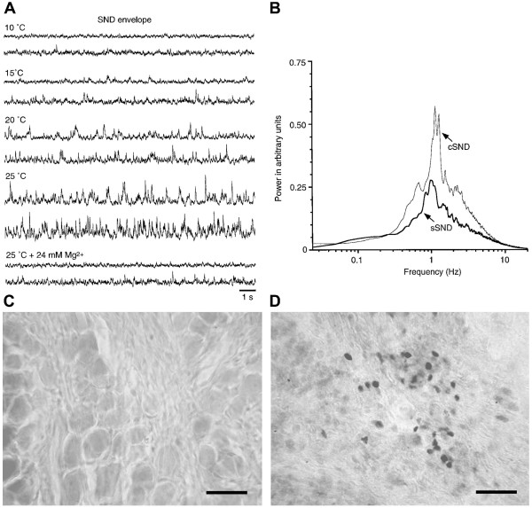
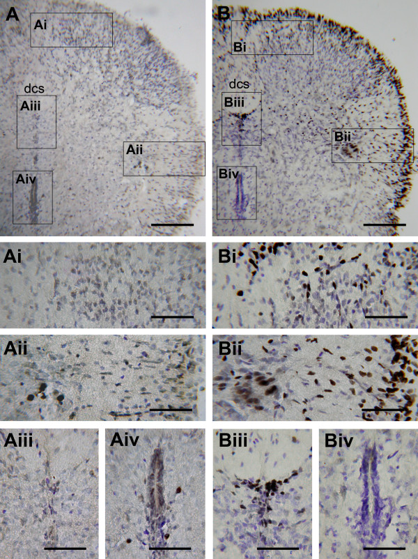
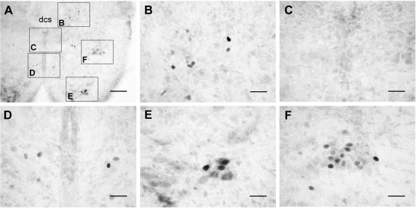
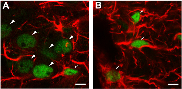
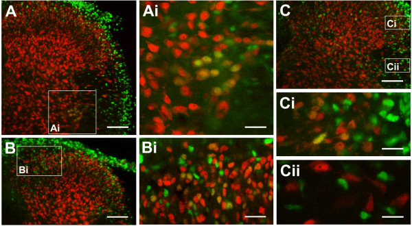
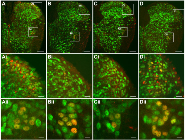
An isolated thoracic spinal cord of the neonatal rat in vitro spontaneously generates sympathetic nerve discharge (SND) at ~25 degrees C, but it fails in SND genesis at < or = 10 degrees C. Basal levels of the c-Fos expression in the spinal cords incubated at < or = 10 degrees C and ~25 degrees C were compared to determine the anatomical substrates that might participate in SND genesis. Cells that exhibited c-Fos immunoreactivity were virtually absent in the spinal cords incubated at < or = 10 degrees C. However, in the spinal cords incubated at ~25 degrees C, c-Fos-positive cells were found in the dorsal laminae, the white matter, lamina X, and the intermediolateral cell column (IML). Cell identities were verified by double labeling of c-Fos with neuron-specific nuclear protein (NeuN), glial fibrillary acidic protein (GFAP), or choline acetyltransferase (ChAT). The c-Fos-positive cells distributed in the white matter and lamina X were NeuN-negative or GFAP-positive and were glial cells. Endogenously active neurons showing c-Fos and NeuN double labeling were scattered in the dorsal laminae and concentrated in the IML. Double labeling of c-Fos and ChAT confirmed the presence of active sympathetic preganglionic neurons (SPNs) in the IML. Suppression of SND genesis by tetrodotoxin (TTX) or mecamylamine (MECA, nicotinic receptor blocker) almost abolished c-Fos expression in dorsal laminae, but only mildly affected c-Fos expression in the SPNs. Therefore, c-Fos expression in some SPNs does not require synaptic activation. Our results suggest that spinal SND genesis is initiated from some spontaneously active SPNs, which are capable of TTX- or MECA-resistant c-Fos expression.


Figures

Figure 1
Figure 1
Temperature-dependent features for in vitro nerve-cord preparations to generate sympathetic nerve discharge (SND). (A) Original traces of the SND envelope. Incubation temperature is indicated at the top of each panel. Upper and lower traces in each panel show envelopes of cervical SND (cSND) and splanchnic SND (sSND), respectively. SNDs were discernible at ≥ 20°C. Adding 24 mM Mg2+ to the bath solution at 25°C reduced SNDs to the level at 10°C. (B) Power spectra of cSND and sSND envelopes at 25°C. Spectra were averaged from 21 experiments. Power spectra of both SNDs were similar, showing a peak at ~1 Hz. Photomicrographs in (C) and (D) show c-Fos protein expression in the dorsal root ganglion and celiac ganglion, respectively. The observation was obtained from a nerve-cord-ganglion preparation that incubated under optimal in vitro conditions for SND genesis (25°C normal aCSF, 3 h). c-Fos-positive cells were absent in the dorsal root ganglion (C), but present in the celiac sympathetic ganglion (D). Scale bars = 30 μm in (C, D).
Figure 2
Figure 2
Time course of c-Fos expression in the cords incubated in 25°C normal aCSF. Photomicrographs show transverse sections of T3 from three preparations with different incubation time in hours: 1.5 (A), 3 (B), and 6 (C). After 1.5 h incubation, limited cells with intense c-Fos immunoreactivity (IR) were found in the IML (Ai). As the incubation time was prolonged to 3 h, c-Fos-positive cells in the IML increased (Bi), and were comparably abundant in the cord after the 6 h incubation (Ci). Note the presence of c-Fos-positive cells in the ventral horn and around the cc in (A) and the absence of these cells in (B, C). Also note the time-dependent spread of cell distribution to medial ventrolateral regions of the cord. Histological abbreviations in this and following figures: cc, central canal; dcs, dorsal corticospinal tract; IML, intermediolateral cell column; lfu, lateral funiculus. Scale bars = 150 μm in (A-C) and 30 μm in (Ai-Ci). Detailed distributions of c-Fos-positive cells in the cord are shown in following figures.
Figure 3
Figure 3
Temperature-dependent in vitro c-Fos expression in the cord. Tissue sections were counterstained with cresyl violet. Photomicrographs show the dorsal quadrant of transverse sections of T3 from the cord with 3 h incubation in ≤ 10°C (A) or 25°C (B) normal aCSF. In the cord incubated at ≤ 10°C, only a few c-Fos-positive cells were found in the IML (Aii) and around the cc (Aiv). In the cord incubated at 25°C, abundant c-Fos-positive cells concentrated in the IML (Bii) and the lamina X (Biii); some were scattered throughout the dorsal laminae (Bi) or the lfu (Bii), and accumulated in the outer rim of the white matter (B, Bii). A few c-Fos positive cells scattered around the cc were observed in the cord incubated at ≤ 10°C (Aiv), which were absent when the cord was incubated at 25°C (Biv). Scale bars = 150 μm in (A, B) and 60 μm in (Ai-iv, Bi-iv).
Figure 4
Figure 4
In vivo c-Fos expression in the cord. Basal levels of c-Fos expression under normal physiological conditions were promptly preserved by a transcardial perfusion of fixative. (A) Photomicrograph of a transverse section of the T3 spinal cord segment. Insets in (A) are magnified as indicated. c-Fos-positive nuclei were distributed in the dorsal laminae near the dcs (B), around the cc (D), in the ventral horn (E), and in the IML (F). No c-Fos-positive cells were found in lamina X (C), the lfu, or the outer rim of the white matter (A). Scale bars = 100 μm in (A) and 25 μm in (B-F).
Figure 5
Figure 5
Confocal images of a transverse section of T6 spinal cord segment, showing c-Fos (FITC) and GFAP (rhodamine) labeling in the IML (A) and the lfu (B). Arrowheads: neuronal nuclei, indicating isolated nuclear c-Fos-IR not surrounded by fibrillary GFAP-IR. Arrows: neuroglial nuclei, indicating nuclear c-Fos-IR in close proximity to fibrillary GFAP-IR. (A) Concentrated c-Fos-positive neurons with large round nuclei (arrowheads) in the IML. The neuroglial cells have small slender nuclei (arrows; one at the upper right is only faintly labeled for c-Fos-IR). (B) c-Fos-positive neuroglial cells in the lfu. Here, nuclear c-Fos-IR is intimately embedded in GFAP-IR (arrows). Scale bars = 10 μm in (A, B).
Figure 6
Figure 6
Distribution of c-Fos-positive neurons in the dorsal laminae and the IML. Photomicrographs show transverse sections of T3 (A, B) and T9 (C) spinal cord segments. Nuclei with double labeling for c-Fos-IR (FITC) and NeuN-IR (rhodamine) were found in the IML (A, Ai), the internal dorsal laminae (B, Bi) and the external dorsal laminae (C, Ci). All c-Fos-positive nuclei in the lfu and the outer rim of the white matter were NeuN-negative (Cii). Scale bars = 100 μm in (A-C), 35 μm in (Ai, Bi), and 20 μm in (Ci-ii).
Figure 7
Figure 7
Colocalization of c-Fos-IR (rhodamine) and ChAT-IR (FITC) to demonstrate that c-Fos-positive neurons in the IML are cholinergic sympathetic preganglionic neurons (SPNs). Insets in (A, B) are magnified as indicated. (A) Low magnification photomicrograph of the dorsal quadrant of a transverse section of T3 spinal cord segment. A triangular cluster of IML cells showed intense ChAT-IR that embraced nuclei labeled with c-Fos-IR (Ai). Distribution of ChAT-positive cells extended dorsomedially toward the intermediate zone of lamina X (Aii), where localizations of ChAT-IR and c-Fos-IR were separate. In (Aii), open arrowheads on Top indicate c-Fos-positive ChAT-negative cells in the dorsal lamina X; filled arrowheads at Bottom indicate nuclei with mild c-Fos-IR surrounded by diffusive ChAT-IR in the ventral lamina X. Arrows indicate ChAT-positive c-Fos-negative cells in the intermediate zone. (B) Low magnification photomicrograph of the ventral quadrant of a transverse section of T5 spinal cord segment. ChAT-IR was present in the IML and the ventral horn; only in the IML, ChAT-IR was colocalized with c-Fos-IR (Bi). (C) High magnification photomicrograph of a horizontal section of T7-T8 spinal cord segments, showing c-Fos-positive nuclei in close proximity to cytoplasmic-like ChAT-IR in the IML. Scale bars = 100 μm in (A, B), 25 μm in (Ai-ii), and 20 μm in (Bi, C).
Figure 8
Figure 8
Distribution of c-Fos-positive neurons in the cords incubated in normal aCSF (A) or aCSF containing 0.5 μM TTX (B), 20 μM MECA (C), or 800 μM KYN (D). c-Fos-positive neurons are revealed by double labeling of NeuN-IR (FITC) and c-Fos-IR (rhodamine). Compared with the control (Ai), c-Fos expression in the dorsal laminae neurons was apparently reduced by TTX (Bi) or MECA (Ci) but not by KYN treatment (Di). In the IML, the abundance of c-Fos-positive neurons was diminished only by MECA (Cii), but not by TTX (Bii) or KYN treatment (Dii). Scale bars = 100 μm in (A-D), 20 μm in (Ai-Di), and 10 μm in (Aii-Dii).
Figure 9
Figure 9
Drug-induced changes in SND genesis and in the numbers of c-Fos-positive IML neurons. The spinal cords were incubated in normal aCSF (control) or aCSF containing 0.5 μM TTX, 20 μM MECA, or 800 μM KYN. (A) Original traces of the sSND envelope. sSND was abolished or reduced in cords incubated in aCSF containing TTX or MECA, but not apparently affected by application of KYN. (B) Numbers of c-Fos-positive neurons in the IML. The numbers were obtained from averaging counts of IML cells double labeled for c-Fos-IR and NeuN-IR in transverse sections of T3 spinal cord segments (30-μm-thickness). Compared with the control (8.2 ± 1.1; n = 6), the number of c-Fos-positive neurons was significantly reduced by application of MECA (3.8 ± 0.5; n = 5; P < 0.01), but not by application of TTX (6.2 ± 1.3; n = 5) or KYN (8.5 ± 0.6; n = 8).

Similar articles

Cited by

References

    1. Barman SM, Orer HS, Gebber GL. The role of the medullary lateral tegmental field in the generation and baroreceptor reflex control of sympathetic nerve discharge in the cat. Ann N Y Acad Sci. 2001;940:270–85. - PubMed
    1. Lipski J, Lin J, Teo MY, van Wyk M. The network vs. pacemaker theory of the activity of RVL presympathetic neurons – a comparison with another putative pacemaker system. Auton Neurosci. 2002;98:85–89. - PubMed
    1. Dampney RA, Horiuchi J, Tagawa T, Fontes MA, Potts PD, Polson JW. Medullary and supramedullary mechanisms regulating sympathetic vasomotor tone. Acta Physiol Scand. 2003;177(3):209–18. - PubMed
    1. Guyenet PG. The sympathetic control of blood pressure. Nat Rev Neurosci. 2006;7(5):335–46. - PubMed
    1. Su C-K, Phoon S-L, Yen C-T. Identification of active thoracic spinal segments responsible for tonic and bursting sympathetic discharge in neonatal rats. Brain Res. 2003;966:288–99. - PubMed

Publication types