

Dopaminergic projections to the medial preoptic area of postpartum rats
- PMID: 19409227
- PMCID: PMC2888488
- DOI: 10.1016/j.neuroscience.2009.01.060
Dopaminergic projections to the medial preoptic area of postpartum rats
Abstract
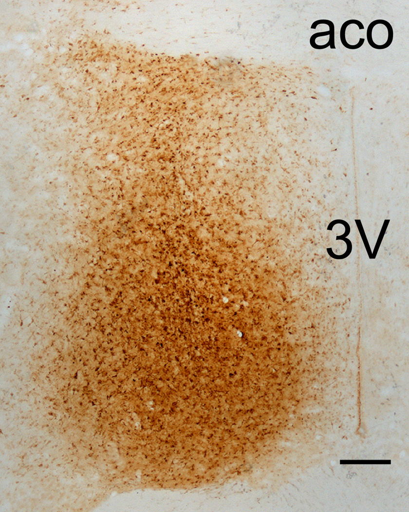
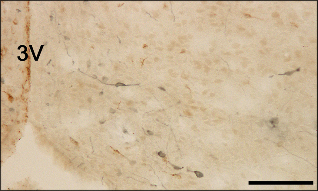
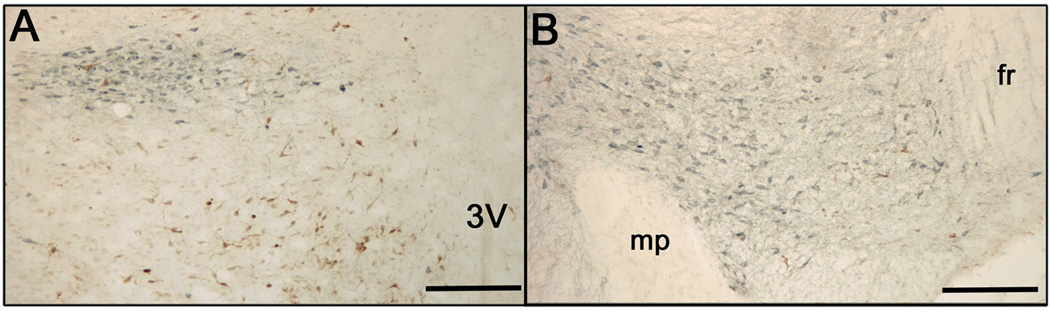
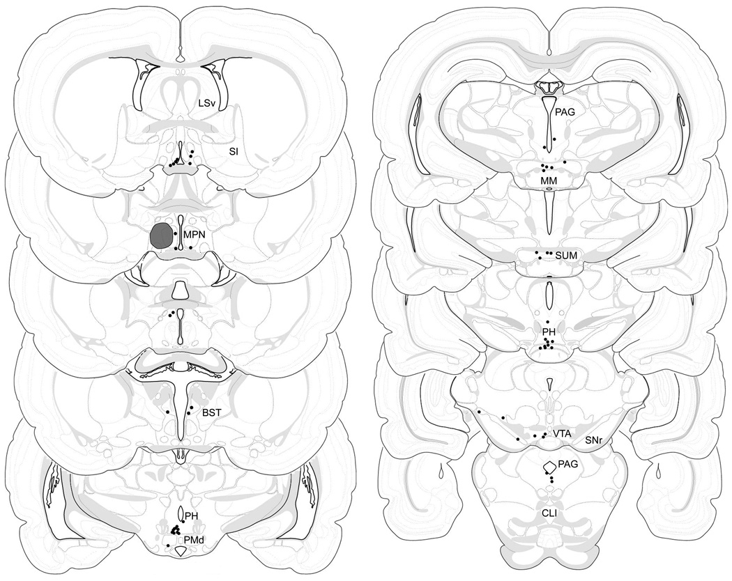
Dopamine receptor activity in the rodent medial preoptic area (mPOA) is crucial for the display of maternal behaviors, as well as numerous other physiological and behavioral functions. However, the origin of dopaminergic input to the mPOA has not been identified through neuroanatomical tracing. To accomplish this, the retrograde tracer Fluorogold was iontophoretically applied to the mPOA of postpartum laboratory rats, and dual-label immunocytochemistry for Fluorogold and tyrosine hydroxylase later performed to identify dopaminergic cells of the forebrain and midbrain projecting to the mPOA. Results indicate that the number of dopaminergic cells projecting to the mPOA is moderate ( approximately 90 cells to one hemisphere), and that these cells have an unexpectedly wide distribution. Even so, more than half of the dual-labeled cells were found in either what has been considered extensions of the A10 dopamine group (particularly the ventrocaudal posterior hypothalamus and adjacent medial supramammillary nucleus), or in the A10 group of the ventral tegmental area. The rostral hypothalamus and surrounding region also contained numerous dual-labeled cells, with the greatest number found within the mPOA itself (including in the anteroventral preoptic area and preoptic periventricular nucleus). Notably, dual-labeled cells were rare in the zona incerta (A13), a site previously suggested to provide dopaminergic input to the mPOA. This study is the first to use anatomical tracing to detail the dopaminergic projections to the mPOA in the laboratory rat, and indicates that much of this projection originates more caudally than previously suggested.
Figures


Similar articles
-
Immunocytochemical investigation of nuclear progestin receptor expression within dopaminergic neurones of the female rat brain.J Neuroendocrinol. 2004 Jun;16(6):534-43. doi: 10.1111/j.1365-2826.2004.01198.x. J Neuroendocrinol. 2004. PMID: 15189328
-
Sex-induced fos in the medial preoptic area: projections to the midbrain.Neuroreport. 2001 Oct 8;12(14):3065-8. doi: 10.1097/00001756-200110080-00016. Neuroreport. 2001. PMID: 11568637
-
Projection sites of medial preoptic area and ventral bed nucleus of the stria terminalis neurons that express Fos during maternal behavior in female rats.J Neuroendocrinol. 1997 May;9(5):369-84. doi: 10.1046/j.1365-2826.1997.t01-1-00597.x. J Neuroendocrinol. 1997. PMID: 9181491
-
Getting his act together: roles of glutamate, nitric oxide, and dopamine in the medial preoptic area.Brain Res. 2006 Dec 18;1126(1):66-75. doi: 10.1016/j.brainres.2006.08.031. Epub 2006 Sep 11. Brain Res. 2006. PMID: 16963001 Review.
-
The dopaminergic mesencephalic projections to the hippocampal formation in the rat.Prog Neuropsychopharmacol Biol Psychiatry. 1997 Jan;21(1):1-22. doi: 10.1016/s0278-5846(96)00157-1. Prog Neuropsychopharmacol Biol Psychiatry. 1997. PMID: 9075256 Review.
Cited by
-
Tyrosine Hydroxylase Knockdown at the Hypothalamic Supramammillary Nucleus Area Induces Obesity and Glucose Intolerance.Neuroendocrinology. 2024;114(5):483-510. doi: 10.1159/000535944. Epub 2023 Dec 21. Neuroendocrinology. 2024. PMID: 38128505 Free PMC article.
-
Plasticity of paternity: Effects of fatherhood on synaptic, intrinsic and morphological characteristics of neurons in the medial preoptic area of male California mice.Behav Brain Res. 2019 Jun 3;365:89-102. doi: 10.1016/j.bbr.2019.02.029. Epub 2019 Feb 22. Behav Brain Res. 2019. PMID: 30802534 Free PMC article.
-
Decreased mesolimbic dopaminergic signaling underlies the waning of maternal caregiving across the postpartum period in rats.Psychopharmacology (Berl). 2020 Apr;237(4):1107-1119. doi: 10.1007/s00213-019-05441-7. Epub 2020 Jan 11. Psychopharmacology (Berl). 2020. PMID: 31927604
-
Functions of medial hypothalamic and mesolimbic dopamine circuitries in aggression.Curr Opin Behav Sci. 2018 Dec;24:104-112. doi: 10.1016/j.cobeha.2018.06.011. Epub 2018 Jul 3. Curr Opin Behav Sci. 2018. PMID: 30746430 Free PMC article.
-
Brain neuronal activation induced by flibanserin treatment in female rats.Psychopharmacology (Berl). 2013 Dec;230(4):639-52. doi: 10.1007/s00213-013-3194-6. Epub 2013 Jul 16. Psychopharmacology (Berl). 2013. PMID: 23857113
References
-
- Abrahamson EE, Moore RY. The posterior hypothalamic area: chemoarchitecture and afferent connections. Brain Res. 2001;889:1–22. - PubMed
-
- Aranda L, Santín LJ, Begega A, Aquirre JA, Arias JL. Supramammillary and adjacent nuclei lesions impair spatial working memory and induce anxiolitic-like behavior. Behavioural Brain Res. 2006;167:156–164. - PubMed
-
- Bakowska JC, Morrell JI. Atlas of the neurons that express mRNA for the long form of the prolactin receptor in the forebrain of the female rat. J Comp Neurol. 1997;386:161–177. - PubMed
-
- Balthazart J, Absil P. Identification of catecholaminergic inputs to and outputs from aromatase-containing brain areas of the Japanese quail by tract tracing combined with tyrosine hydroxylase immunocytochemistry. J Comp Neurol. 1997;382(3):401–428. - PubMed
Publication types
MeSH terms
Substances
Grants and funding
LinkOut - more resources
Full Text Sources
Medical
